Feeds:
Posts
Comments

Posts Tagged ‘myocardial infarction’

Reporter: Aviva Lev-Ari, PhD, RN

SHOCK II: IABP Use Questioned

When myocardial infarction (MI) is complicated by cardiogenic shock, use of intraaortic balloon counterpulsation (IABP) did not reduce mortality among patients scheduled for revascularization–a finding that calls into question current guidelines for treating cardiogenic shock in this population.

 SHOCK II: IABP Use Questioned

By Peggy Peck, Editor-in-Chief, MedPage Today
Published: August 27, 2012

Reviewed by Robert Jasmer, MD; Associate Clinical Professor of Medicine, University of California, San Francisco

Action Points


Action Points


  • When myocardial infarction (MI) is complicated by cardiogenic shock, use of intraaortic balloon counterpulsation (IABP) did not reduce mortality among patients scheduled for revascularization, a finding that calls into question current guidelines for treating cardiogenic shock in this population.
  • Note that authors of an editorial wrote that data from IABP-SHOCK II, and a number of recent meta-analyses, “do not support the routine use of IABP in patients with acute myocardial infarction complicated by cardiogenic shock, and the level I guideline recommendation is now strongly challenged.”

When myocardial infarction (MI) is complicated by cardiogenic shock, use of intraaortic balloon counterpulsation (IABP) did not reduce mortality among patients scheduled for revascularization — a finding that calls into question current guidelines for treating cardiogenic shock in this population.

At 30 days, only 60% of the patients treated with IABP were still alive, a mortality that was no different from the rate in the control group (39.7% versus 41.3% relative risk 0.96, 95% CI 0.79-1.17, P=0.69), according to findings from the IABP-SHOCK II trial reported online by the New England Journal of Medicine.

The findings were simultaneously reported as a Hot Line presentation at the European Society of Cardiology meeting in Munich.

Holger Thiele, MD, from University of Leipzig-Heart Center, Leipzig, Germany, and colleagues recruited 600 patients for a randomized, prospective, open-label, multicenter trial and assigned 300 to IABP.

 While there was no mortality benefit for IABP, there also was no apparent harm:

Rates of major bleeding: 3.3% versus 4.4% in controls (P=0.53)
Rates of sepsis: 15.7% versus 20.5% (P=0.15)
Rates of stroke: 0.7% versus 1.7% (P=0.28)
Rates of peripheral ischemic complications: 4.3% versus 3.4% (P=0.53)
Current American College of Cardiology/American Heart Association guidelines for treatment of STEMI support use of IABP in this population, but that recommendation comes from a trial “that did not address this question, it really looked at the question of revascularization of these patients,” said Mariell Jessup, MD, of the University of Pennsylvania Perelman School of Medicine in Philadelphia.

The earlier trial, called SHOCK (Should We Emergently Revascularize Occluded Coronaries for Cardiogenic Shock), “was really about bypass surgery in these patients,” she said.

Jessup, who is president-elect of the American Heart Association, told MedPage Today, that the results of the IABP-SHOCK II trial “may very well be the most important finding to be reported at this meeting.”

She said the current Class I recommendation is for use of IABP when the patient is not stable. “It is possible that this [IABP-SHOCK II] could completely change this guideline.”

Jessup noted that use of IABP has become the norm for treating these patients and she suggested that physicians will find it hard to resist using IABP because “it is hard for physicians to not do something for these patients.”

Christopher O’Connor, MD, and Joseph Rogers, MD, echoed Jessup’s view in an NEJM editorial. They are from Duke University in Durham, N.C.

Under the title, “Evidence for Overturning the Guidelines in Cardiogenic Shock” O’Connor and Rogers wrote that data from IABP-SHOCK II, and a number of recent meta-analyses, “do not support the routine use of IABP in patients with acute myocardial infarction complicated by cardiogenic shock, and the level I guideline recommendation is now strongly challenged. Members of guideline committees and clinicians should take note of another example of a recommendation that is based on insufficient data.”

Patients in the IAPB-SHOCK trial were recruited from June 16, 2009 through March 3, 2012 at 37 centers in Germany.

Thirty of the 299 patients assigned to the control group did eventually undergo IABP, usually within 24 hours of randomization, and 26 of those patients were classified as protocol violations. Likewise, 13 patients assigned to IABP did not undergo the treatment, with death being the most common reason.

The authors noted a number of limitations, starting with lack of blinding, and the failure to obtain “hemodynamic measurements or assess laboratory inflammatory markers other than blood pressure, heart rate, and C-reactive protein levels.”

Also, the mortality rate in both arms was lower than anticipated — 40% versus a range of 42% to 48% in other studies — suggesting that most patients in this study had mild or moderate cardiogenic shock, which could limit the generalizability of these results, they cautioned.

“Finally, we do no yet have any information about longer-term outcomes. Since a balloon intraaortic counterpulsation was used for a median of only 3 days, it seems unlikely that any beneficial effect will become evident later than 30 days,” they wrote.

The trial was supported by the German Research Foundation, the German Heart Foundation, the German Cardiac Society, Arbeitsgemeinschaft Leitende Kardiologische Krankenhausärzte, the University of Leipzig-Heart Center, Marquet Cardiopulmonary, and Teleflex Medical.

Thiele disclosed financial support from Eli Lilly, Terumo, AstraZeneca, Boehringer Ingelheim, Daiichi Sankyo, Eli Lilly, and the Medicines Company.

Primary source: New England Journal of Medicine

Source reference:

Thiele H, et al. “Intraaortic balloon support for myocardial infarction with cardiogenic shock” N Engl J Med 2012; DOI: 10.1056/NEJMoal208410.

Additional source: New England Journal of Medicine

Source reference:
O’Connor CM, Rogers JG. “Evidence for overturning the guidelines in cardiogenic shock” N Engl J Med 2012; DOI: 10.1056/NEJMel209601.

http://www.medpagetoday.com/Cardiology/PCI/34387

Read Full Post »

New Drug-Eluting Stent Works Well in STEMI

Reporter: Aviva Lev-Ari, PhD, RN

UPDATED on 8/8/2013

Meta-analysis makes case for drug-eluting stents in STEMI

AUGUST 7, 2013 

New York, NY – Newer-generation drug-eluting stents, particularly the everolimus-eluting stent (Xience V, Abbot; Promus, Boston Scientific), significantly reduce the risk of target vessel revascularization (TVR) in patients with ST-segment-elevation MI (STEMI) without increasing the risk of adverse safety outcomes, including rates of stent thrombosis, when compared with bare-metal stents [1].

These are the principal findings of a new meta-analysis of 28 randomized, controlled clinical trials involving more than 34 000 patient-years of follow-up.

Published online August 6, 2013 in Circulation: Cardiovascular Interventions, the analysis showed that compared with the sirolimus-eluting stent (Cypher, Cordis), the paclitaxel-eluting stent (Taxus, Boston Scientific), and bare-metal stents, the use of an everolimus-eluting stent reduced the relative risk of stent thrombosis 62%, 61%, and 58%, respectively.

“I would make a strong argument to say that the current guidelines should change,” lead investigator Dr Sripal Bangalore (New York University School of Medicine) told heartwire. “The reduction in TVR is not surprising. We know that drug-eluting stents compared with bare-metal stents reduce TVR, but the biggest thing we were able show was that stent thrombosis is also reduced when compared with a bare-metal stent, as well as compared with first-generation drug-eluting stents.”

The current American College of Cardiology/American Heart Association (ACC/AHA) and European Society of Cardiology (ESC) clinical guidelines state that drug-eluting-stent implantation is a class IIa indication in STEMI patients. The recommendations are based on concerns about an increased risk of stent thrombosis with the drug-eluting stents compared with their bare-metal counterparts. Bangalore said concerns have also been raised about the risk of stent thrombosis beyond one year with the first-generation drug-eluting stents, a time point when dual antiplatelet therapy is stopped.

The newer-generation drug-eluting stents, however, have been shown in various studies to be as safe asbare-metal stents in the STEMI setting. For this reason, they conducted a meta-analysis of randomized, controlled trials comparing the sirolimus-, paclitaxel-, everolimus-, and zotarolimus-eluting stents against each other and against bare-metal stents.

When compared with bare-metal stents, the sirolimus-, paclitaxel-, and everolimus-eluting stent reduced the relative risk of TVR by 53%, 31%, and 57%, respectively. The sirolimus-eluting stent was significantly more effective than the paclitaxel-eluting stent at reducing TVR, as was the everolimus-eluting stent. Overall, there was a 67% probability that the Endeavor Resolute zotarolimus-eluting stent (Medtronic) had the lowest risk of TVR, although the data are based on just one trial with 281 patients, note the investigators.

Median rate of efficacy and definite/probable stent thrombosis

Stent type TVR rate (per 1000 patient-years of follow-up) Definite/probable stent thrombosis (per 1000 patient-years of follow-up)
Bare metal 64.00 16.60
Sirolimus 28.93 15.75
Paclitaxel 44.38 18.46
Everolimus 26.55 6.54
Zotarolimus 59.01 11.41
Zotarolimus (Resolute) 14.76 NA*


*For stent thrombosis, there were no available data on the Resolute stent

When compared with bare-metal stents, the everolimus-eluting stent reduced the risk of any stent thrombosis by 58%. The Xience stent was also associated with a statistically significant 62% and 61% reduction in the risk of stent thrombosis compared with the first-generation Cypher and Taxus stents.

Bangalore said that a previous patient-level analysis conducted by Dr Giuseppe De Luca (Ospedale Maggiore della Carità, Novara, Italy), reported by heartwire at that time, showed there was a significant 50% increase in the risk of late (more than one year) reinfarction with drug-eluting stents and an almost doubling of very late stent thrombosis with first-generation stents. In this newest meta-analysis, however, the researchers did not observe a similarly increased risk of very late stent thrombosis with the everolimus-eluting stent.

“Based on the totality of data, I would say that it’s time the guidelines make drug-eluting stents and especially the everolimus-eluting stent a class I indication in STEMI patients who can take dual antiplatelet therapy,” said Bangalore.

Source

  1. Bangalore S, Amoroso N, Fusaro M, Kumar S, Feit F. Outcomes with various drug-eluting or bare-metal stents in patients with ST-segment elevation myocardial infarction. Circ Cardiovasc Interv 2013; DOI:10.1161/CIRCINTERVENTIONS.113.000415. Available at: http://circinterventions.ahajournals.org.


New Drug-Eluting Stent Works Well in STEMI

By Michael Smith, North American Correspondent, MedPage Today

Published: August 21, 2012

Reviewed by Robert Jasmer, MD; Associate Clinical Professor of Medicine, University of California, San Francisco and Dorothy Caputo, MA, BSN, RN, Nurse Planner

 Watch Video

 A new-generation biodegradable drug-eluting stent had a lower rate of major cardiac events than similar bare-metal devices, researchers reported.

In a randomized trial, patients with ST-segment elevation myocardial infarction (STEMI) needed fewer revascularization procedures and had a lower risk of a new heart attack in the target blood vessel, according to Stephan Windecker, MD, of Bern University Hospital in Bern, Switzerland, and colleagues.

On the other hand, rates of cardiac death were not significantly different, Windecker and colleagues reported in the Aug. 22/29 issue of the Journal of the American Medical Association.

Drug-eluting stents have been shown to reduce the need for repeat revascularization, compared with bare-metal stents, but at the cost of delayed healing, chronic inflammation, and late stent thrombosis, the researchers noted.

The long-term effects result from the persistence of the polymer, Windecker and colleagues noted — something that might be avoided by using a biodegradable polymer.

The biodegradable BioMatrix Flex stent, which delivers the immunosuppressant drug biolimus, was non-inferior in a 4-year trial to the sirolimus-eluting Cypher stent, which does not break down over time.

But it had not been tested against bare-metal stents. To help fill the gap, Windecker and colleagues studied 1-year outcomes in 1,161 STEMI patients randomly assigned to get either the biolimus-eluting biodegradable stent or a similar bare-metal device.

The primary endpoint of the trial was the 1-year rate of major adverse cardiac events — a composite of cardiac death, target vessel-related re-infarction, and ischemia-driven target-lesion revascularization.

Windecker and colleagues found that 24 patients (4.3%) with biodegradable stents had a major adverse cardiac event at 1 year, compared with 49 (8.7%) who were given the bare-metal devices (HR 0.49, 95% CI 0.30 to 0.80, P=0.004).

The difference was driven by a lower risk of two of the elements of the combined endpoint: target vessel-related reinfarction and ischemia-driven target-lesion revascularization. Specifically:

  • Three patients getting the biodegradable stent (0.5%) had a re-infarction related to the target vessel, compared with 15 (2.7%) of those with bare-metal devices (HR 0.20, 95% CI 0.06 to 0.69, P=0.01).
  • Nine patients (1.6%) with biodegradable stents and 32 (5.7%) with bare-metal devices needed target-lesion revascularization (HR 0.28, 95% CI 0.13 to 0.59, P<0.001).
  • Rates of cardiac death were numerically lower, but not significantly so, in the biodegradable stent patients — 16 deaths, or 2.9%, versus 20, or 3.5%.

Definite stent thrombosis occurred in five patients treated with the drug-eluting stents and 12 patients with bare-metal stents, but the difference did not reach significance.

The findings should be “reassuring” to both doctors and patients, Windecker said in a video released by the journal.

The study is “a well-done trial with convincing results regarding its primary end point,” commented Salvatore Cassese, MD, and Adnan Kastrati, MD, both of the Technische Universitat in Munich, Germany.

But, in an accompanying editorial, they argued that it still may not settle the question of long-term complications.

Despite “positive signals,” they wrote, the study has “neither the required sample size nor the sufficient length of follow-up to provide the definitive answer about the long-term safety” of the new biodegradable drug-eluting stents.

The researchers cautioned that the biodegradable drug-eluting stent is not yet approved in the U.S., although European authorities have given it the nod.

They also noted that the study, while demonstrating superiority on the overall endpoint, did not have sufficient statistical power to address the individual components definitively.

The study had support from the Swiss National Science Foundation and Biosensors Europe SA. Windecker reported financial links through his institution with Abbott, Boston Scientific, Biosensors, Biotronik, Cordis, Medtronic, and St. Jude Medical.

The editorial authors reported support from the European Commission. Kastrati reported holding a patent related to polymer-free sirolimus and probucol coating, as well as financial links with Abbott, Biosensors, Cordis, and Medtronic.

From the American Heart Association:

Related Articles in Heart.org

Related links

Read Full Post »

Scale‑Free Diagnosis of AMI from Clinical Laboratory Values

Author: Larry H. Bernstein, MD, FCAP


Scale‑Free Diagnosis of AMI from Clinical Laboratory Values

William P. Fisher, Jr., Larry H. Bernstein, Thomas A Naegele, Arden

Forrey, Asadullah Qamar, Joseph Babb, Eugene W. Rypka, Donna Yasick

Objective. Clinicians are often challenged with interpreting myriads of laboratory test results with few resources for knowing which values are most relevant, when any given value indicates a need for action, or how urgent the need for action is. The arrival of the electronic health record creates a context in which computational resources for meeting these challenges will be readily available. The purpose of this study was to evaluate the feasibility of employing probabilistic conjoint (Rasch) measurement models for creating the needed scale‑free standard measures and data quality standards.

Methods. Pathology data from 144 clients suspected of suffering myocardial infarctions were obtained. Thirty indicators were converted from their original values to ratings indicating a worsening of condition. These conversions took advantage of the fact that serial measurement of creatine kinase (CK; EC 2.7.3.2) isoenzyme MB (CK‑MB) and lactic dehydrogenase (LD; EC 1.1.1.27) isoenzyme 1 (LD‑1) in serum have characteristic evolutions in acute myocardial infarction (AMI). CK‑MB concentration begins to rise within 4 to 8 hours, peaks at 12 to 24 hours, and returns to normal within 48 to 72 hours. LD‑1 becomes elevated as early as 8 to 24 hours after infarction, and reaches a peak in 48 to 72 hours. However, the ratio of serum activity of LD‑1/total LD may be more definitive than LD‑1 activity itself. While these are most important in ECG negative AMI, they are not by themselves a “gold standard” for diagnosis.

The additional information and functionality required for such standards, including probabilistic estimates of scale parameters whose values do not depend on the calibrating sample and the capacity to deal with missing data, were sought by fitting the data to a Rasch partial credit model. This model estimates separate rating step values for each group of items sharing a common rating structure, en route to testing the hypothesis that the items work together to delineate a unidimensional measurement continuum defined by the repetition of a single unit quantity.

Results. Twenty of the 30 items were identified as delineating a unidimensional continuum.  Client measurement reliability was 0.90, and item calibration reliability was 0.96. Overall model fit is indicated by the client information‑ weighted mean square fit (infit) statistic (mean = .94, SD = .34) and  outlier‑ sensitive mean square fit (outfit) statistic (mean = 1.02, SD = .72), and the item infit (mean = .99, SD = .41) and outfit (mean = 1.04, SD = .72). The data‑to‑ model global fit is also indicated by the chi‑square of 3094.5, with 164 maximum independent parameters, 2766 maximum degrees of  freedom, and a probability (statistical significance) of less than .01 that this ora greater chi‑square would be observed with perfect data‑model fit.

Discussion. The analysis identified the 20 values most relevant to the diagnosis of AMI; these data may also support the construction of a unidimensional measure of AMI severity. If the construct supports both diagnostic and severity inferences, then the clinical action needed and its urgency will be indicated by the client’s measure. Similar analyses of data from other diagnostic groups will determine the extent to which lab value item relevance and hierarchies vary across diagnoses; such variation will be crucial to determining computer‑based decision support algorithms, which will match individual clients’ data with specific diagnostic profiles. Further analyses will also demonstrate the extent to which diagnosis is affected by missing data.


Read Full Post »

Human embryonic pluripotent stem cells and healing post-myocardial infarction

Curator: Larry H. Bernstein, MD, FCAP
I present a followup based on several recent posts related to the promise of using induced human pluripotent stem cells for repair of ischemia damaged myocardium postinfarct and related effect of heart failure (HF).  There has been a change in the concept of cardiovascular risk related to the emergent knowledge of the biology underlying oxidative stress.  The more recent discovery of the relationship between ongoing inflammation and clinical outcomes has led to a variety of blood-based assays which may impart additional knowledge about an individual’s propensity for future cardiovascular events (1). Vascular injury and repair are significantly mediated by circulating endothelial progenitor cells (1).  Circulating progenitor endothelial cells are defined by co-expression of the markers CD34, CD309 (VEGFR-2/KDR) which are measured by pre-enrichment flow cytometry with specific identification of cell markers (CD34, CD133) and endothelial cell antigens (KDR/VEGFR-2, CD31) (2), used in the assessment of various diseases and physiological states.  Improvements in flow cytometry include the Attune® cytometer, which enables the collection of more than 4,000,000 live white blood cell (WBC) events in just 35 minutes (3). Using these methods of analyses, it became evident that circulating endothelial progenitor cells have angiogenic potential.

Activators and inhibitors have been tested for their ability to modulate angiogenesis in early phase clinical trials, and in the case of anti-Flk1 antibodies clinical utility has been demonstrated for anti-tumor strategies (4). Extending this concept further, we pose that just as the progenitor role invoked for angiogenesis, transcriptional networks and interactions are involved in the morphogenesis of the developing vertebrate heart. The identities of crucial regulators involved in defined events in cardio-genesis are being uncovered at a rapid rate. Tissue development and regeneration involve tightly coordinated and integrated processes: selective proliferation of resident stem and precursor cells, differentiation into target somatic cell type, and spatial morphological organization. (4, 5, 6). However, our ability to cross the divide between knowledge and change has not been easy, as reported by Aviva Lev-Ari (7).  In a two-day-old mouse, a heart attack causes active stem cells to grow new heart cells; a few months later, the heart is mostly repaired. But in an adult mouse, recovery from such an attack leads to classic after-effects: scar tissue, permanent loss of function and life-threatening arrhythmias (7, 8).

Myocardial cell replacement therapies are hampered by a paucity of sources for human cardiomyocytes and by the expected immune rejection of allogeneic cell grafts. The success using dermal fibroblasts from HF patients reprogrammed by retroviral delivery of Oct4, Sox2, and Klf4 or by using an excisable polycistronic lentiviral vector resulted in HF-hiPSCs induced to differentiate into cardiomyocytes (HF-hiPSC-CMs)(9). Multi-electrode array recordings revealed adequate responses to stimulation.  Further study with in vivo transplantation in the rat heart revealed the ability of the HF-hiPSC-CMs to engraft, survive, and structurally integrate with host cardiomyocytes and within 48 hours the tissues were beating together. Human-induced pluripotent stem cells thus can be established from patients with advanced heart failure and coaxed to differentiate into cardiomyocytes, which can integrate with host cardiac tissue (10).  The success of the approach rests on modifying the myocardial electro-physiological substrate using cell grafts genetically engineered to express specific ionic channels (11). The expressed potassium channels alter the local myocardial electrophysiological properties by reducing cardiac automaticity and prolonging refractoriness.  The key feature involves reprogramming a patient’s own skin cells by delivering three genes followed by a small molecule called valproic acid to the cell nucleus (12).

An alternative approach avoiding the caveats of limited graft survival, is to stimulate a resident source, restricted homing to the site of injury and host immune rejection (13). Thymosin β4 restores vascular potential to adult epicardial-derived progenitor cells with injury.  Specifically, it activates adult progenitors to re-express a key embryonic epicardial gene, Wilm’s tumour 1 (Wt1).  It was inferred that embryonic reprogramming would mobilize this cell population and differentiation would give rise to de novo cardiomyocytes. Delivery of Tβ4, in conjunction with GMT (an acronym for three genes that normally guide embryonic heart development), into the damaged region resulted in reduction of scar area and improvement in cardiac function compared to GMT or Tβ4 alone. Thymosin-beta4 facilitates cardiac repair after infarction by promoting cell migration and myocyte survival. Additionally, the tetra peptide Ac-SDKP was reported to reduce left ventricular fibrosis in hypertensive rats, reverse fibrosis and inflammation in rats with MI, and stimulate both in vitro and in vivo angiogenesis. Effects of Ac-SDKP, such as the enhancement of angiogenesis and the decrease in inflammation and collagenase activity, are similar to those described for thymosin-beta4. However, there are conflicting reports (14-18).

There are other studies that show promise.  There has been the first infusion of stem cells into the coronary artery (19). This result was at least as effective as intramyocardial injection in limiting LV remodeling and improving both regional and global LV function. The intracoronary route appears to be superior in terms of uniformity of cell distribution, myocyte regeneration, and amount of viable tissue in the risk region. Another finds that down regulation of leukocyte HIF-1? Expression resulted in decreased recruitment of WBC to the sites of inflammation and improvement in cardiac function following MI (20).  Irradiated 6-to 8-week-old C57/BL6J mice received 50 000 GFP HIF-1? or scramble siRNA transfected hematopoietic stem cells. Down regulation of HIF-1? suppressed WBC cytokine receptors CCR1,-2, and-4, which are necessary for WBC mobilization and recruitment to inflammatory cytokines following MI.  There also have been cited limitations to success in older patients (21). The findings suggest that coronary artery disease and cardiac remodeling in chronic ischemia has a significant negative correlation between the age of the patient and the number of migrated ckit-positive cells.

Lymphocytes infiltrate and react with ischemia damaged heart tissue, which can impair proper tissue healing.  In a study with isoproterenol induced myocardial necrosis TNF-α, IFN-γ and CCL-5, but not FOXP3 + expression, was increased in draining lymph nodes, indicating that the observed lymphocyte population that proliferated in response to cardiac components presented a pro-inflammatory and pro-fibrotic profile.  The group was rendered tolerant by myocardial gavage and expressed cardiac FOXP3 + earlier than did the control group, and showed a milder inflammatory infiltrate, lower MMP-9 expression, less collagen deposition, and improved cardiac performance when compared to animals that received only isoproterenol administration (22).  Patients with acute myocardial infarction show high circulating levels of neuropeptide Substance P (SP) and NK1-positive cells that co express Progenitor Cell (PC) antigen, such as CD34, KDR, and CXCR4. Moreover, NK1-expressing PC is abundant in infarcted hearts, highlighting the role of SP in reparative neovascularization (23). Do CD4 + T cells become activated and influence wound healing after experimental MI?   To study the role of CD4 + T cells in wound healing and remodeling, CD4 + T-cell- deficient mice (CD4 knockout [KO], MHCII) and T-cell receptor-transgenic OT-II. Within the infarcted myocardium, CD4 KO mice displayed higher total numbers of leukocytes and proinflammatory monocytes (18.3±3.0 104/mg WT versus 75.7±17.0 10 4/mg CD4 KO, P<0.05), and MHCII and OT-II mice displayed significantly greater mortality. Collagen matrix formation in the infarct zone was severely disturbed in CD4 KO and MHCII mice, as well as in OT-II mice (24).

Thus, it appears that CD4T cells become activated after MI and facilitate wound healing of the myocardium. Inflammation and immune responses are integral components in he healing process after myocardial infarction. Importantly, dendritic cell (DC) infiltration occurs in the infarcted heart.  In concert with the previous two studies, DC-ablated infarcts had enhanced monocyte/ macrophage recruitment. Among these cells, marked infiltration of proinflammatory Ly6C high monocytes and F4/80 + CD206 – M1 macrophages and, conversely, impaired recruitment of anti-inflammatory Ly6C low monocytes and F4/80 + CD206 + M2 macrophages in the infarcted myocardium were identified in the DC-ablated group compared with the control group (25). Thus, the DC is a potent immunoprotective regulator during the post-infarction healing process via its control of monocyte/macrophage homeostasis.  Despite the recent successes, there are a number of interlocking and possibly explanatory processes to control in the mix.

What about medical therapies?  Here too there is a factor in engaging eNOS or iNOS activity as detailed in the presentation by Aviva Lev-Ari (26).  60–70% of major cardiovascular events cannot be prevented with current approaches focused on LDL, such as statin therapy, and low HDL levels are particularly common in males with early-onset atherosclerosis.  She makes the point that there is compelling evidence that HDL is not solely a marker of lower risk of cardiovascular disease but instead is a mediator of vascular health.

Aviva Lev-Ari examines the phytoestrogen, Genistein, and other drugs. Genistein acutely stimulates Nitric Oxide synthesis in vascular Endothelial cells by a cyclic adenosine 5′-monophosphate-dependent mechanism (Liu et al., 2004). The intracellular signaling pathways for activation of eNOS by genistein were shown independent of PI3K/Akt or ERK/MAPK but depended on the cAMP/PKA cascade. In addition, the genistein action on eNOS was not inhibited by an ER antagonist and was unrelated to tyrosine kinase inhibition. Furthermore, genistein has antiatherogenic effects and inhibits proliferation of vascular endothelial and smooth muscle cells, and in vitro studies suggest a protective role of genistein in the vasculature.  In Liu et al., (2004) study, genistein acted directly on BAECs and HUVECs to activate eNOS and NO production through nongenomic mechanisms in whole vascular endothelial cells.  In addition, 5-hydroxytryptamine evokes endothelial nitric oxide synthase activation.  In this example, eNOS co-localizes with PECAM-1, but not with VE-cadherin and plakoglobin at the intercellular junctions of the endothelium.

Finally, activation of endothelial nitric oxide synthase (eNOS) resulted in the production of nitric oxide (NO) that mediates the vasorelaxing properties of endothelial cells.  The responses were effectively blocked by a 5-HT1B receptor antagonist, a 5-HT1B/5-HT2 receptor antagonist, and eNOS selective antagonists, L-Nomega -monomethyl-L-arginine (L-NMMA) and L-N omega-iminoethyl-L-ornithine (L-NIO). This lends credence to a 5-HT1B receptor/eNOS pathway, accounting in part for the activation of eNOS by 5-HT.  Finally, a third-generation ß-blocker augments vascular Nitric Oxide release. Nebivolol increases vascular NO productionby causing a rise in endothelial free [Ca2+]i and endothelial NO synthase–dependent NO production. It is a ß1-selective adrenergic receptor antagonist with proposed nitric oxide (NO)–mediated vasodilating properties. Nevertheless, it appears that not nebivolol, but its metabolites augment NO production (Broeders et al., 2000).  These findings reveal new insights into interaction with eNOS in vascular therapy: [1] new indications for TDZs as stimulators of eNOS; [2] new indications for beta blockers as NO stimulant. Nebivolol is a vasodilator, thus functions as an antihypertensive.

References:

1.  Saha S. Innovations in Bio-instrumentation for Measurement of Circulating Progenitor Endothelial Cells in Human Blood.  Pharma Intell. July 8, 2012. http://pharmaceuticalintelligence.com/2012/07/08/innovations-in-bio-instrumentation-for-measurement-of-circulating-progenitor-endothelial-cells-in-human-blood/

(http://www.ncbi.nlm.nih.gov/pubmed/19124422)

2.  Ibid (http://www.ncbi.nlm.nih.gov/pubmed/20381496).

3.  Ibid (http://zh.invitrogen.com/etc/medialib/files/Cell-Analysis/PDFs.Par.54318.File.tmp/CO24210-Human-CEC_cancer.pdf)

4. Saha S. Endothelial Differentiation and Morphogenesis of Cardiac Precursors. Pharma Intelligence. July 17, 2012.

5. Ibid (http://circres.ahajournals.org/content/90/5/509.full).

6. Ibid (http://www.ncbi.nlm.nih.gov/pubmed/22669846).

7.  Aviva-Lev-Ari.  Stem cells create new heart cells in baby mice, but not in adults, study shows.Aug 3, 2012. Pharma Intelligence.

8.  Krishna Ramanujan http://www.news.cornell.edu/stories/July12/HeartStemCells.html

9. Saha S. Human Embryonic-Derived Cardiac Progenitor Cells for Myocardial Repair.  Pharma Intelligence. Aug 1, 2012.

10.  Zwi-Dantsis L, Huber I, Habib M, Winterstern A, (..), Gepstein L. Derivation and cardiomyocyte differentiation of induced pluripotent stem cells from heart failure patients. Eur Heart J. 2012 May 22. [Epub ahead of print]  (VBL RX, Inc. Tel Aviv, http://www.vblrx.com).

11.  Yankelson L, Feld Y, Bressler-Stramer T, Itzhaki I,(..), Gepstein L. Cell therapy for modification of the myocardial electrophysiological substrate. Circulation. 2008 Feb 12; 117(6):720-31. Epub 2008 Jan 22.

12.  Huber I, Itzhaki I, Caspi O, Arbel G, (..), Gepstein L. Identification and selection of cardiomyocytes during human embryonic stem cell differentiation. FASEB J. 2007 Aug; 21(10):2551-63. Epub 2007 Apr 13.

13.  Aviva Lev-Ari. Resident-cell-based Therapy in Human Ischaemic Heart Disease: Evolution in the PROMISE of Thymosin beta4 for Cardiac Repair. Pharma Intelligence. April 30, 2012.

14.  Ibid. Shrivastava S, Srivastava D, Olson EN, DiMaio JM, Bock-Marquette I.

Ann N Y Acad Sci. 2010 Apr; 1194:87-96.

15.  Ibid.  Smart N, Risebro CA, Clark JE, Ehler E, (..), Riley PR, Thymosin beta4 facilitates epicardial neovascularization of the injured adult heart.  Ann N Y Acad Sci. 2010 Apr;1194:97-104

16.   Ibid. Smart N, Bollini S, Dubé KN, Vieira JM, (..) Riley PR. Nature. 2011 Jun 8; 474(7353):640-4 

17.   Ibid. Zhou B, Honor LB, Ma Q, Oh JH, (..), Pu WT. Thymosin beta 4 treatment after myocardial infarction does not reprogram epicardial cells into cardiomyocytes.  J Mol Cell Cardiol. 2012 Jan; 52(1):43-7. Epub 2011 Aug 26.

18.   Ibid. Scientists Report that Process of Converting Non-Beating Heart Cells into Functional, Beating Heart Cells is Enhanced Using Thymosin Beta 4 in Conjunction with Gene Therapy.  Regenerx Biopharmaceuticals, Inc. Nature. Apr. 18, 2012

19.  Li, Q., Guo, Y., Ou, Q., Chen, N., (…), Bolli, R. Intracoronary administration of cardiac stem cells in mice: A new, improved technique for cell therapy in murine models.  Basic Research in Cardiology 2011; 106 (5), pp. 849-864.

20. Dong, F., Khalil, M.,Kiedrowski, M.,O’Connor, C.,(..) ,Penn, M.S. Critical role for leukocyte hypoxia inducible factor-1α expression in post-myocardial infarction left ventricular remodeling.  Circulation Research   2010; 106 (3) , pp. 601-610

21. Aghila Rani, K.G.,Jayakumar, K.,Sarma, P.S., Kartha, C.C. Clinical determinants of ckit-positive cardiac cell yield in coronary disease. Asian Cardiovascular and Thoracic Annals 2009; 17 (2), pp. 139-142.

22. Ramos, G.C., Dalbó, S., Leite, D.P.,Goldfeder, E. ,(..), Assreuy, J. The autoimmune nature of post-infarct myocardial healing: Oral tolerance to cardiac antigens as a novel strategy to improve cardiac healing. Autoimmunity 2012; 45 (3), pp. 233-244.

23.  Amadesi, S., Reni, C., Katare, R., Meloni, M., (…),Madeddu, P. Role for substance P-based nociceptive signaling in progenitor cell activation and angiogenesis during ischemia in mice and in human subjects. Circulation 2012; 125 (14) , pp. 1774-1786.

24. Hofmann, U.,Beyersdorf, N.,Weirather, J.,Podolskaya, A.(..), Frantz, S. Activation of CD4 + T lymphocytes improves wound healing and survival after experimental myocardial infarction in mice. Circulation 2012; 125 (13) , pp. 1652-1663.

25. Anzai, A., Anzai, T.,Nagai, S., Maekawa, Y., (…), Fukuda, K. Regulatory role of dendritic cells in postinfarction healing and left ventricular remodeling. Circulation 2012; 125 (10), pp. 1234-1245

26. Lev-Ari A. Cardiovascular Disease (CVD) and the Role of agent alternatives in endothelial Nitric Oxide Synthase (eNOS) Activation and Nitric Oxide Production. Pharma Intelligence. July 19, 2012.

27.  Ibid. Li AC, Binder CJ, Gutierrez A, Brown KK, (..), Glass CK. Differential inhibition of macrophage foam-cell formation and atherosclerosis in mice by PPAR-alpha, Beta/delta, and gamma.  J Clin Invest 2004; 114:1564-1576.

28.  Ibid. Broeders MAW, Doevendans PA, Bekkers BCAM, (…), van der Zee R. Nebivolol: A Third-Generation ß-Blocker That Augments Vascular Nitric Oxide Release, Endothelial ß2-Adrenergic Receptor–Mediated Nitric Oxide Production. Circulation 2000; 102:677.

Read Full Post »

Curated by: Dr. V. S. Karra, Ph.D.

Pain is a major symptom in many medical conditions, and can significantly interfere with a person’s quality of life and general functioning.[1]. It is often caused by intense or damaging stimuli, such as stubbing a toe, burning a finger, putting alcohol on a cut, and bumping the “funny bone.”

English: Illustration of the pain pathway in R...

Pain is an absolutely unpleasant one. Knowing the time of onset, location, intensity, pattern of occurrence (continuous, intermittent, etc.), exacerbating and relieving factors, and quality (burning, sharp, etc.) of the pain will help the examining physician to accurately diagnose the problem. For example, chest pain described as extreme heaviness may indicate myocardial infarction, while chest pain described as tearing may indicate aortic dissection.

Acute pain is usually managed with medications such as analgesics and anesthetics. Management of chronic pain, however, is much more difficult and may require an interdisciplinary approach for treating or easing the suffering and improving the quality of life. Psychological factors such as social support, hypnotic suggestion, excitement, or distraction can significantly modulate pain’s intensity or unpleasantness.

The International Association for the Study of Pain (IASP) states that “Pain is an unpleasant sensory and emotional experience associated with actual or potential tissue damage, or described in terms of such damage”.[2].

Following is the IASP’s classification of pain:

(1) region of the body involved (e.g., abdomen, lower limbs),

(2) system whose dysfunction may be causing the pain (e.g., nervous, gastrointestinal),

(3) duration and pattern of occurrence,

(4) intensity and time since onset, and

(5) etiology

This system has been criticized by Clifford J. Woolf and others as inadequate for guiding research and treatment.

According to Woolf, there are three classes of pain :

Nociceptive pain: is caused by stimulation of peripheral nerve fibers and the stimulants could be Thermal, Mechanical and/ or Chemical. For example “heat or cold” (thermal), “crushing, tearing, etc.” (mechanical) and “iodine in a cut, chili powder in the eyes” (chemical).

Inflammatory pain: is associated with tissue damage and the infiltration of immune cells, and

Pathological pain: is a disease state caused by damage to the nervous system (neuropathic pain) or by its abnormal function (dysfunctional pain, like in fibromyalgia, irritable bowel syndrome, tension type headache, etc.).[3]

Pain will have a very detrimental effect on the quality of life. Experimental subjects challenged by acute pain and patients in chronic pain experience impairments in attention control, working memory, mental flexibility, problem solving, and information processing speed.[4]. Acute and chronic pain are also associated with increased depression, anxiety, fear, and anger.[5].

Patients who often have a background level of pain controlled by medications and whos pain periodically “breaks through” the medication is called breathrough pain and it is common in cancer patients . The characteristics of breakthrough cancer pain vary from person to person and according to the cause.

Harold Merskey said: “If I have matters right, the consequences of pain will include direct physical distress, unemployment, financial difficulties, marital disharmony, and difficulties in concentration and attention…”

Pain perception (point at which the stimulus begins to hurt) and tolerance thresholds (point at which the individual can’t tolerate the pain any more and when the subject acts to stop the pain) are not the same. The perception of pain is influenced by a multitude of variables including gender, age, mood, ethnicity and genetic factors [6],

Thus it is important to:

  • understand mechanisms of susceptibility to (chronic) pain,
  • Explore the genetics, emphasizing the conservation of pain-related genes, their functions and their advantages if any
  • Understand the role of gene polymorphisms in normal and pathological modulation of pain in models, humans, and as future drug targets
  • Explore the latest findings from human genome-wide investigation of genomic variability and gene expression on pain
  • Understand genetic and genomic techniques to study genetic contribution to (human) pain.
  • Study the progress of cutting-edge clinical trials and translate research findings to clinical practice
  • develop preventative approaches and novel treatment strategies

Advances in molecular, statistical and behavioral methodologies have suddenly allowed genetic investigations of complex biological phenomena, including pain. Genetic studies of pain are already showing their power to identify new molecular targets for drug development and create new animal models of pain pathology, says Jeffrey S. Mogil, PhD who is currently the E.P. Taylor Professor of Pain Studies and the Canada Research Chair in the Genetics of Pain and wrote a book on “The Genetics of Pain“.

Pain genetics can explain why we’re not all alike with respect to pain – why some people hurt more, and receive less benefit from existing analgesics. The knowledge gained holds the promise of allowing truly individualized pain therapy, says Mogil.

Algorithms for accessing and integrating available public data to examine disease-relevant mechanisms are of growing interest as publically available data sets grow at an ever-increasing rate. A meta-analysis of publicly available microarray data from rodents exposed to neuropathic or inflammatory pain was able to efficiently prioritize pain-related genes [7].

A similar approach using human gene expression data could be highly beneficial in generating data-driven hypotheses for pain genetics.

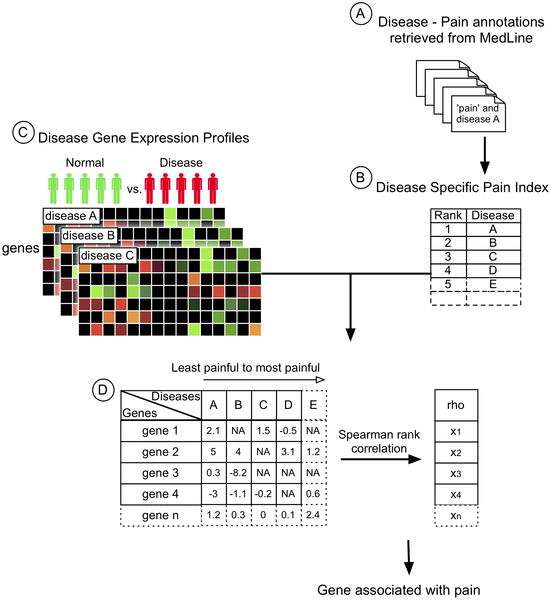
Most recent article, published on June 7, 2012, in open access journal  PLoS Computational Biology, on “Integrative Approach to Pain Genetics Identifies Pain Sensitivity Loci across Diseases” presented a novel integrative approach that combines publicly available molecular data and automatically extracted knowledge regarding pain contained in the literature to assist the discovery of novel pain genes. This study was approved by the Institutional Review Boards of Stanford University and SRI International.

In this meta-analysis, they took advantage of the vast amount of existing disease-related clinical literature and gene expression microarray data stored in large international repositories and

  • Ranked thousands of diseases according to the Figure shown below.

  • Obtained gene expression profiles of 121 of these human diseases from public sources.
  • Selected ‘genes with expression variation significantly correlated with DSPI across diseases’ as candidate pain genes.
  • Genotyped selected candidate pain genes in an independent human cohort, and finally
  • Evaluated for significant association between variants and measures of pain sensitivity.

In this study, the genes were chosen based on their high correlation with the DSPI and plausible biology as assessed by the available literature and human expression profile across tissue using The Scripps Research Institute BioGPS database [8].

The selected genes were:

  • ABLIM3 (actin binding LIM protein family, member 3),
  • PDE2A (phosphodiesterase 2A, cGMP-stimulated),
  • CREB1 (cAMP responsive element binding protein 1),
  • NAALAD2 (N-acetylated alpha-linked acidic dipeptidase 2), and
  • NCALD (neurocalcin delta).

These genes were selected from the candidate list and were prospectively tested for variants that may be associated with differential pain sensitivity in an independent human cohort.

ABLIM3 was selected as the top candidate as it showed the highest correlation with the DSPI. ABLIM3 is a newly characterized protein-coding gene. ABLIM3 is expressed in various tissues, most prominently in muscle and neuronal tissue [9], [10].

Polymorphisms in ABLIM3 (rs4512126) and NCALD (rs12548828, rs7826700, and rs1075791) showed significant association with the cold pressor pain threshold

“The strongest signal was with rs4512126 (5q32, ABLIM3, P = 1.3×10⁻¹⁰)  for the sensitivity to cold pressor pain in males, but not in females – a sex-specific association.”

Significant associations were also observed with rs12548828, rs7826700 and rs1075791 on 8q22.2 within NCALD (P = 1.7×10⁻⁴, 1.8×10⁻⁴, and 2.2×10⁻⁴ respectively).

Authors said that, “This data-derived list of pain gene candidates enables additional focused and efficient biological studies validating additional candidates.”

Authors have demonstrated the utility of a novel paradigm that integrates publicly available disease-specific gene expression data with clinical data curated from MEDLINE to facilitate the discovery of pain-relevant genes. This approach was validated through a targeted genetic association study in an independent human cohort, where variants of selected pain gene candidates were evaluated for associations with experimental pain sensitivity measures in humans.

Authors hope that “the outlined approach can complement existing research efforts by assisting the formulation of data-driven hypotheses, and may serve as a template to discover genetic components of other clinically important phenotypes.“

Further Reading:

Pain Gene Database (PGD)[11]

MeSH: Medical Subject Heading is a comprehensive vocabulary thesaurus organized in a hierarchical structure allowing the indexing of publications with various levels of specificity.

The 20 diseases with the highest disease-pain ratio from the DSPI are listed out of a total of 2962 diseases are

 .

Curated by: Dr. V. S. Karra, Ph.D.

Read Full Post »

« Newer Posts