Healthcare analytics, AI solutions for biological big data, providing an AI platform for the biotech, life sciences, medical and pharmaceutical industries, as well as for related technological approaches, i.e., curation and text analysis with machine learning and other activities related to AI applications to these industries.
(Left): Control rabbit brain, showing neuropil near the CA1 band in the hippocampus. (Right): Vitrified rabbit brain, same location. Synapses, vesicles, and microfilaments are clear. The myelinated axon shows excellent preservation. (credit: Robert L. McIntyre and Gregory M. Fahy/Cryobiology)
The Brain Preservation Foundation (BPF) has announced that a team at 21st Century Medicine led by Robert McIntyre, PhD., has won the Small Mammal Brain Preservation Prize, which carries an award of $26,735.
The Small Mammalian Brain Preservation Prize was awarded after the determination that the protocol developed by McIntyre, termed Aldehyde-Stabilized Cryopreservation, was able to preserve an entire rabbit brain with well-preserved ultrastructure, including cell membranes, synapses, and intracellular structures such as synaptic vesicles (full protocol here).
The judges for the prize were Kenneth Hayworth, PhD., Brain Preservation Foundation President and neuroscientist at the Howard Hughes Medical Institute; and Prof.Sebastian Seung, PhD., Princeton Neuroscience Institute and Computer Science Department.
First preservation of the connectome
“This is a milestone in the development of brain preservation techniques: it is the first time that the preservation of the connectome has been demonstrated in a whole brain (prior to this only small parts of brains have been preserved to this level of detail),” said the BPF announcement.
“Current models of the brain suggest that the connectome contains all the information necessary for personal identity (i.e., memory and personality). This technique is not the same as conventional cryonics (rapidly freezing the brain), which has never demonstrated preservation of the ultrastructure of the brain. Thus the winning of this prize represents a significant advance in the methods available for large scale studies of the connectome and could lead to procedures that preserve a complete human brain.
Kenneth Hayworth (KH) (President of the Brain Preservation Foundation (BPF)) and Michael Shermer (member of BPF advisory board) witnessed (on Sept. 25, 2015) the full Aldehyde Stabilized Cryopreservation surgical procedure performed on this rabbit at the laboratories of 21 Century Medicine under the direction of 21CM lead researcher Robert McIntyre. This included the live rabbit’s carotid arteries being perfused with glutaraldehyde and subsequent perfusion with cryoprotectant agent (CPA). KH witnessed this rabbit brain being put in -135 degrees C storage, removal from storage the following day (verifying that it had vitrified solid), and KH witnessed all subsequent tissue processing steps involved in the evaluation process. (credit: The Brain Preservation Foundation)
“The key breakthrough was the rapid perfusion of a deadly chemical fixative (glutaraldehyde) through the brain’s vascular system, instantly stopping metabolic decay and fixing all proteins in place by covalent crosslinks. This stabilized the tissue and vasculature so that cryoprotectant could be perfused at an optimal temperature and rate. The result was an intact rabbit brain filled with such a high concentration of cryoprotectants that it could be stored as a solid ‘vitrified’ block at a temperature of -135 degrees Celsius.”
Winning the award also required that the procedure be published in a peer reviewed scientific publication. McIntyre satisfied this requirement and published the protocol in an open-access paper in the Journal of Cryobiology.
3D microscope evaluation of the rabbit brain tissue preservation (credit: Brain Preservation Foundation)
The Brain Preservation Foundation plans to continue to promote the idea that brain preservation following legal death, by using scientifically validated techniques, is a reasonable choice for consenting individuals to make. Focus now shifts to the final Large Mammal phase of the contest, which requires an intact pig brain to be preserved with similar fidelity in a manner that could be directly adapted to terminal patients in a hospital setting.
The 21st Century Medicine team has recently submitted to the BPF such a preserved pig brain for official evaluation. Lead researcher Robert McIntyre has started Nectome to further develop this method.
“Of course, [the demonstrated brain preservation procedure] is only useful if you think all the relevant information is preserved in the fixation,” said Anders Sandberg, PhD., of the Future of Humanity Institute/Oxford Martin School. “Protein states and small molecule chemical information may be messed up.”
Proponents of cryonics have long sought a technique that could put terminal patients into longterm stasis, the goal being a form of medical time travel in which patients are stabilized against decay with the hope of being biologically revived and cured by future technologies. Despite decades of research, this goal of reversible cryopreservation remains far out of reach — too much damage occurs during the cryopreservation itself.
This has led a new generation of researchers to focus on a more achievable and demonstrable goal–preservation of brain structure only. Specifically preservation of the delicate pattern of synaptic connections (the “connectome”) which neuroscience contends encodes a person’s memory and identity. Instead of biological revival, these new researchers often envision a future “synthetic revival” comprising nanometer-scale scanning of the preserved brain to serve as the basis for mind uploading.
This shift in focus toward “synthetic” revival has completely transformed the cryonics debate, opening up new avenues of research and bringing it squarely within the purview of today’s scientific investigation. Hundreds of neuroscience papers have detailed how memory and personality are encoded structurally in synaptic connections, and recent advances in connectome imaging and brain simulation can be seen as a preview of the synthetic revival technologies to come.
Until now, the crucial unanswered questions were “How well does cryonics preserve the brain’s connectome?” and “Are there alternatives/modifications to cryonics that might preserve the connectome better and in a manner that could be demonstrated today?” The Brain Preservation Prize was put forward in 2010 to spur research that could definitively answer these questions. Now, five years later, these questions have been answered: Traditional cryonics procedures were not able to demonstrate (to the BPF’s satisfaction) preservation of the connectome, but the newly invented “Aldehyde-Stabilized Cryopreservation” technique was.
This result directly answers what has for decades been the main skeptical and scientific criticism against cryonics –that it does not provably preserve the delicate synaptic circuitry of the brain. As such, this research sets the stage for renewed interest within the scientific community, and offers a potential challenge to medical researchers to develop a human surgical procedure based on these successful animal experiments.
Abstract of Aldehyde-stabilized cryopreservation
We describe here a new cryobiological and neurobiological technique, aldehyde-stabilized cryopreservation (ASC), which demonstrates the relevance and utility of advanced cryopreservation science for the neurobiological research community. ASC is a new brain-banking technique designed to facilitate neuroanatomic research such as connectomics research, and has the unique ability to combine stable long term ice-free sample storage with excellent anatomical resolution. To demonstrate the feasibility of ASC, we perfuse-fixed rabbit and pig brains with a glutaraldehyde-based fixative, then slowly perfused increasing concentrations of ethylene glycol over several hours in a manner similar to techniques used for whole organ cryopreservation. Once 65% w/v ethylene glycol was reached, we vitrified brains at −135 °C for indefinite long-term storage. Vitrified brains were rewarmed and the cryoprotectant removed either by perfusion or gradual diffusion from brain slices. We evaluated ASC-processed brains by electron microscopy of multiple regions across the whole brain and by Focused Ion Beam Milling and Scanning Electron Microscopy (FIB-SEM) imaging of selected brain volumes. Preservation was uniformly excellent: processes were easily traceable and synapses were crisp in both species. Aldehyde-stabilized cryopreservation has many advantages over other brain-banking techniques: chemicals are delivered via perfusion, which enables easy scaling to brains of any size; vitrification ensures that the ultrastructure of the brain will not degrade even over very long storage times; and the cryoprotectant can be removed, yielding a perfusable aldehyde-preserved brain which is suitable for a wide variety of brain assays.
Totally weird – IOW those “covalent bonds” act like a preservation matrix. So this brain indeed has been “fixed” – just at a smaller scale and level.
A couple of other factors:
* Quite a lot of the brain that counts (memory) may be on a larger scale than this – and may be preserved. While it is not, per the Connetome idea, at the macro axon scale – it is a general idea that at the molecular scale, something “plays” through the consciousness mechanism (Search = Hameroff Memory.)
I personally suspect a DNA like encoding in an as yet unproven language software. Perhaps even multiple “scale” functionality that would be a combination of organelle specialization (perhaps time perception) and THEN the inter-connectedness.
* As for personality, I know that that is entirely reproducible – in spite of such extreme complexity – but that is a proof for another day.
Just for kicks, note how the “search” code above results in prefabricated libraries being sent to your mind.
Gorden Russell –
You had me until I got to this part: “…a deadly chemical fixative (glutaraldehyde) through the brain’s vascular system…”
So this process perfectly preserves your brain after killer it dead.
So in the future it can be scanned and printed out into a perfect copy — but the copy won’t be you, it’ll be somebody else who is just like you. You will still be dead.
I’d rather be a live brain in a jar atop a robot wired into the spinal column so that I could still have all of my senses while awaiting the time a human body can be regrown.
CT
We have to differentiate on how we define “me” or “you”. Do we mean our memories (data) or consciousness (process). Our memories, personality, knowledge… alone (e.g. while we sleep and are unconscious)… are like fixed data until the brain (or a computer) begins to run and consciousness comes into existence .
We could copy the data to a computer (through scanning), which in the next step (after the simulation is beginning to operate) would create consciousness as well (defining itself as “me” or “you”). It wouldn’t be the same consciousness (process) due to other environmental inputs (and over time other memory/data- background). But the same is true for a biological based consciousness. My consciousness right now is not the consciousness anymore I had last year. It’s always a unique set-up.
From my point of view, the sentiment that there is some kind of metaphysical soul over an entire lifetime is an illusion based on the fact that we have memories, knowledge and personality (which we would have after the scanning process of our brain as well), that were formed in the past, and we are able to (subjectively altered) recreate it (and remember it) in our current state of consciousness. As a result we conclude, that we are/ have the same state of consciousness as the past me, which is (as I see it) an illusion.
So if we would be able to make a perfect copy of our brain that is able to create consciousness (in any kind of computer substrate, digital, analog or quantum) it wouldn’t be more or less the me (the consciousness) at the present than my future me in 5 minutes or years would be (in its biological form). From my point of view, the status quo wouldn’t change.
It is a copy because maybe one day they can do it without killing the original. The only way out of this conundrum was explained to me on this web site a while back in comments: if they substituted every neuron in my brain one at a time over a certain timescale so that eventually my brain would be synthetic, ‘”I” probably wouldn’t even notice.
But you are dreaming during your sleep.
Glutaraldehyde will put an end to all of your dreams.
A printed copy of you may have similar dreams, but not your dreams.
Alternative CRISPR discovered @MIT, Volume 2 (Volume Two: Latest in Genomics Methodologies for Therapeutics: Gene Editing, NGS and BioInformatics, Simulations and the Genome Ontology), Part 2: CRISPR for Gene Editing and DNA Repair
Alternative CRISPR Discovered @MIT
Reporter & Curator: Larry H. Bernstein, MD, FCAP
New breakthrough! – A better alternative CRISPR system just identified
CRISPR-Cas9 system has revolutionized the field of genome editing since its first application in human cells was reported in 2012. A recent publication in Cell reported the identification of a different CRISPR system with the potential for even simpler and more precise genome editing. The newly identified CRISPR-Cpf1 system mediates robust DNA interference with features different from Cas9. Cpf1 possesses several advantages over the currently used Cas9 system.
The Cpf1 system is simpler than Cas9 system as it requires only a single RNA for its DNA-cutting enzymatic activity.
Cpf1 cut has shot overhangs on the exposed ends, allowing more efficient and precise genome engineering; while Cas9 cut produces blunt ends that often undergo mutations when rejoined.
Cpf1 is smaller than Cas9, thus easier to deliver into the cells or tissues.
Cpf1 cut is far away from the recognition site, leaving space for further editing if mutation occurred at the cutting site.
The Cpf1 complex recognize very different PAM sequences than those of Cas9, adding more flexibility in choosing target sites.
These properties of Cpf1 and its potential with more precise gene editing expanded the application scope of CRISPR, from gene knock-out and knock-ins, genomic deletions, to even gene therapy.
•Cpf1 is a CRISPR-associated two-component RNA-programmable DNA nuclease
•Targeted DNA is cleaved as a 5-nt staggered cut distal to a 5′ T-rich PAM
•Two Cpf1 orthologs exhibit robust nuclease activity in human cells
Summary
The microbial adaptive immune system CRISPR mediates defense against foreign genetic elements through two classes of RNA-guided nuclease effectors. Class 1 effectors utilize multi-protein complexes, whereas class 2 effectors rely on single-component effector proteins such as the well-characterized Cas9. Here, we report characterization of Cpf1, a putative class 2 CRISPR effector. We demonstrate that Cpf1 mediates robust DNA interference with features distinct from Cas9. Cpf1 is a single RNA-guided endonuclease lacking tracrRNA, and it utilizes a T-rich protospacer-adjacent motif. Moreover, Cpf1 cleaves DNA via a staggered DNA double-stranded break. Out of 16 Cpf1-family proteins, we identified two candidate enzymes from Acidaminococcus and Lachnospiraceae, with efficient genome-editing activity in human cells. Identifying this mechanism of interference broadens our understanding of CRISPR-Cas systems and advances their genome editing applications.
Almost all archaea and many bacteria achieve adaptive immunity through a diverse set of CRISPR-Cas (clustered regularly interspaced short palindromicrepeats and CRISPR-associated proteins) systems, each of which consists of a combination of Cas effector proteins and CRISPR RNAs (crRNAs) (Makarova et al., 2011, Makarova et al., 2015). The defense activity of the CRISPR-Cas systems includes three stages: (1) adaptation, when a complex of Cas proteins excises a segment of the target DNA (known as a protospacer) and inserts it into the CRISPR array (where this sequence becomes a spacer); (2) expression and processing of the precursor CRISPR (pre-cr) RNA resulting in the formation of mature crRNAs; and (3) interference, when the effector module—either another Cas protein complex or a single large protein—is guided by a crRNA to recognize and cleave target DNA (or in some cases, RNA) (Horvath and Barrangou, 2010,Sorek et al., 2013, Barrangou and Marraffini, 2014). The adaptation stage is mediated by the complex of the Cas1 and Cas2 proteins, which are shared by all known CRISPR-Cas systems, and sometimes involves additional Cas proteins. Diversity is observed at the level of processing of the pre-crRNA to mature crRNA guides, proceeding via either a Cas6-related ribonuclease or a housekeeping RNaseIII that specifically cleaves double-stranded RNA hybrids of pre-crRNA and tracrRNA. Moreover, the effector modules differ substantially among the CRISPR-Cas systems (Makarova et al., 2011, Makarova et al., 2015,Charpentier et al., 2015). In the latest classification, the diverse CRISPR-Cas systems are divided into two classes according to the configuration of their effector modules: class 1 CRISPR systems utilize several Cas proteins and the crRNA to form an effector complex, whereas class 2 CRISPR systems employ a large single-component Cas protein in conjunction with crRNAs to mediate interference (Makarova et al., 2015).
Multiple class 1 CRISPR-Cas systems, which include the type I and type III systems, have been identified and functionally characterized in detail, revealing the complex architecture and dynamics of the effector complexes (Brouns et al., 2008, Marraffini and Sontheimer, 2008, Hale et al., 2009, Sinkunas et al., 2013,Jackson et al., 2014, Mulepati et al., 2014). Several class 2 CRISPR-Cas systems have also been identified and experimentally characterized, but they are all type II and employ homologous RNA-guided endonucleases of the Cas9 family as effectors (Barrangou et al., 2007, Garneau et al., 2010, Deltcheva et al., 2011, Sapranauskas et al., 2011, Jinek et al., 2012, Gasiunas et al., 2012). A second, putative class 2 CRISPR system, tentatively assigned to type V, has been recently identified in several bacterial genomes (http://www.jcvi.org/cgi-bin/tigrfams/HmmReportPage.cgi?acc=TIGR04330) (Schunder et al., 2013, Vestergaard et al., 2014, Makarova et al., 2015). The putative type V CRISPR-Cas systems contain a large, ∼1,300 amino acid protein called Cpf1 (CRISPR from Prevotella and Francisella 1). It remains unknown, however, whether Cpf1-containing CRISPR loci indeed represent functional CRISPR systems. Given the broad applications of Cas9 as a genome-engineering tool (Hsu et al., 2014, Jiang and Marraffini, 2015), we sought to explore the function of Cpf1-based putative CRISPR systems.
Here, we show that Cpf1-containing CRISPR-Cas loci of Francisella novicida U112 encode functional defense systems capable of mediating plasmid interference in bacterial cells guided by the CRISPR spacers. Unlike Cas9 systems, Cpf1-containing CRISPR systems have three features. First, Cpf1-associated CRISPR arrays are processed into mature crRNAs without the requirement of an additional trans-activating crRNA (tracrRNA) (Deltcheva et al., 2011, Chylinski et al., 2013). Second, Cpf1-crRNA complexes efficiently cleave target DNA proceeded by a short T-rich protospacer-adjacent motif (PAM), in contrast to the G-rich PAM following the target DNA for Cas9 systems. Third, Cpf1 introduces a staggered DNA double-stranded break with a 4 or 5-nt 5′ overhang.
To explore the suitability of Cpf1 for genome-editing applications, we characterized the RNA-guided DNA-targeting requirements for 16 Cpf1-family proteins from diverse bacteria, and we identified two Cpf1 enzymes fromAcidaminococcus sp. BV3L6 and Lachnospiraceae bacterium ND2006 that are capable of mediating robust genome editing in human cells. Collectively, these results establish Cpf1 as a class 2 CRISPR-Cas system that includes an effective single RNA-guided endonuclease with distinct properties that has the potential to substantially advance our ability to manipulate eukaryotic genomes.
Results
Figure 1
The Francisella novicida U112 Cpf1 CRISPR Locus Provides Immunity against Transformation of Plasmids Containing Protospacers Flanked by a 5′-TTN PAM
(A) Organization of two CRISPR loci found in Francisella novicida U112 (NC_008601). The domain architectures of FnCas9 and FnCpf1 are compared.
(B) Schematic illustrating the plasmid depletion assay for discovering the PAM position and identity. Competent E. coliharboring either the heterologous FnCpf1 locus plasmid (pFnCpf1) or the empty vector control were transformed with a library of plasmids containing the matching protospacer flanked by randomized 5′ or 3′ PAM sequences and selected with antibiotic to deplete plasmids carrying successfully targeted PAM. Plasmids from surviving colonies were extracted and sequenced to determine depleted PAM sequences.
(C and D) Sequence logo for the FnCpf1 PAM as determined by the plasmid depletion assay. Letter height at each position is measured by information content (C) or frequency (D); error bars show 95% Bayesian confidence interval.
(E) E. coli harboring pFnCpf1 provides robust interference against plasmids carrying 5′-TTN PAMs (n = 3; error bars represent mean ± SEM).
Cpf1-Containing CRISPR Loci Are Active Bacterial Immune Systems
The Cpf1-Associated CRISPR Array Is Processed Independent of TracrRNA
Cpf1 Is a Single crRNA-Guided Endonuclease
The RuvC-like Domain of Cpf1 Mediates RNA-Guided DNA Cleavage
Sequence and Structural Requirements for the Cpf1 crRNA
Cpf1-Family Proteins from Diverse Bacteria Share Common crRNA Structures and PAMs
Cpf1 Can Be Harnessed to Facilitate Genome Editing in Human Cells
In this work, we characterize Cpf1-containing class 2 CRISPR systems, classified as type V, and show that its effector protein, Cpf1, is a single RNA-guided endonuclease. Cpf1 substantially differs from Cas9—to date, the only other experimentally characterized class 2 effector—in terms of structure and function and might provide important advantages for genome-editing applications. Specifically, Cpf1 contains a single identified nuclease domain, in contrast to the two nuclease domains present in Cas9. The results presented here show that, in FnCpf1, inactivation of RuvC-like domain abolishes cleavage of both DNA strands. Conceivably, FnCpf1 forms a homodimer (Figure S2B), with the RuvC-like domains of each of the two subunits cleaving one DNA strand. However, we cannot rule out that FnCpf1 contains a second yet-to-be-identified nuclease domain. Structural characterization of Cpf1-RNA-DNA complexes will allow testing of these hypotheses and elucidation of the cleavage mechanism.
February 8, 2016 | When a geneticist stares down the 3 billion DNA base pairs of the human genome, searching for a clue to what’s gone awry in a single patient, it helps to narrow the field. One of the most popular places to look is the exome, the tiny fraction of our DNA―less than 2%―that actually codes for proteins. For patients with rare genetic diseases, which might be fully explained by one key mutation, many studies sequence the whole exome and leave all the noncoding DNA out. Similarly, personalized cancer tests, which can help bring to light unexpected treatment options, often sequence the tumor exome, or a smaller panel of protein-coding genes.
Unfortunately, we know that’s not the whole picture. “There are a substantial number of noncoding regions that are just as effective at turning off a gene as a mutation in the gene itself,” says Richard Sherwood, a geneticist at Brigham and Women’s Hospital in Boston. “Exome sequencing is not going to be a good proxy for what genes are working.”
Sherwood studies regulatory DNA, the vast segment of the genome that governs which genes are turned on or off in any cell at a given time. It’s a confounding area of genetics; we don’t even know how much of the genome is made up of these regulatory elements. While genes can be recognized by the presence of “start” and “stop” codons―sequences of three DNA letters that tell the cell’s molecular machinery which stretches of DNA to transcribe into RNA, and eventually into protein―there are no definite signs like this for regulatory DNA.
Instead, studies to discover new regulatory elements have been somewhat trial-and-error. If you suspect a gene’s activity might be regulated by a nearby DNA element, you can inhibit that element in a living cell, and see if your gene shuts down with it.
With these painstaking experiments, scientists can slowly work their way through potential regulatory regions―but they can’t sweep across the genome with the kind of high-throughput testing that other areas of genetics thrive on. “Previously, you couldn’t do these sorts of tests in a large form, like 4,000 of them at once,” says David Gifford, a computational biologist at MIT. “You would really need to have a more hypothesis-directed methodology.”
Recently, Gifford and Sherwood collaborated on a paper, published in Nature Biotechnology, which presents a new method for testing thousands of DNA loci for regulatory activity at once. Their assay, called MERA (multiplexed editing regulatory assay), is built on the recent technology boom in CRISPR-Cas9 gene editing, which lets scientists quickly and easily cut specific sequences of DNA out of the genome.
So far, their team, including lead author Nisha Rajagopal from Gifford’s lab, has used MERA to study the regulation of four genes involved in the development of embryonic stem cells. Already, the results have defied the accepted wisdom about regulatory DNA. Many areas of the genome flagged by MERA as important factors in gene expression do not fall into any known categories of regulatory elements, and would likely never have been tested with previous-generation methods.
“Our approach allows you to look away from the lampposts,” says Sherwood. “The more unbiased you can be, the more we’ll actually know.”
A New Kind of CRISPR Screen
In the past three years, CRISPR-Cas9 experiments have taken all areas of molecular biology by storm, and Sherwood and Gifford are far from the first to use the technology to run large numbers of tests in parallel. CRISPR screens are an excellent way to learn which genes are involved in a cellular process, like tumor growth or drug resistance. In these assays, scientists knock out entire genes, one by one, and see what happens to cells without them.
This kind of CRISPR screen, however, operates on too small a scale to study the regulatory genome. For each gene knocked out in a CRISPR screen, you have to engineer a strain of virus to deliver a “guide RNA” into the cellular genome, showing the vicelike Cas9 molecule which DNA region to cut. That works well if you know exactly where a gene lies and only need to cut it once—but in a high-throughput regulatory test, you would want to blanket vast stretches of DNA with cuts, not knowing which areas will turn out to contain regulatory elements. Creating a new virus for each of these cuts is hugely impractical.
The insight behind MERA is that, with the right preparation, most of the genetic engineering can be done in advance. Gifford and Sherwood’s team used a standard viral vector to put a “dummy” guide RNA sequence, one that wouldn’t tell Cas9 to cut anything, into an embryonic stem cell’s genome. Then they grew plenty of cells with this prebuilt CRISPR system inside, and attacked each one with a Cas9 molecule targeted to the dummy sequence, chopping out the fake guide.
Normally, the result would just be a gap in the CRISPR system where the guide once was. But along with Cas9, the researchers also exposed the cells to new, “real” guide RNA sequences. Through a DNA repair mechanism called homologous recombination, the cells dutifully patched over the gaps with new guides, whose sequences were very similar to the missing dummy code. At the end of the process, each cell had a unique guide sequence ready to make cuts at a specific DNA locus—just like in a standard CRISPR screen, but with much less hands-on engineering.
By using a large enough library of guide RNA molecules, a MERA screen can include thousands of cuts that completely tile a broad region of the genome, providing an agnostic look at anywhere regulatory elements might be hiding. “It’s a lot easier [than a typical CRISPR screen],” says Sherwood. “The day the library comes in, you just perform one PCR reaction, and the cells do the rest of the work.”
In the team’s first batch of MERA screens, they created almost 4,000 guide RNAs for each gene they studied, covering roughly 40,000 DNA bases of the “cis-regulatory region,” or the area surrounding the gene where most regulatory elements are thought to lie. It’s unclear just how large any gene’s cis-regulatory region is, but 40,000 bases is a big leap from the highly targeted assays that have come before.
“We’re now starting to do follow-up studies where we increase the number of guide RNAs,” Sherwood adds. “Eventually, what you’d like is to be able to tile an entire chromosome.”
Far From the Lampposts
Sherwood and Gifford tried to focus their assays on regions that would be rich in regulatory elements. To that end, they made sure their guide RNAs covered parts of the genome with well-known signs of regulatory activity, like histone markers and transcription factor binding sites. For many of these areas, Cas9 cuts did, in fact, shut down gene expression in the MERA screens.
But the study also targeted regions around each gene that were empty of any known regulatory features. “We tiled some other regions that we thought might serve as negative controls,” explains Gifford. “But they turned out not to be negative at all.”
The study’s most surprising finding was that several cuts to seemingly random areas of the genome caused genes to become nonfunctional. The authors named these DNA regions “unmarked regulatory elements,” or UREs. They were especially prevalent around the genes Tdgf1 and Zfp42, and in many cases, seemed to be every bit as necessary to gene activity as more predictable hits on the MERA screen.
These results caught the researchers so off guard that it was natural to wonder if MERA screens are prone to false positives. Yet follow-up experiments strongly supported the existence of UREs. Switching the guide RNAs from aTdgf1 MERA screen and aZfp42 screen, for example, produced almost no positive results: the UREs’ regulatory effects were indeed specific to the genes near them.
In a more specific test, the researchers chose a particular URE connected to Tdgf1, and cut it out of a brand new population of cells for a closer look. “We showed that, if we deleted that region from the genome, the cells lost expression of the gene,” says Sherwood. “And then when we put it back in, the gene became expressed again. Which was good proof to us that the URE itself was responsible.”
From these results, it seems likely that follow-up MERA screens will find even more unknown stretches of regulatory DNA. Gifford and Sherwood’s experiments didn’t try to cover as much ground around their target genes as they might have, because the researchers assumed that MERA would mostly confirm what was already known. At best, they hoped MERA would rule out some suspected regulatory regions, and help show which regulatory elements have the biggest effect on gene expression.
“We tended to prioritize regions that had been known before,” Sherwood says. “Unfortunately, in the end, our datasets weren’t ideally suited to discovering these UREs.”
Getting to Basic Principles
MERA could open up huge swaths of the regulatory genome to investigation. Compared to an ordinary CRISPR screen, says Sherwood, “there’s only upside,” as MERA is cheaper, easier, and faster to run.
Still, interpreting the results is not trivial. Like other CRISPR screens, MERA makes cuts at precise points in the genome, but does not tell cells to repair those cuts in any particular way. As a result, a population of cells all carrying the same guide RNA can have a huge variety of different gaps and scars in their genomes, typically deletions in the range of 10 to 100 bases long. Gifford and Sherwood created up to 100 cells for each of their guides, and sometimes found that gene expression was affected in some but not all of them; only sequencing the genomes of their mutated cells could reveal exactly what changes had been made.
By repeating these experiments many times, and learning which mutations affect gene expression, it will eventually be possible to pin down the exact DNA bases that make up each regulatory element. Future studies might even be able to distinguish between regulatory elements with small and large effects on gene expression. In Gifford and Sherwood’s MERA screens, the target genes were altered to produce a green fluorescent protein, so the results were read in terms of whether cells gave off fluorescent light. But a more precise, though expensive, approach would be to perform RNA sequencing, to learn which cuts reduced the cell’s ability to transcribe a gene into RNA, and by how much.
A MERA screen offers a rich volume of data on the behavior of the regulatory genome. Yet, as with so much else in genetics, there are few robust principles to let scientists know where they should be focusing their efforts. Histone markers provide only a very rough sketch of regulatory elements, often proving to be red herrings on closer examination. And the existence of UREs, if confirmed by future experiments, shows that we don’t yet even know which areas of the genome to rule out in the hunt for regulatory regions.
“Every dataset we get comes closer and closer to computational principles that let us predict these regions,” says Sherwood. As more studies are conducted, patterns may emerge in the DNA sequences of regulatory elements that link UREs together, or reveal which histone markers truly point toward regulatory effects. There might also be functional clues hidden in these sequences, hinting at what is happening on a molecular level as regulatory elements turn genes on and off in the course of a cell’s development.
For now, however, the data is still rough and disorganized. For better and for worse, high-throughput tools like MERA are becoming the foundation for most discoveries in genetics—and that means there is a lot more work to do before the regulatory genome begins to come into focus.
CORRECTED 2/9/16: Originally, this story incorrectly stated that only certain cell types could be assayed with MERA for reasons related to homologous recombination. In fact, the authors see no reason MERA could not be applied to any in vitro cell line, and hope to perform screens in a wide range of cell types. The text has been edited to correct the error.
Gene Editing for Exon 51: Why CRISPR Snipping might be better than Exon Skipping for DMD
Why CRISPR might be better than exon skipping for DMD: Snipping vs. skipping for DMD
By Lauren Martz, Senior Writer
Published on Thursday, January 21, 2016
As if to preempt the regulatory setbacks in Duchenne muscular dystrophy (DMD) that last week disappointed the field, a trio of preclinical studies emerged two weeks earlier showing that cutting out DMD mutations with gene editing might offer a viable alternative to the exon-skipping strategies that have dominated the pipeline. Now, the question is whether there’s reason to believe the mouse studies will translate any better to the clinic.
The studies, published Dec. 31 in Science, provide in vivo proof of concept for the first time that CRISPR-Cas9 used postnatally can have a disease-modifying effect. Despite the hype around its therapeutic promise, the technology has so far proved itself primarily in research applications, for example, in modifying cells for in vitro screening or creating animal models of disease.
RNA interference (RNAi) silences, or knocks down, the translation of a gene by inducing degradation of a gene target’s transcript. To advance RNAi applications, Thermo Fisher Scientific has developed two types of small RNA molecules: short interfering RNAs and microRNAs. The company also offers products for RNAi analysis in vitro and in vivo, including libraries for high-throughput applications.
Genes can be knocked down with RNA interference (RNAi) or knocked out with CRISPR-Cas9. RNAi, the screening workhorse, knocks down the translation of genes by inducing rapid degradation of a gene target’s transcript.
CRISPR-Cas9, the new but already celebrated genome-editing technology, cleaves specific DNA sequences to render genes inoperative. Although mechanistically different, the two techniques complement one another, and when used together facilitate discovery and validation of scientific findings.
RNAi technologies along with other developments in functional genomics screening were discussed by industry leaders at the recent Discovery on Target conference. The conference, which emphasized the identification and validation of novel drug targets and the exploration of unknown cellular pathways, included a symposium on the development of CRISPR-based therapies.
RNAi screening can be performed in either pooled or arrayed formats. Pooled screening provides an affordable benchtop option, but requires back-end deconvolution and deep sequencing to identify the shRNA causing the specific phenotype. Targets are much easier to identify using the arrayed format since each shRNA clone is in an individual well.
“CRISPR complements RNAi screens,” commented Ryan Raver, Ph.D., global product manager of functional genomics at Sigma-Aldrich. “You can do a whole genome screen with either small interfering RNA (siRNA) or small hairpin RNA (shRNA), then validate with individual CRISPRs to ensure it is a true result.”
A powerful and useful validation method for knockdown or knockout studies is to use lentiviral open reading frames (ORFs) for gene re-expression, for rescue experiments, or to detect whether the wild-type phenotype is restored. However, the ORF randomly integrates into the genome. Also, with this validation technique, gene expression is not acting under the endogenous promoter. Accordingly, physiologically relevant levels of the gene may not be expressed unless controlled for via an inducible system.
In the future, CRISPR activators may provide more efficient ways to express not only wild-type but also mutant forms of genes under the endogenous promoter.
Choice of screening technique depends on the researcher and the research question. Whole gene knockout may be necessary to observe a phenotype, while partial knockdown might be required to investigate functions of essential or lethal genes. Use of both techniques is recommended to identify all potential targets.
For example, recently, a whole genome siRNA screen on a human glioblastoma cell line identified a gene, known as FAT1, as a negative regulator of apoptosis. A CRISPR-mediated knockout of the gene also conferred sensitivity to death receptor–induced apoptosis with an even stronger phenotype, thereby validating FAT1’s new role and link to extrinsic apoptosis, a new model system.
Dr. Raver indicated that next-generation RNAi libraries that are microRNA-adapted might have a more robust knockdown of gene expression, up to 90–95% in some cases. Ultracomplex shRNA libraries help to minimize both false-negative and false-positive rates by targeting each gene with ~25 independent shRNAs and by including thousands of negative-control shRNAs.
Recently, a relevant paper emerged from the laboratory of Jonathan Weissman, Ph.D., a professor of cellular and molecular pharmacology at the University of California, San Francisco. The paper described how a new ultracomplex pooled shRNA library was optimized by means of a microRNA-adapted system. This system, which was able to achieve high specificity in the detection of hit genes, produced robust results. In fact, they were comparable to results obtained with a CRISPR pooled screen. Members of the Weissman group systematically optimized the promoter and microRNA contexts for shRNA expression along with a selection of guide strands.
Using a sublibrary of proteostasis genes (targeting 2,933 genes), the investigators compared CRISPR and RNAi pooled screens. Data showed 48 hits unique to RNAi, 40 unique to CRISPR, and an overlap of 21 hits (with a 5% false discovery rate cut-off). Together, the technologies provided a more complete research story.
Arrayed CRISPR Screens
Click Image To Enlarge +
Synthetic crRNA:tracrRNA reagents can be used in a similar manner to siRNA reagents for assessment of phenotypes in a cell population. Top row: A reporter cell line stably expressing Cas9 nuclease was transfected with GE Dharmacon’s Edit-R synthetic crRNA:tracrRNA system, which was used to target three positive control genes (PSMD7, PSMD14, and VCP) and a negative control gene (PPIB). Green cells indicate EGFP signaling occuring as a result of proteasome pathway disruption. Bottom row: A siGENOME siRNA pool targeting the same genes was used in the same reporter cell line.
“RNA screens are well accepted and will continue to be used, but it is important biologically that researchers step away from the RNA mechanism to further study and validate their hits to eliminate potential bias,” explained Louise Baskin, senior product manager, Dharmacon, part of GE Healthcare. “The natural progression is to adopt CRISPR in the later stages.”
RNAi uses the cell’s endogenous mechanism. All of the components required for gene knockdown are already within the cell, and the delivery of the siRNA starts the process. With the CRISPR gene-editing system, which is derived from a bacterial immune defense system, delivery of both the guide RNA and the Cas9 nuclease, often the rate limiter in terms of knockout efficiency, are required.
In pooled approaches, the cell has to either drop out or overexpress so that it is sortable, limiting the types of addressable biological questions. A CRISPR-arrayed approach opens up the door for use of other analytical tools, such as high-content imaging, to identify hits of interest.
To facilitate use of CRISPR, GE recently introduced Dharmacon Edit-R synthetic CRISPR RNA (crRNA) libraries that can be used to carry out high-throughput arrayed analysis of multiple genes. Rather than a vector- or plasmid-based gRNA to guide the targeting of the Cas9 cleavage, a synthetic crRNA and tracrRNA are used. These algorithm-designed crRNA reagents can be delivered into the cells very much like siRNA, opening up the capability to screen multiple target regions for many different genes simultaneously.
GE showed a very strong overlap between CRISPR and RNAi using this arrayed approach to validate RNAi screen hits with synthetic crRNA. The data concluded that CRISPR can be used for medium- or high-throughput validation of knockdown studies.
“We will continue to see a lot of cooperation between RNAi and gene editing,” declared Baskin. “Using the CRISPR mechanism to knockin could introduce mutations to help understand gene function at a much deeper level, including a more thorough functional analysis of noncoding genes.
“These regulatory RNAs often act in the nucleus to control translation and transcription, so to knockdown these genes with RNAi would require export to the cytoplasm. Precision gene editing, which takes place in the nucleus, will help us understand the noncoding transcriptome and dive deeper into how those genes regulate disease progression, cellular development and other aspects of human health and biology.”
Tool Selection
Click Image To Enlarge +
Schematic of a pooled shRNA screening workflow developed by Transomic Technologies. Cells are transduced, and positive or negative selection screens are performed. PCR amplification and sequencing of the shRNA integrated into the target cell genome allows the determination of shRNA representation in the population.
The functional genomics tool should fit the specific biology; the biology should not be forced to fit the tool. Points to consider include the regulation of expression, the cell line or model system, as well as assay scale and design. For example, there may be a need for regulatable expression. There may be limitations around the cell line or model system. And assay scale and design may include delivery conditions and timing to optimally complete perturbation and reporting.
“Both RNAi- and CRISPR-based gene modulation strategies have pros and cons that should be considered based on the biology of the genes being studied,” commented Gwen Fewell, Ph.D., chief commercial officer, Transomic Technologies.
RNAi reagents, which can produce hypomorphic or transient gene-suppression states, are well known for their use in probing drug targets. In addition, these reagents are enriching studies of gene function. CRISPR-Cas9 reagents, which produce clean heterozygous and null mutations, are important for studying tumor suppressors and other genes where complete loss of function is desired.
Timing to readout the effects of gene perturbation must be considered to ensure that the biological assay is feasible. RNAi gene knockdown effects can be seen in as little as 24–72 hours, and inducible and reversible gene knockdown can be realized. CRISPR-based gene knockout effects may become complete and permanent only after 10 days.
Both RNAi and CRISPR reagents work well for pooled positive selection screens; however, for negative selection screens, RNAi is the more mature tool. Current versions of CRISPR pooled reagents can produce mixed populations containing a fraction of non-null mutations, which can reduce the overall accuracy of the readout.
To meet the needs of varied and complex biological questions, Transomic Technologies has developed both RNAi and CRISPR tools with options for optimal expression, selection, and assay scale. For example, the company’s shERWOOD-UltramiR shRNA reagents incorporate advances in design and small RNA processing to produce increased potency and specificity of knockdown, particularly important for pooled screens.
Sequence-verified pooled shRNA screening libraries provide flexibility in promoter choice, in vitro formats, in vivo formats, and a choice of viral vectors for optimal delivery and expression in biologically relevant cell lines, primary cells or in vivo.
The company’s line of lentiviral-based CRISPR-Cas9 reagents has variable selectable markers such that guide RNA- and Cas9-expressing vectors, including inducible Cas9, can be co-delivered and selected for in the same cell to increase editing efficiency. Promoter options are available to ensure expression across a range of cell types.
“Researchers are using RNAi and CRISPR reagents individually and in combination as cross-validation tools, or to engineer CRISPR-based models to perform RNAi-based assays,” informs Dr. Fewell. “Most exciting are parallel CRISPR and RNAi screens that have tremendous potential to uncover novel biology.”
Converging Technologies
The convergence of RNAi technology with genome-editing tools, such as CRISPR-Cas9 and transcription activator-like effector nucleases, combined with next-generation sequencing will allow researchers to dissect biological systems in a way not previously possible.
“From a purely technical standpoint, the challenges for traditional RNAi screens come down to efficient delivery of the RNAi reagents and having those reagents provide significant, consistent, and lasting knockdown of the target mRNAs,” states Ross Whittaker, Ph.D., a product manager for genome editing products at Thermo Fisher Scientific. “We have approached these challenges with a series of reagents and siRNA libraries designed to increase the success of RNAi screens.”
Thermo Fisher’ provides lipid-transfection RNAiMax reagents, which effectively deliver siRNA. In addition, the company’s Silencer and Silencer Select siRNA libraries provide consistent and significant knockdown of the target mRNAs. These siRNA libraries utilize highly stringent bioinformatic designs that ensure accurate and potent targeting for gene-silencing studies. The Silencer Select technology adds a higher level of efficacy and specificity due to chemical modifications with locked nucleic acid (LNA) chemistry.
The libraries alleviate concerns for false-positive or false-negative data. The high potency allows less reagent use; thus, more screens or validations can be conducted per library.
Dr. Whittaker believes that researchers will migrate regularly between RNAi and CRISPR-Cas9 technology in the future. CRISPR-Cas9 will be used to create engineered cell lines not only to validate RNAi hits but also to follow up on the underlying mechanisms. Cell lines engineered with CRISPR-Cas9 will be utilized in RNAi screens. In the long term, CRISPR-Cas9 screening will likely replace RNAi screening in many cases, especially with the introduction of arrayed CRISPR libraries.
Validating Antibodies with RNAi
Unreliable antibody specificity is a widespread problem for researchers, but RNAi is assuaging scientists’ concerns as a validation method.
The procedure introduces short hairpin RNAs (shRNAs) to reduce expression levels of a targeted protein. The associated antibody follows. With its protein knocked down, a truly specific antibody shows dramatically reduced or no signal on a Western blot. Short of knockout animal models, RNAi is arguably the most effective method of validating research antibodies.
The method is not common among antibody suppliers—time and cost being the chief barriers to its adoption, although some companies are beginning to embrace RNAi validation.
“In the interest of fostering better science, Proteintech felt it was necessary to implement this practice,” said Jason Li, Ph.D., founder and CEO of Proteintech Group, which made RNAi standard protocol in February 2015. “When researchers can depend on reproducibility, they execute more thorough experiments and advance the treatment of human diseases and conditions.”
Junk DNA Kept in Good Repair by Nuclear Membrane
Heterochromatin has the dubious distinction of being called the “dark matter” of DNA, and it has even suffered the indignity of being dismissed as “junk DNA.” But it seems to get more respectful treatment inside the nucleus, where it has the benefit of a special repair mechanism. This mechanism, discovered by scientists based at the University of Southern California (USC), transports broken heterochromatin sequences from the hurly-burly of the heterochromatin domain so that they can be repaired in the relative peace and quiet of the nuclear periphery.
This finding suggests that the nuclear membrane is more versatile than is generally appreciated. Yes, it serves as a protective container for nuclear material, and it uses its pores to manage the transport of molecules in and out of the nucleus. But it may also play a special role in maintaining the integrity of heterochromatin, which tends to be overlooked because it consists largely of noncoding DNA, including repetitive stretches of no apparent function.
“Scientists are now starting to pay a lot of attention to this mysterious component of the genome,” said Irene E. Chiolo, Ph.D., an assistant professor at USC. “Heterochromatin is not only essential for chromosome maintenance during cell division; it also poses specific threats to genome stability. Heterochromatin is potentially one of the most powerful driving forces for cancer formation, but it is the ‘dark matter’ of the genome. We are just beginning to unravel how repair works here.”
Dr. Chilo led an effort to understand how heterochromatin stays in good repair, even though it is particularly vulnerable to a kind of repair error called ectopic recombination. This kind of error is apt to occur when flaws in repeated sequences undergo homologous recombination (HR) by means of double-strand break (DSB) repair. Specifically, repeated sequences tend to recombine with each other during DNA repair.
Working with the fruit fly Drosophila melanogaster, Dr. Chilo’s team observed that breaks in heterochromatin are repaired after damaged sequences move away from the rest of the chromosome to the inner wall of the nuclear membrane. There, a trio of proteins mends the break in a safe environment, where it cannot accidentally get tangled up with incorrect chromosomes.
The details appeared October 26 in Nature Cell Biology, in an article entitled, “Heterochromatic breaks move to the nuclear periphery to continue recombinational repair.”
“[Heterochromatic] DSBs move to the nuclear periphery to continue HR repair,” the authors wrote. “Relocalization depends on nuclear pores and inner nuclear membrane proteins (INMPs) that anchor repair sites to the nuclear periphery through the Smc5/6-interacting proteins STUbL/RENi. Both the initial block to HR progression inside the heterochromatin domain, and the targeting of repair sites to the nuclear periphery, rely on SUMO and SUMO E3 ligases.”
“We knew that nuclear membrane dysfunctions are common in cancer cells,” Dr. Chiolo said. “Our studies now suggest how these dysfunctions can affect heterochromatin repair and have a causative role in cancer progression.”
This study may help reveal how and why organisms become more predisposed to cancer as they age—the nuclear membrane progressively deteriorates as an organism ages, removing this bulwark against genome instability.
Next, Dr. Chiolo and her team will explore how the movement of broken sequences is accomplished and regulated, and what happens in cells and organisms when this membrane-based repair mechanism fails. Their ultimate goal is to understand how this mechanism functions in human cells and identify new strategies to prevent their catastrophic failure and cancer formation.
Gene Found that Regulates Stem Cell Number Production
The gene Prkci promotes the generation of differentiated cells (red). However if Prkci activity is reduced or absent, neural stem cells (green) are promoted. [In Kyoung Mah]
A scientific team from the University of Southern California (USC) and the University of California, San Diego have described an important gene that maintains a critical balance between producing too many and too few stem cells. Called Prkci, the gene influences whether stem cells self-renew to produce more stem cells, or differentiate into more specialized cell types, such as blood or nerves.
When it comes to stem cells, too much of a good thing isn’t necessarily a benefit: producing too many new stem cells may lead to cancer; making too few inhibits the repair and maintenance of the body.
In their experiments, the researchers grew mouse embryonic stem cells, which lacked Prkci, into embryo-like structures in the laboratory. Without Prkci, the stem cells favored self-renewal, generating large numbers of stem cells and, subsequently, an abundance of secondary structures.
Upon closer inspection, the stem cells lacking Prkci had many activated genes typical of stem cells, and some activated genes typical of neural, cardiac, and blood-forming cells. Therefore, the loss of Prkci can also encourage stem cells to differentiate into the progenitor cells that form neurons, heart muscle, and blood.
Prkci achieves these effects by activating or deactivating a well-known group of interacting genes that are part of the Notch signaling pathway. In the absence of Prkci, the Notch pathway produces a protein that signals to stem cells to make more stem cells. In the presence of Prkci, the Notch pathway remains silent, and stem cells differentiate into specific cell types.
These findings have implications for developing patient therapies. Even though Prkci can be active in certain skin cancers, inhibiting it might lead to unintended consequences, such as tumor overgrowth. However, for patients with certain injuries or diseases, it could be therapeutic to use small molecule inhibitors to block the activity of Prkci, thus boosting stem cell production.
“We expect that our findings will be applicable in diverse contexts and make it possible to easily generate stem cells that have typically been difficult to generate,” said Francesca Mariani, Ph.D., principal investigator at the Eli and Edythe Broad Center for Regenerative Medicine and Stem Cell Research at USC.
Their study (“Atypical PKC-iota Controls Stem Cell Expansion via Regulation of the Notch Pathway”) was published in a Stem Cell Reports.
Atypical PKC-iota Controls Stem Cell Expansion via Regulation of the Notch Pathway
In Kyoung Mah,1 Rachel Soloff,2,3 Stephen M. Hedrick,2 and Francesca V. Mariani1, *
The number of stem/progenitor cells available can profoundly impact tissue homeostasis and the response to injury or disease. Here, we propose that an atypical PKC, Prkci, is a key player in regulating the switch from an expansion to a differentiation/maintenance phase via regulation of Notch, thus linking the polarity pathway with the control of stem cell self-renewal. Prkci is known to influence symmetric cell division in invertebrates; however a definitive role in mammals has not yet emerged. Using a genetic approach, we find that loss of Prkci results in a marked increase in the number of various stem/progenitor cells. The mechanism used likely involves inactivation and symmetric localization of NUMB, leading to the activation of NOTCH1 and its downstream effectors. Inhibition of atypical PKCs may be useful for boosting the production of pluripotent stem cells, multipotent stem cells, or possibly even primordial germ cells by promoting the stem cell/progenitor fate.
The control of asymmetric versus symmetric cell division in stem and progenitor cells balances self-renewal and differentiation to mediate tissue homeostasis and repair and involves key proteins that control cell polarity. In the case of excess symmetric division, too many stem-cell-like daughter cells are generated that can lead to tumor initiation and growth. Conversely, excess asymmetric cell division can severely limit the number of cells available for homeostasis and repair (Go´mez-Lo´pez et al., 2014; Inaba and Yamashita, 2012). The Notch pathway has been implicated in controlling stem cell self-renewal in a number of different contexts (Hori et al., 2013). However, how cell polarity, asymmetric cell division, and the activation of determinants ultimately impinges upon the control of stem cell expansion and maintenance is not fully understood. In this study, we examine the role of an atypical protein kinase C (aPKC), PRKCi, in stem cell self-renewal and, in particular, determine whether PRKCi acts via the Notch pathway. PKCs are serine-threonine kinases that control many basic cellular processes and are typically classified into three subgroups—conventional, novel, and the aPKCs iota and zeta, which, in contrast to the others, are not activated by diacylglyceride or calcium. The aPKC proteins are best known for being central components of an evolutionarily conserved Par3-Par6-aPKC trimeric complex that controls cell polarity in C. elegans, Drosophila, Xenopus, zebrafish, and mammalian cells (Suzuki and Ohno, 2006).
Before Notch influences stem cell self-renewal, the regulation of cell polarity, asymmetric versus symmetric cell division, and the segregation of cell fate determinants such as NUMB may first be required (Knoblich, 2008). For example, mutational analysis in Drosophila has demonstrated that the aPKC-containing trimeric complex is required for maintaining polarity and for mediating asymmetric cell division during neurogenesis via activation and segregation of NUMB (Wirtz-Peitz et al., 2008). NUMB then functions as a cell fate determinant by inhibiting Notch signaling and preventing self-renewal (Wang et al., 2006). In mammals, the PAR3-PAR6-aPKC complex also can bind and phosphorylate NUMB in epithelial cells and can regulate the unequal distribution of Numb during asymmetric cell division (Smith et al., 2007). During mammalian neurogenesis, asymmetric division is also thought to involve the PAR3-PAR6-aPKC complex, NUMB segregation, and NOTCH activation (Bultje et al., 2009).
Mice deficient in Prkcz are grossly normal, with mild defects in secondary lymphoid organs (Leitges et al., 2001). In contrast, deficiency of the Prkci isozyme results in early embryonic lethality at embryonic day (E)9.5 (Seidl et al., 2013; Soloff et al., 2004). A few studies have investigated the conditional inactivation of Prkci; however, no dramatic changes in progenitor generation were detected in hematopoietic stem cells (HSCs) or the brain (Imai et al., 2006; Sengupta et al., 2011), although one study found evidence of a role for Prkci in controlling asymmetric cell division in the skin (Niessen et al., 2013). Analysis may be complicated by functional redundancy between the iota and zeta isoforms and/or because further studies perturbing aPKCs in specific cell lineages and/or at specific developmental stages are needed.
Here, we investigate the requirement of Prkci in mouse cells using an in vitro system that bypasses early embryonic lethality. Embryonic stem (ES) cells are used to make embryoid bodies (EBs) that develop like the early post-implantation embryo in terms of lineage specification and morphology and can also be maintained in culture long enough to observe advanced stages of cellular differentiation (Desbaillets et al., 2000). Using this approach, we provide genetic evidence that inactivation of Prkci signaling leads to enhanced generation of pluripotent cells and some types of multipotent stem cells, including cells with primordial germ cell (PGC) characteristics. In addition, we provide evidence that aPKCs ultimately regulate stem cell fate via the Notch pathway.
Figure 1. Prkci/ EBs Contain Cells with Pluripotency Characteristics (A and A0 ) Day (d) 12 heterozygous EBs have few OCT4/E-CAD+ cells, while null EBs contain many in clusters at the EB periphery. Inset: OCT4 (nucleus)/E-CAD (cytoplasm) double-positive cells. (B and B0 ) Adjacent sections in a null EB show that OCT4+ cells are likely also SSEA1+. (C) Dissociated day-12 Prkci/ EBs contain five to six times more OCT4+ and approximately three times more SSEA1+ cells than heterozygous EBs (three independent experiments). (D and D0 ) After 2 days in ES cell culture, no colonies are visible in null SSEA1 cultures while present in null SSEA1+ cultures (red arrows). (E–E00) SSEA1+ sorted cells can be maintained for many passages, 27+. (E) Prkci+/ sorted cells make colonies with differentiated cells at the outer edges (n = 27/35). (E0 ) Null cells form colonies with distinct edges (n = 39/45). (E00) The percentage of undifferentiated colonies is shown. ***p < 0.001. (F) Sorted null cells express stem cell and differentiation markers at similar levels to normal ES cells (versus heterozygous EBs) (three independent experiments). (G) EBs made from null SSEA1+ sorted cells express germ layer marker genes at the indicated days. Error bars indicate mean ± SEM, three independent experiments. Scale bars, 100 mm in (A, D, and E); 25 mm in (B). See also Figure S1.
RESULTS
Prkci/ Cultures Have More Pluripotent Cells Even under Differentiation Conditions First, we compared Prkci null EB development to that of Prkci/ embryos. Consistent with another null allele (Seidl et al., 2013), both null embryos and EBs fail to properly cavitate (Figures S1A and S1B). The failure to cavitate is unlikely to be due to the inability to form one of the three germ layers, as null EBs express germ-layer-specific genes (Figure S1E). A failure of cavitation could alternatively be caused by an accumulation of pluripotent cells. For example, EBs generated from Timeless knockdown cells do not cavitate and contain large numbers of OCT4-expressing cells (O’Reilly et al., 2011). In addition, EBs generated with Prkcz isoform knockdown cells contain OCT4+ cells under differentiation conditions (Dutta et al., 2011; Rajendran et al., 2013). Thus, we first evaluated ES colony differentiation by alkaline phosphatase (AP) staining. After 4 days without leukemia inhibitory factor (LIF), Prkci/ ES cell colonies retained crisp boundaries and strong AP staining. In contrast, Prkci+/ colonies had uneven colony boundaries with diffuse AP staining (Figures S1F–S1F00). To definitively detect pluripotent cells, day-12 EBs were assayed for OCT4 and E-CADHERIN (E-CAD) protein expression. Prkci+/ EBs had very few OCT4/E-CAD double-positive cells (Figure 1A); however, null EBs contained large clusters of OCT4/E-CAD double-positive cells, concentrated in a peripheral zone (Figure 1A0 ). By examining adjacent sections, we found that OCT4+ cells could also be positive for stage-specific embryonic antigen 1 (SSEA1) (Figures 1B and 1B0 ). Quantification by fluorescence-activated cell sorting (FACS) analysis showed that day-12 Prkci/ EBs had more OCT4+ and SSEA1+ cells than Prkci+/ EBs (Figure 1C). We did not find any difference between heterozygous and wild-type cells with respect to the number of OCT4+ or SSEA1+ cells or in their levels of expression for Oct4, Nanog, and Sox2 (Figures S1I, S1I0 and S1J). However, we did find that Oct4, Nanog, and Sox2 were highly upregulated in OCT4+ null cells (Figure S1G). Thus, together, these data indicate that Prkci/ EBs contain large numbers of pluripotent stem cells, despite being cultured under differentiation conditions.
Functional Pluripotency Tests If primary EBs have a pluripotent population with the capacity to undergo self-renewal, they can easily form secondary EBs (O’Reilly et al., 2011). Using this assay, we found that more secondary EBs could be generated from Prkci/ versus Prkci+/ EBs, especially at days 6, 10, and 16; even when plated at a low density to control for aggregation (Figure S1H). To test whether SSEA1+ cells could maintain pluripotency long term, FACS-sorted Prkci/ SSEA1+ and SSEA1 cells were plated at a low density and maintained under ES cell culture conditions. SSEA1 cells were never able to form identifiable colonies and could not be maintained in culture (Figure 1D). SSEA1+ cells, however, formed many distinct colonies after 2 days of culture, and these cells could be maintained for over 27 passages (Figures 1D0 , 1E0 , and 1E00). Prkci+/ SSEA1+ cells formed colonies that easily differentiated at the outer edge, even in the presence of LIF (Figure 1E). In contrast Prkci/ SSEA1+ cells maintained distinct round colonies (Figure 1E0 ). Next, we determined whether null SSEA1+ cells expressed pluripotency and differentiation markers similarly to normal ES cells. Indeed, we found that Oct4, Nanog, and Sox2 were upregulated in both null SSEA1+ EB cells and heterozygous ES cells. In addition, differentiated markers (Fgf5, T, Wnt3, and Afp) and tissue stem/progenitor cell markers (neural: Nestin, Sox1, and NeuroD; cardiac: Nkx2-5 and Isl1; and hematopoietic: Gata1 and Hba-x) were downregulated in both SSEA1+ cells and heterozygous ES cells (Figure 1F). SSEA1+ cells likely have a wide range of potential, since EBs generated from these cells expressed markers for all three germ layers (Figure 1G).
Figure 2. Prkci and Pluripotency Pathways (A) ERK1/2 phosphorylation (Y202/Y204) is reduced in null ES cells and early day (d)-6 null EBs compared to heterozygous EBs and strongly increased at later stages. The first lane shows ES cells activated (A) by serum treatment 1 day after serum depletion. (B) Quantification of pERK1/2 normalized to non-phosphorylated ERK1/2 (three independent experiments; mean ± SEM; **p < 0.01). (C) pERK1/2 Y202/Y204 is strongly expressed in the columnar epithelium of heterozygous EBs that have just cavitated. Null EBs have lower expression. OCT4 and pERK1/2 expression do not co-localize. Scale bar, 100 mm. (D) pERK1/2Y202/Y204 levels are lower in null SSEA1+ sorted cells than in heterozygous or in null day-12 EBs that have undergone further differentiation. pSTAT3 and STAT levels are unchanged. See also Figure S2.
ERK1/2 Signaling during EB Development Stem cell self-renewal has been shown to require the activation of the JAK/STAT3 and PI3K/AKT pathways and the inhibition of ERK1/2 and GSK3 pathways (Kunath et al., 2007; Niwa et al., 1998; Sato et al., 2004; Watanabe et al., 2006). We found that both STAT3 and phosphorylated STAT3 levels were not grossly altered and that the p-STAT3/STAT3 ratio was similar between heterozygous and null ES cells and EBs (Figures S2A and S2B). In addition we did not see any difference in AKT, pAKT, or b-CATENIN levels when comparing heterozygous to null ES cells or EBs (Figures S2A and S2C). Thus, the effects observed by the loss of Prkci are unlikely to be due to a significant alteration in the JAK/STAT3, PI3K/AKT, or GSK3 pathways.
Next, we investigated ERK1/2 expression and activation. Consistent with other studies showing ERK1/2 activation to be downstream of Prkci in some mammalian cell types (Boeckeler et al., 2010; Litherland et al., 2010), pERK1/2 was markedly inactivated in Prkci null versus heterozygous ES cells. In addition, during differentiation, null EBs displayed strong pERK1/2 inhibition early (until day 6). Later, pERK1/2 was activated strongly, as the EB began differentiating (Figures 2A and 2B). By immunofluorescence, pERK1/2 was strongly enriched in the columnar epithelium of control EBs, while overall levels were much lower in Prkci/ EBs (Figure 2C). In addition, high OCT4 expression correlated with a marked inactivation of pERK1/2 (Figure 2C). Next, we examined Prkci/ SSEA1+ cells by western blot. We found that SSEA1+ cells isolated from day-12 null EBs had pSTAT3 expression levels similar to whole EBs, while pERK1/2 levels were low (Figure 2D). Thus, these experiments indicate that the higher numbers of pluripotent cells in null EBs correlate with a strong inactivation of ERK1/2.
Neural Stem Cell Fate Is Favored in Prkci/ EBs It is well known that ERK/MEK inhibition is not sufficient for pluripotent stem cell maintenance (Ying et al., 2008); thus, other pathways are likely involved. Therefore, we used a TaqMan Mouse Stem Cell Pluripotency Panel (#4385363) on an OpenArray platform to investigate the mechanism of Prkci action. Day 13 and day 20 Prkci/ EBs expressed high levels of pluripotency and stemness markers versus heterozygous EBs, including Oct4, Utf1, Nodal, Xist, Fgf4, Gal, Lefty1, and Lefty2. However, interestingly, EBs also expressed markers for differentiated cell types and tissue stem cells, including Sst, Syp, and Sycp3 (neural-related genes), Isl1 (cardiac progenitor marker), Hba-x, and Cd34 (hematopoietic markers). Based on this first-pass test, we sought to determine whether loss of Prkci might favor the generation of neural, cardiac, and hematopoietic cell types and/or their progenitors.
Figure 3. Neural Stem Cell Populations Are Increased in Null EBs (A–C0 ) Prkci/ EBs (B) have more NESTINpositive cells than Prkci+/ EBs (A). (C and C0 ) MAP2 and TUJ1 are expressed in null EBs, similarly to heterozygous EBs (data not shown). (D) EBs were assessed for PAX6 expression, and the images were used for quantification (Figures S3A and S3B). The pixel count ratio of PAX6+ cells in null EBs (green) is substantially higher than that found in heterozygous EBs (black) (three independent experiments; mean ± SEM; *p < 0.05). (E–F000) Day 4 after RA treatment, Prkci/ EBs have more NESTIN- than TUJ1-positive neurons (E and F). However, null cells can still terminally differentiate into NEUROD-, NEUN-, and MAP2-positive cells (F0 –F000). Scale bars, 25 mm in (A and C) and 50 mm in (E). See also Figure S3. Ste
The Generation of Cardiomyocyte and Erythrocyte Progenitors Is Also Favored Next, we examined ISL1 expression (a cardiac stem cell marker) by immunofluorescence and found that Prkci/ EBs contained larger ISL1 clusters compared with Prkci+/ EBs; this was confirmed using an image quantification assay (Figures 4A, 4A0 , and 4C). Differentiated cardiac cells and ventral spinal neurons can also express ISL1 (Ericson et al., 1992); therefore, we also examined Nkx2-5 expression, a better stem cell marker and regulator of cardiac progenitor determination (Brown et al., 2004), by RT-PCR and immunofluorescence. In null EBs, Nkx2-5 was upregulated (Figure 4D). In addition, in response to RA, which can promote cardiac fates in vitro (Niebruegge et al., 2008), cells expressing NKX2-5 were more prevalent in null versus heterozygous EBs (Figures 4B and 4B0 ).The abundant cardiac progenitors found in null EBs were still capable of undergoing differentiation (Figures 4E–4F0 ).
Figure 4. Cardiomyocyte and Erythrocyte Progenitors Are Increased in Prkci/ EBs (A–F0 ) In (A, A0 , E, and E0 ), Prkci/ EBs cultured without LIF have more ISL1 (cardiac progenitor marker) and a-ACTININ-positive cells compared to heterozygous EBs. (C) At day (d) 9, the pixel count ratio for ISL1 expression indicates that null EBs (green) have larger ISL1 populations than heterozygous EBs (black) (three independent experiments, n = 20 heterozygous EBs, 21 null EBs total; mean ± SEM; *p < 0.05). In (B, B0 , D, F, and F0 ), RA treatment induces more NKX2-5 (both nuclear and cytoplasmic) and a-ACTININ expression in null EBs. Arrows point to fibers in (F0 ). (G) Null EBs (green) generate more beating EBs with RA treatment compared to heterozygous EBs (black) (four independent experiments; mean ± SEM; *p < 0.05, ***p < 0.001). (H) Dissociated null EBs of different stages (green) generate more erythrocytes in a colony-forming assay (CFU-E) (four independent experiments; mean ± SEM; **p < 0.01). (I) Examples of red colonies. (J) Gene expression for primitive HSC markers is upregulated in null EBs (relative to heterozygous EBs) (three independent experiments; mean ± SEM). Scale bars, 50 mm in (A, B, and E); 100 mm in (F), and 25 mm in (I). See also Figure S4. 6
Hba-x expression is restricted to yolk sac blood islands and primitive erythrocyte populations (Lux et al., 2008; Trimborn et al., 1999). Cd34 is also a primitive HSC marker (Sutherland et al., 1992). Next, we determined whether the elevated expression of these markers observed with OpenArray might represent higher numbers of primitive hematopoietic progenitors. Using a colony-forming assay (Baum et al., 1992), we found that red colonies (indicative of erythrocyte differentiation; examples in Figure 4I) were produced significantly earlier and more readily from cells isolated from null versus heterozygous EBs (Figure 4H). By quantitative real-time PCR, upregulation of Hba-x and Cd34 genes confirmed the OpenArray results (Figure 4J). In addition, we found Gata1, an erythropoiesis-specific factor, and Epor, an erythropoietin receptor that mediates erythroid cell proliferation and differentiation (Chiba et al., 1991), to be highly upregulated in null versus heterozygous EBs (Figure 4J). These data suggest that the loss of Prkci promotes the generation of primitive erythroid progenitors that can differentiate into erythrocytes.
To determine whether the aforementioned tissue stem cells identified were represented in the OCT4+ population that we described earlier, we examined the expression of PAX6, ISL1, and OCT4 in adjacent EB sections. We found that cells expressing OCT4 appeared to represent a distinct population from those expressing PAX6 and ISL1 (although some cells were PAX6 and ISL1 double-positive) (Figures S4A–S4C).
Prkci/ Cells Are More Likely to Inherit NUMB/aNOTCH1 Symmetrically The enhanced production of both pluripotent and tissue stem cells suggests that the mechanism underlying the action of Prkci in these different contexts is fundamentally similar. Because the Notch pathway controls stem cell self-renewal in many contexts (Hori et al., 2013), and because previous studies implicated a connection between PRKCi function and the Notch pathway (Bultje et al., 2009; Smith et al., 2007), we examined the localization and activation of a key player in the Notch pathway, NUMB, (Inaba and Yamashita, 2012). Differences in NUMB expression were first evident in whole EBs, where polarized expression was evident in the ectodermal and endodermal epithelia of heterozygous EBs, while Prkci/ EBs exhibited a more even distribution (Figures 5A–5B0 ). To more definitively determine the inheritance of NUMB during cell division, doublets undergoing telophase or cytokinesis were scored for symmetric (evenly distributed in both cells) or asymmetric (unequally distributed) NUMB localization (examples: Figures 5C and 5C0 ).
Because NUMB can be directly phosphorylated by aPKCs (both PRKCi and PRKCz) (Smith et al., 2007; Zhou et al., 2011), loss of Prkci might be expected to lead to decreased NUMB phosphorylation. Three NUMB phosphorylation sites—Ser7, Ser276, and Ser295—could be aPKC mediated (Smith et al., 2007). By immunofluorescence, we found that one of the most well-characterized sites (Ser276), was strongly inactivated in null versus heterozygous EBs, especially in the core (Figures 5F and 5G). Western analysis also confirmed that the levels of pNUMB (Ser276) were decreased in null versus heterozygous EBs (Figure S5F). Thus, genetic inactivation of Prkci leads to a marked decrease in the phosphorylation status of NUMB.
Notch pathway inhibition by NUMB has been observed in flies and mammals (Berdnik et al., 2002; French et al., 2002). Therefore, we investigated whether reduced Numb activity in Prkci/ EBs might lead to enhanced NOTCH1 activity and the upregulation of the downstream transcriptional readouts (Meier-Stiegen et al., 2010). An overall increase in NOTCH1 activation was supported by western blot analysis showing that the level of activated NOTCH1 (aNOTCH1) was strongly increased in day 6 and day 10 null versus heterozygous EBs (Figure S5G). This was supported by immunofluorescence in EBs, where widespread strong expression of aNOTCH1 was seen in most null cells (Figures 5I and 5I0 ), while in heterozygous EBs, this pattern was observed only in the OCT4+ cells (Figures 5H and 5H0 ).
Figure 5. Prkci/ Cells Preferentially Inherit Symmetric Localization of NUMB and aNOTCH1 and Notch Signaling Is Required for Stem Cell Self-Renewal in Null Cells (A–B0 ) In (A and B), day (d)-7 heterozygous EBs have polarized NUMB localization within epithelia and strong expression in the endoderm, while null EBs have a more even distribution. (A0 and B0 ) Enlarged views. (C and C0 ) Asymmetric and symmetric NUMB expression examples. (D) Doublets from day-10 null EBs have more symmetric inheritance when compared to day-10 heterozygous doublets (three independent experiments; mean ± SEM; **p < 0.01). A red line indicates a ratio of 1 (equal percent symmetric and asymmetric). (E) CD24high null doublets exhibited more symmetric NUMB inheritance than CD24high heterozygous doublets (three independent experiments; mean ± SEM; *p < 0.05). A red line indicates where the ratio is 1. (F and G) Decreased pNUMB (Ser276) is evident in the core of null versus heterozygous EBs (n = 10 of each genotype). (H–I0 ) In (H and I), aNOTCH1 is strongly expressed in heterozygous EBs, including both OCT4+ and OCT4 cells, while strong aNOTCH1 expression is predominant in OCT4+ cells of null EBs (n = 10 of each genotype)). (H0 and I0 ) Enlarged views of boxed regions. OCT4+ cells are demarcated with dotted lines. (J and J0 ) OCT4+ cells express HES5 strongly in the nucleus (three independent experiments). (K) Null doublets from dissociated EBs have more symmetric aNOTCH1 inheritance compared to heterozygous doublets (three independent experiments; mean ± SEM; **p < 0.01). A red line indicates where the ratio is 1. (L) CD24high Prkci/ doublets exhibit more symmetric aNOTCH1 than CD24high heterozygous doublets (three independent experiments; mean ± SEM; *p < 0.05). A red line indicates where the ratio is 1. (M and M0 ) Examples of asymmetric and symmetric aNOTCH1 localization. (N and O) Day-3 DMSO-treated null ES colonies show strong AP staining all the way to the colony edge in (N). Treatment with 3 mM DAPT led to more differentiation in (O). (P–R) OCT4 is strongly expressed in day-4 DMSO-treated null ES cultures (P). With DAPT (Q,R), OCT4 expression is decreased. (S) Working model: In daughter cells that undergo differentiation, PRKCi can associate with PAR3 and PAR6. NUMB is recruited and directly phosphorylated. The activation of NUMB then leads to an inhibition in NOTCH1 activation and stimulation of a differentiation/maintenance program. In the absence of Prkci, the PAR3/PAR6 complex cannot assemble (although it may do so minimally with Prkcz). NUMB asymmetric localization and phosphorylation is reduced. Low levels of pNUMB are not sufficient to block NOTCH1 activation, and activated NOTCH1 preserves the stem cell self-renewal program. We suggest that PRKCi functions to drive differentiation by pushing the switch from an expansion phase that is symmetric to a differentiation and/or maintenance phase that is predominantly asymmetric. In situations of low or absent PRKCi, we propose that the expansion phase is prolonged. Scale bars, 50 mm in (A, B, F, G, H, I, J, J0 , P–R); 200 mm in (A0 and B0 ); 25 mm in (C, C0 , M, and M0 ); and 100 mm in (H0 , I0 , N, and O). See also Figure S5.
Figure 6. Additional Inhibition of PRKCz Results in an Even Higher Percentage of OCT4-, SSEA1-, and STELLA-Positive Cells (A and A0 ) After day 4 without LIF, heterozygous ES cells undergo differentiation in the presence of Go¨6983, while null ES cells stay as distinct colonies in (A0 ). (B and B0 ) Go¨6983 stimulates an increase in OCT4+ populations in heterozygous EBs and an even larger OCT4+ population in null EBs in (B0 , insets: green and red channels separately). (C–D0 ) An even higher percentage of cells are OCT4+ (C and C0 ) and SSEA1+ (D and D0 ) with Go¨6983 treatment (day 12, three independent experiments). (E and F) More STELLA+ clusters containing a larger number of cells are present in drugtreated heterozygous EBs. (G and H) Null EBs also have more STELLA+ clusters and cells. Drug-treated null EBs exhibit a dramatic increase in the number of STELLA+ cells. (I–K) Some cells are double positive for STELLA and VASA in drug-treated null EBs (yellow arrows). There are also VASAonly (green arrows) and STELLA-only cells (red arrows) (three independent experiments). (L–P) Treatment with ZIP results in an increase in OCT4+ and STELLA+ cells. ZIP treatment also results in more cells that are VASA+ (three independent experiments); n = 11 for Prkci+/, and n = 13 for Prkci+/ + ZIP; n = 14 for Prkci/, and n = 20 for Prkci/ + ZIP; eight EBs assayed for both STELLA and VASA expression). Scale bars, 100 mm in (A and A0 ); 50 mm in (B and B0 ); and 25 mm in (E, I, and L).
DISCUSSION In this report, we suggest that Prkci controls the balance between stem cell expansion and differentiation/maintenance by regulating the activation of NUMB, NOTCH1, and Hes /Hey downstream effector genes. In the absence of Prkci, the pluripotent cell fate is favored, even without LIF, yet cells still retain a broad capacity to differentiate. In addition, loss of Prkci results in enhanced generation of tissue progenitors such as neural stem cells and cardiomyocyte and erythrocyte progenitors. In contrast to recent findings on Prkcz (Dutta et al., 2011), loss of Prkci does not appear to influence STAT3, AKT, or GSK3 signaling but results in decreased ERK1/2 activation. We hypothesize that, in the absence of Prkci, although ERK1/2 inhibition may be involved, it is the decreased NUMB phosphorylation and increased NOTCH1 activation that promotes stem and progenitor cell fate. Thus, we conclude that PRKCi, a protein known to be required for cell polarity, also plays an essential role in controlling stem cell fate and generation via regulating NOTCH1 activation.
Notch Activation Drives the Decision to Self-Renew versus Differentiate Notch plays an important role in balancing stem cell selfrenewal and differentiation in a variety of stem cell types and may be one of the key downstream effectors of Prkci signaling. Sustained Notch1 activity in embryonic neural progenitors has been shown to maintain their undifferentiated state (Jadhav et al., 2006). Similarly, sustained constitutive activation of NOTCH1 stimulates the proliferation of immature cardiomyocytes in the rat myocardium (Collesi et al., 2008). In HSCs, overexpression of constitutively active NOTCH1 in hematopoietic progenitors and stem cells supports both primitive and definitive HSC selfrenewal (Stier et al., 2002). Together, these studies suggest that activation and/or sustained Notch signaling can lead to an increase in certain tissue stem cell populations. Thus, a working model for how tissue stem cell populations are favored in the absence of Prkci involves a sequence of events that ultimately leads to Notch activation. Recent studies have shown that aPKCs can be found in a complex with NUMB in both Drosophila and mammalian cells (Smith et al., 2007; Zhou et al., 2011); hence, in our working model (Figure 5S), we propose that the localization and phosphorylation of NUMB is highly dependent on the activity of PRKCi. When Prkci is downregulated or absent (as shown here), cell polarity is not promoted, leading to diffuse distribution and decreased phosphorylation of NUMB. Without active NUMB, NOTCH1 activation is enhanced, Hes/Hey genes are upregulated, and stem/progenitor fate generation is favored. To initiate differentiation, polarization could be stochastically determined but could also be dependent on external cues such as the presentation of certain ligands or extracellular matrix (ECM) proteins (Habib et al., 2013). When PRKCi is active and the cell becomes polarized, a trimeric complex is formed with PRKCi, PAR3, and PAR6. Numb is then recruited and phosphorylated, leading to Notch inactivation, the repression of downstream Hes/Hey genes, and differentiation is favored (see Figure 5S). Support for this working model comes from studies in Drosophila showing that the aPKC complex is essential for Numb activation and asymmetric localization (Knoblich, 2008; Smith et al., 2007; Wang et al., 2006). Additional studies on mouse neural progenitors show that regulating Numb localization and Notch activation is critical for maintaining the proper number of stem/progenitor cells in balance with differentiation (Bultje et al., 2009). Thus, an important function for PRKCi may be to regulate the switch between symmetric expansion of stem/progenitor cells to an asymmetric differentiation/maintenance phase. In situations of low or absent PRKCi, we propose that the expansion phase is favored. Thus, temporarily blocking either, or both, of the aPKC isozymes may be a powerful approach for expanding specific stem/progenitor populations for use in basic research or for therapeutic applications.
Although we do not see changes in the activation status of the STAT3, AKT, or GSK3 pathway, loss of Prkci results in an inhibition of ERK1/2 (Figures 2A and 2B). This result is consistent with the findings that ERK1/2 inhibition is both correlated with and directly increases ES cell selfrenewal (Burdon et al., 1999). Modulation of ERK1/2 activity by Prkci has been observed in cancer cells and chondrocytes (Litherland et al., 2010; Murray et al., 2011). Although it is not clear whether a direct interaction exists between Prkci and ERK1/2, Prkcz directly interacts with ERK1/2 in the mouse liver and in hypoxia-exposed cells (Das et al., 2008; Peng et al., 2008). The Prkcz isozyme is still expressed in Prkci null cells but evidently cannot suf- ficiently compensate and activate the pathway normally. Furthermore, knocking down Prkcz function in ES cells does not result in ERK1/2 inhibition, suggesting that this isozyme does not impact ERK1/2 signaling in ES cells (Dutta et al., 2011). Therefore, although PRKCi may interact with ERK1/2 and be directly required for its activation, ERK1/2 inhibition could also be a readout for cells that are more stem-like. Further studies will be needed to address this question.
Utility of Inhibiting aPKC Function Loss of Prkci resulted in EBs that contained slightly more STELLA+ cells than EBs made from +/ cells. Furthermore, inhibition of both aPKC isozymes by treating Prkci null cells with the PKC inhibitor Go¨6983 or the more specific inhibitor, ZIP, strongly promoted the generation of large clusters of STELLA+ and VASA+ cells, suggesting that inhibition of both isozymes is important for PGC progenitor expansion (Figure 6). It is unclear what the mechanism for this might be; however, one possibility is that blocking both aPKCs is necessary to promote NOTCH1 activation in PGCs or in PGC progenitor cells that may ordinarily have strong inhibitions to expansion (Feng et al., 2014). Regardless of mechanism, the ability to generate PGC-like cells in culture is notoriously challenging, and our results provide a method for future studies on PGC specification and differentiation. Expansion of stem/progenitor pools may not be desirable in the context of cancer. Prkci has been characterized as a human oncogene, a useful prognostic cancer marker, and a therapeutic target for cancer treatment. Overexpression of Prkci is found in epithelial cancers (Fields and Regala, 2007), and Prkci inhibitors are being evaluated as candidate cancer therapies (Atwood et al., 2013; Mansfield et al., 2013). However, because our results show that Prkci inhibition leads to enhanced stem cell production in vitro, Prkci inhibitor treatment as a cancer therapy might lead to unintended consequences (tumor overgrowth), depending on the context and treatment regimen. Thus, extending our findings to human stem and cancer stem cells is needed.
In summary, here, we demonstrate that loss of Prkci leads to the generation of abundant pluripotent cells, even under differentiation conditions. In addition, we show that tissue stem cells such as neural stem cells, primitive erythrocytes, and cardiomyocyte progenitors can also be abundantly produced in the absence of Prkci. These increases in stem cell production correlate with decreased NUMB activation and symmetric NUMB localization and require Notch signaling. Further inhibition of Prkcz may have an additive effect and can enhance the production of PGC-like cells. Thus, Prkci (along with Prkcz) may play key roles in stem cell self-renewal and differentiation by regulating the Notch pathway. Furthermore, inhibition of Prkci and or Prkcz activity with specific small-molecule inhibitors might be a powerful method to boost stem cell production in the context of injury or disease.
New technology uncovered the stem cell niche in the bone marrow
Hematopoietic stem cells (HSCs) are so rare that it’s difficult to comprehensively localize dividing and non-dividing HSCs. Thus, there has controversy about their specific location in the bone marrow. A recent Nature publication reported that the HSCs resides mainly in perisinusoidal niches through out the bone marrow and there are no spatially distinct niches for dividing and non-dividing blood-forming stem cells. This group of researchers at UT Southwestern Medical Center started the generation of a GFP knock-in for the gene Ctnnal1, a generic marker for HSCs in mice (α-catulinGFP mice) and confirmed that α-catulin-GFP+c-kit+ cells represent blood-forming HSCs by showing that α-catulin-GFP+c-kit+ cells gave long term multi-lineage reconstitution of irradiated mice. Using a tissue-clearing technique and deep confocal imaging, they were able to image thousands of α-catulin-GFP+c-kit+ cells and see their relation to other cells. This publication improved the understanding of the microenvironment of HSCs in the bone marrow, which would significantly improve the safety and effectiveness of bone marrow transplantation.
Haematopoietic stem cells (HSCs) reside in a perivascular niche but the specific location of this niche remains controversial1. HSCs are rare and few can be found in thin tissue sections2, 3 or upon live imaging4, making it difficult to comprehensively localize dividing and non-dividing HSCs. Here, using a green fluorescent protein (GFP) knock-in for the gene Ctnnal1 in mice (hereafter denoted as α–catulinGFP), we discover that α–catulinGFP is expressed by only 0.02% of bone marrow haematopoietic cells, including almost all HSCs. We find that approximately 30% of α–catulin−GFP+c-kit+ cells give long-term multilineage reconstitution of irradiated mice, indicating thatα–catulin−GFP+c-kit+ cells are comparable in HSC purity to cells obtained using the best markers currently available. We optically cleared the bone marrow to perform deep confocal imaging, allowing us to image thousands of α–catulin–GFP+c-kit+ cells and to digitally reconstruct large segments of bone marrow. The distribution of α–catulin–GFP+c-kit+ cells indicated that HSCs were more common in central marrow than near bone surfaces, and in the diaphysis relative to the metaphysis. Nearly all HSCs contacted leptin receptor positive (Lepr+) and Cxcl12high niche cells, and approximately 85% of HSCs were within 10 μm of a sinusoidal blood vessel. Most HSCs, both dividing (Ki-67+) and non-dividing (Ki-67−), were distant from arterioles, transition zone vessels, and bone surfaces. Dividing and non-dividing HSCs thus reside mainly in perisinusoidal niches with Lepr+Cxcl12high cells throughout the bone marrow.
Figure 1: Deep imaging of α–catulin−GFP+ HSCs in digitally reconstructed bone marrow.close
a, Only 0.021 ± 0.006% of α–catulinGFP/+ bone marrow cells were GFP+ (n = 14 mice in 11 independent experiments). b, Nearly allα–catulin−GFP+c-kit+ bone marrow cells were CD150+CD48− (n = 9 mice in 3 independent experiments;
Extended Data Figure 3: α–catulin−GFP expression among haematopoietic cells is highly restricted to HSCs.
a, The frequency of α–catulin−GFP+ bone marrow cells in negative control α–catulin+/+ (WT) mice and α-catulinGFP/+ mice (n = 14 mice per genotype in 11 independent experiments). In all cases in this figure, percentages refer to the frequency of each population as a percentage of WBM cells. b, α–catulin−GFP+c-kit+ cells from Fig. 1b are shown (blue dots) along with all other bone marrow cells in the same sample (red dots). c, CD150+CD48−LSK HSCs express α–catulin−GFP but CD150−CD48−LSK MPPs do not (n = 17 mice in 12 independent experiments). A minority of the α–catulin−GFP+c-kit+ cells had high forward scatter, lacked reconstituting potential, and were gated out when isolating HSCs by flow cytometry and when identifying HSCs during imaging (see Extended Data Fig. 5for further explanation). d, Lin−c-kitlowSca-1lowCD127+CD135+ common lymphoid progenitors (CLPs), Lin−c-kit+Sca-1−CD34+CD16/32− common myeloid progenitors (CMPs), Lin−c-kit+Sca-1−CD34+CD16/32+ granulocyte-macrophage progenitors (GMPs), and Lin−c-kit+Sca-1−CD34−CD16/32− megakaryocyte-erythroid progenitors (MEPs) did not express α–catulin−GFP. α–catulinGFP/+ and control cell populations had similar levels of background GFP signals that accounted for fewer than 1% of the cells in each population (n = 9 mice per genotype in 2 independent experiments).
Extended Data Figure 7: HSC density is higher in the diaphysis as compared to the metaphysis.
a, Schematic of a femur showing the separation of epiphysis/metaphysis from diaphysis. We divided metaphysis from diaphysis at the point where the central sinus branched (see red line in panels a, f,and i). This is also the point at wh…
Extended Data Figure 9: Bone marrow blood vessel types can be distinguished based on vessel diameter, continuity of basal lamina, morphology, and position; and no difference in the distribution of HSCs in the bone marrow of male and female mice was detected.close
a, b, Schematic (a) and properties (b) of blood vessels in the bone marrow. Blood enters the marrow through arterioles that branch as they become smaller in diameter and approach the endosteum, where they connect to smaller diameter tra…
Since the first immune checkpoint–blocking monoclonal antibody was approved in the United States in 2011 for the treatment of advanced cancer, the rate of progress in the field of cancer immunotherapy has only accelerated. This mode of cancer treatment has yielded durable complete responses in a subset of patients with metastatic cancer for whom no other treatment was effective. It is a class of therapy that is not inherently cancer type–specific, and investigators are only beginning to understand why some cancers, such as melanoma, are more sensitive to immunotherapy than others. Although immunotherapy is not yet approved for the treatment of gastrointestinal cancers, it is already clear that many gastrointestinal cancers can be sensitive to it. We will review recent clinical trial results demonstrating this, and offer our perspective on the role that immunotherapy might play in the treatment of advanced gastrointestinal malignancies in the years ahead.
Introduction Immunotherapy can be defined as a therapeutic intervention that is focused on the immune system, as opposed to the cancer itself. Thus, it becomes the patient’s own immune response, rather than an exogenous drug, that acts directly against the disease. This approach to the treatment of cancer is viewed by many as a modern paradigm shift in oncology, in part because of recent successes of immune checkpoint blockade in diverse cancers.[1-3] It is important to keep in mind, however, that attempts to recruit the immune system in the effort against cancer are not new, and there is much to learn from early experiences in the field.
Immunotherapy has long been part of the standard treatment for early-stage cancers. For example, the intravesical Bacillus Calmette-Guérin vaccine and topical imiquimod are used to treat non–muscle-invasive bladder cancer and superficial basal cell carcinoma, respectively. Both of these agents are immunostimulants that function by activating immune cells in an antigen-nonspecific manner.[4,5] Their efficacy suggests that directing the immune response to a specific target is unnecessary in some cases, presaging disappointing efforts in therapeutic cancer vaccination designed to direct the immune system to targets associated with malignant cells.[6,7]
The experience with systemic immunotherapy for cancer in prior decades has been more controversial. High-dose interleukin (IL)-2 treatment for renal cell carcinoma and melanoma has led to extremely durable responses for a minority of patients, but has also led to excessive toxicity for others.[8] Without evidence of improved overall survival (OS) in a large randomized clinical trial, the precise setting for this therapy in patient care has been disputed. Nevertheless, IL-2 allowed the oncology community to glimpse both the potential efficacy and the potential harms of using the immune system to treat metastatic cancer.
Immune Checkpoint Blockade
Immune checkpoint blockade represents a class of anticancer agents that function by blocking inhibitory immune cell receptors. Among the most important members of this category are monoclonal antibodies (mAbs) that block cytotoxic T-lymphocyte–associated antigen 4 (CTLA-4) and programmed death 1 (PD-1) or its ligand PD-L1. After an antigen-presenting cell (APC) captures a tumor-associated antigen, it presents a portion of the antigen as a peptide to naive T cells in the context of a so-called immunologic synapse. Both stimulatory and inhibitory signaling between the T cell and the APC occur at this synapse. One inhibitory T-cell receptor that functions in this context is CTLA-4; therapeutically blocking CTLA-4 strengthens the immunogenic signal that the APC transmits to the T cell. Once the T cell is activated by the APC, it can then encounter a malignant cell presenting a cognate peptide and mediate its lysis. It is at this phase that the T cell encounters another set of inhibitory signals, including PD-L1 and PD-L2, which are both recognized by PD-1 on T cells. Anti–PD-1 mAbs block this interaction and thus enhance the ability of the activated T cell to lyse its target cell.
Immune checkpoint blockade as a means of treating cancer rose to prominence in 2010 when the anti–CTLA-4 mAb ipilimumab was found to improve median OS for patients with metastatic melanoma from 6.4 to 10 months.[7] This result was important for a number of reasons. First, ipilimumab was the first therapy to improve OS in this patient population in a phase III clinical trial. Second, since an independent study arm incorporated a therapeutic vaccine, it showed that such antigen-directed therapy did not add benefit in this context. Finally, it demonstrated that anti–CTLA-4 therapy can result in durable remissions.[9]
Following the unprecedented activity of CTLA-4 blockade, PD-1 blockade quickly rose to prominence. In fact, anti–PD-1 axis (ie, anti–PD-1 or anti–PD-L1) therapy showed response rates of over 40% in some melanoma studies,[1,10] and it has shown activity in a host of other malignancies, including non–small-cell lung cancer (NSCLC; response rate of 20%),[11,12] bladder cancer (response rate of over 40% in select patients),[3] and gastrointestinal malignancies, as discussed below.
The marked, but non-uniform, responses to checkpoint blockade triggered an international effort to identify biomarkers of response. PD-L1 expression in the tumor, whether on malignant cells or tumor-associated cells, was found to correlate with response to PD-1 axis blockade across a range of malignancies.[3,13,14] It should be noted, however, that a subset of tumors found to be PD-L1–negative did benefit from anti–PD-1 axis therapy, highlighting the fact that PD-L1 should not necessarily be used as a binary biomarker to predict response to therapy.
Although baseline PD-L1 expression correlates with response to PD-1 axis blockade, there is now evidence that genomic alterations may predict for response to checkpoint blockade more broadly. Whole-exome sequencing has demonstrated that mutation burden correlates with response to CTLA-4 blockade in melanoma,[15] and similar work revealed that mutation burden also correlates with response to PD-1 blockade in NSCLC.[16] It is not yet clear, however, that specific mutated sequences (so-called neoepitopes) reliably predict for response to any form of immunotherapy.[17] Such a finding, if prospectively validated, would enable clinicians to administer immunotherapy in much the same way that modern targeted therapies are used—based on the presence of discreet and predefined genetic lesions.
In addition, tumors that were responsive to checkpoint blockade were found to be more inflamed at baseline. For example, tumors rich in infiltrating T cells, and T helper 1 (Th1)-associated cytokines, were found to be particularly responsive.[18,19]
These findings do not only further our understanding of why immunotherapy is effective for some patients, but they also impact how immunotherapy will be used in the future. Therefore, they are of major significance as the field of immunotherapy begins to expand into gastrointestinal malignancies.
Pancreatic Cancer
Despite its historic intransigence, there are multiple lines of evidence indicating that pancreatic cancer can be responsive to immunotherapy. Pancreatic tumors have been found to exclude T cells at baseline in a manner that can be reversed.[20] Combination regimens designed to stimulate T cells with PD-L1 blockade and overcome T-cell exclusion via inhibition of the chemokine C-X-C ligand 12 (CXCL12) mediated tumor regression in an autochthonous animal model of pancreatic ductal adenocarcinoma.[21]
Based on clinical data, considering the paucity of responses to date, it is unlikely that anti–CTLA-4 therapy alone will have a role in the care of pancreatic cancer patients in the future. Nevertheless, there is instructive anecdotal evidence that even single-agent ipilimumab has activity among patients with pancreatic cancer. ….
Gastric Cancer
As with pancreatic cancer, responses to anti–CTLA-4 monotherapy in gastric carcinoma are rare and can be quite delayed. For example, in a phase II study of the anti–CTLA-4 mAb tremelimumab, 1 of 18 gastric cancer patients achieved a PR after 25 months on treatment.[30]
Consistent with other cancers, responses to PD-1 axis blockade in gastric cancer appear to be more frequent than responses to CTLA-4 blockade. Such results were anticipated by preclinical data showing that PD-L1 expression on gastric carcinoma cells, but not healthy gastric tissue or gastric adenomas, could induce T-cell apoptosis in a manner that was reversible with PD-L1–blocking mAbs.[31]
The anti–PD-1 mAb pembrolizumab is currently being tested in an ongoing phase I study of patients with adenocarcinoma of the stomach or gastroesophageal junction.[32] Preliminary results were presented at the European Society for Medical Oncology 2014 Congress. ….
Colorectal Cancer
There is extensive circumstantial data suggesting that colorectal cancer can respond to immune modulation. For example, colorectal cancer is generally associated with a relatively high mutation burden similar to other immune-responsive cancers, such as gastric and head and neck cancers.[33] In addition, there are reports associating immune signatures (eg, increased lymphocytes, especially cytotoxic and Th1 T cells, within the tumor or at the invasive margin) with improved prognosis.[34-36]
It is now apparent that two distinct immunologic subtypes of colorectal cancer exist, according to their mismatch repair (MMR) status. MMR deficiency occurs in approximately 4% of patients with metastatic colorectal cancer.[37] Tumors with MMR deficiency are rich in mutations that may be recognized as neoepitopes when presented to the adaptive immune system.[38,39] As would therefore be expected, MMR-deficient colorectal cancers are enriched for tumor-infiltrating lymphocytes.[40] This immunologic subtype of colorectal cancer represents an inherently sensitive population for T-cell stimulatory therapy. In a recently published phase II study of pembrolizumab,[41] 4 of 10 MMR-deficient patients had an immune-related objective response[23] vs 0 of 18 MMR-proficient patients. In an update presented at the 2015 American Society of Clinical Oncology Annual Meeting, which reported on 13 MMR-deficient and 25 MMR-proficient patients,[42] objective response rates were 62% and 0%, respectively. It is against this background that patients with MMR-deficient colorectal cancer will be evaluated for their response to pembrolizumab in phase II (Clinicaltrials.gov identifier: NCT02460198) and phase III (Clinicaltrials.gov identifier: NCT02563002) clinical trials; as well as for their response to durvalumab in an ongoing phase II study (Clinicaltrials.gov identifier: NCT02227667) we are currently conducting.
The Future of Immunotherapy in Gastrointestinal Cancers
We are optimistic that immunotherapy will become standard of care in at least a subset of gastrointestinal malignancies. In the near term, we anticipate that PD-1 axis blockade will be incorporated into the care of patients with gastroesophageal cancer and MMR-deficient colorectal cancer, and perhaps others, as it has been for patients with NSCLC and melanoma.
CTLA-4 and PD-1 are only two receptors among over a dozen known inhibitory and stimulatory T-cell receptors that can be targeted to augment antitumor T-cell activity.[45] There are thus innumerable combination regimens that can be designed to boost the already notable activity of checkpoint blockade. Furthermore, receptors on other immune cell populations can be activated or blocked to synergize with T-cell stimulatory therapy.[46] For example, current clinical trials are coupling the blockade of an inhibitory killer-cell immunoglobulin-like receptor on natural killer (NK) cells with anti–CTLA-4 (Clinicaltrials.gov identifier: NCT01750580) and anti–PD-1 (Clinicaltrials.gov identifier: NCT01714739) mAbs.
Given that tumor antigen–targeting mAbs (eg, cetuximab, trastuzumab) are approved or in clinical development for several types of gastrointestinal cancers,[47-49] there is interest in enhancing their efficacy through stimulation of immune cells. NK cells represent an attractive target for such a strategy, as they can mediate antibody-dependent cell-mediated cytotoxicity of malignant cells bound by tumor-targeting mAbs. In one such study that includes colorectal cancer patients, cetuximab is being combined with the anti-CD137 agonist mAb urelumab, which is designed to stimulate NK cells, in addition to T cells (Clinicaltrials.gov identifier: NCT02110082). …..
Although adoptive T-cell therapy is not yet ready for widespread clinical application, it has immense potential significance. Tran et al have effectively treated a patient with metastatic cholangiocarcinoma using CD4 T cells selected to recognize the product of a mutation specific to the patient’s tumor.[54] This type of adoptive transfer of selected, but unmodified, T cells has the notable limitation of being restricted to cancer-specific epitopes presented within patient-specific major histocompatibility complex (MHC) molecules. ….
The need for ex vivo manipulation to direct T cells to malignant cells in an MHC-independent manner can be circumvented using so-called bispecific T-cell engager (BiTE) technology. With this approach a therapeutic protein is constructed using mAb fragments specific to CD3 (present on the surface of T cells) and a molecule on the surface of the malignant cell. As with CAR technology, BiTEs have been studied primarily for the treatment of hematologic malignancies.[57] However, BiTEs that recognize the colorectal cancer–associated carcinoembryonic antigen have been developed,[58] and they will soon undergo clinical testing.
Most modern cancer immunotherapy is not inherently disease-specific. Furthermore, such treatments offer patients a chance at durable remissions, something not typically associated with cytotoxic chemotherapy or so-called targeted therapies. For these two reasons it is clear that, despite the remarkable successes to date, we are only at the start of an era in which the patient’s own immune system—with its unique combination of potency, specificity, and memory—begins to take the place of therapies that are designed to be directly toxic to malignant cells.
Lack of microsatellite instability in colon cancer dooms a Combination MEK/PD-L1 Inhibitor Trial
IMblaze370 a ‘great disappointment’ following promise in preclinical models
by Ian Ingram, Deputy Managing Editor, MedPage Today April 24, 2019
An immunotherapy and targeted therapy combination failed to improve survival over standard third-line therapy for patients with chemorefractory metastatic colorectal cancer (CRC) and microsatellite-stable disease, a phase III trial found.
Median overall survival with the PD-L1 inhibitor atezolizumab (Tecentriq) plus MEK inhibitor cobimetinib (Cotellic) was no better than treatment with regorafenib (Stivarga) for these patients (8.9 vs 8.5 months; HR 1.00, 95% Cl 0.73-1.38, P=0.99), reported Fortunato Ciardiello, MD, PhD, of Università degli Studi della Campania Luigi Vanvitelli in Naples, Italy, and colleagues.
And with a median overall survival of 7.1 months, atezolizumab alone was numerically worse than regorafenib (HR 1.19, 95% Cl 0.83-1.71, P=0.34), the researchers wrote in Lancet Oncology.
Median progression-free survival was 1.9 months in each of the atezolizumab arms versus 2.0 months in the regorafenib arm, and objective responses occurred in 3% of patients treated with atezolizumab-cobimetinib and in 2% of patients treated with each of the single agents.
“Although many patients with metastatic colorectal cancer who have tumors with high microsatellite instability benefit from clinical improvement after immune checkpoint inhibitor therapy, patients with microsatellite-stable tumors do not,” Ciardiello’s group wrote.
Only about 3% to 5% of CRC patients have microsatellite instability, a genetic marker for immunotherapy response that led to the FDA approval of the anti-PD-1 agents pembrolizumab (Keytruda) and nivolumab (Opdivo) and the anti–CTLA-4/PD-1 combination of ipilimumab (Yervoy) plus nivolumab for all solid tumor patients who harbor this genetic abnormality and have previously been treated with chemotherapy.
Mouse models of cobimetinib showed anti-tumor activity “while promoting the effector phenotype and longevity of tumor-infiltrating CD8+ T cells,” and an anti-MEK/PD-L1 combination had a synergistic effect that led to durable treatment responses and complete regression in some cases. A phase Ib trial that reported objective responses in 8% of CRC patients with microsatellite stable disease led to development of the phase III IMblaze370 trial.
“Despite the rationale supported by preclinical data, our results suggest that dual inhibition of the PD-L1 immune checkpoint and MAPK-mediated immune suppression is insufficient to generate anti-tumor immune responses in immune-excluded tumors, such as microsatellite-stable metastatic colorectal cancer,” the authors wrote. “This failure to generate a response could be because of alternative mechanisms to bypass the inhibition of the MAPK pathway by a MEK inhibitor.”
In an editorial that accompanied the study, Francesco Sclafani, MD, of the Institut Jules in Brussels, said the findings appear to put an end to the suggestion that MEK inhibition can overcome immune resistance in CRC patients with microsatellite-stable disease.
“There is great disappointment for the negative results of the IMblaze370 trial because of the scientific interest and general enthusiasm for the underlying biological rationale and supportive preliminary clinical findings,” he wrote. “Dwelling on potential reasons for such an unexpected failure is therefore imperative.”
Sclafani noted that the immunomodulatory effects of MEK inhibition are not actually a settled matter, with some data reporting “suppression of T lymphocyte proliferative response and antigen-specific expansion and impairment of antigen processing by dendritic cells,” which could account for the trial’s negative findings.
He also questioned the trial’s lack of a biomarker strategy and said that heterogeneous tumor characteristics in microsatellite-stable CRC may require “distinct immunomodulatory strategies” to restore immunogenicity and generate anti-tumor immune responses.
The investigators noted that a limitation of the study was that it was not designed to examine patient subgroups that may have been more likely to respond to the combination therapy.
From 2016 to 2017, the IMblaze370 study randomized 363 adult CRC patients 2:1:1 to the combination of 840-mg atezolizumab (IV every 2 weeks) plus 60-mg oral cobimetinib daily (days 1-21 of 28-day cycles), 1200-mg atezolizumab monotherapy (IV every 3 weeks), or 160-mg regorafenib monotherapy (days 1-21 of 28-day cycles). Patients were eligible if they had an Eastern Cooperative Oncology Group performance status of 0-1 and had progressed or were intolerant of ≥2 prior lines of systemic therapy. Enrollment of patients with microsatellite instability–high CRC was allowed, but capped at 5%.
Grade 3/4 adverse events (AEs) in the combination arm were twice as frequent as in the atezolizumab monotherapy arm (61% vs 31%, respectively), but similar to the regorafenib arm (58%). Common grade 3/4 AEs (>5%) in the combination arm included diarrhea (11%), increased blood creatine phosphokinase (7%), and anemia (6%).
Serious AEs occurred in 40% of patients in the combination arm versus 23% with regorafenib and 17% with atezolizumab alone. There were two therapy-related deaths with the combination arm due to sepsis and one in the regorafenib arm due to intestinal perforation.
The study was funded by Roche/Genentech.
Ciardiello disclosed financial relationships with Roche/Genentech, Merck Serono, Pfizer, Amgen, Servier, Lilly, Bayer, Bristol-Myers Squibb, and Celgene. Co-authors reported relationships with Roche/Genentech and various other industry entities.
Other posts on the correlation of Microsatellite Instability with PDL1 efficacy on this Open Access Journal include:
Changes in cell metabolism are increasingly recognized as an important way tumors develop and progress, yet these changes are hard to measure and interpret. A new tool designed by MSK scientists allows users to identify metabolic changes in kidney cancer tumors that may one day be targets for therapy.
Much of what we know about cancer comes from studying genes. By sequencing genes in tumors, for example, scientists have learned what mutations are typically found in different cancer types. Genetic methods can also be used to survey which proteins are made in tumors.
Yet this information provides only an indirect measure of how cancer cells operate. To really capture that, you need to know about the dynamic chemical changes occurring in these cells; you need to know about cancer metabolism.
Tracing the products of cell metabolism, known as metabolites, is not easy to do. “Looking at metabolites in cancer has been very difficult because the technology was not available,” says James Hsieh, a physician-scientist at Memorial Sloan Kettering and an expert in kidney cancer. “Until recently, we didn’t have the capacity to look at hundreds, even thousands, of different metabolites inside of cells.”
But with advanced biochemical methods, these myriad metabolites are finally coming into focus. Dr. Hsieh’s team has used such methods to profile metabolic changes in hundreds of kidney cancer tumor samples. What’s more, they’ve developed a new online tool that will help researchers make sense of this vast data pool, highlighting previously unknown connections between metabolism and clear cell renal cell carcinoma — the most common, lethal form of the disease.
Metabolism Explained
Think of a cell as a factory. If genes provide the floor plan for the factory, and proteins make up the built environment, then metabolism is the movement of materials through the factory to make products.
For many years, investigators wanting to understand cancer metabolism looked at enzyme levels — proteins that catalyze chemical reactions. Publically accessible databases, such as those maintained by The Cancer Genome Atlas (TCGA), provide this information. The problem is that enzyme levels don’t necessarily tell you whether, and at what rate, metabolites are actually being made.
“There’s no good way to infer how changes in metabolite levels are connected to enzyme levels,” says Ed Reznik, a postdoctoral fellow in computational biology at the Sloan Kettering Institute who is a co-first author on the study. “You really have to go after the metabolites directly.” (To continue the factory analogy, just because a forklift is present on the shop floor doesn’t mean it’s being used.)
If genes provide the floor plan for the factory, and proteins make up the built environment, then metabolism is the movement of materials through the factory to make products.
To track metabolites, the team obtained samples of tumor tissue and normal tissue from 138 clear cell kidney cancer patients treated at MSK. A surgeon on the team and the paper’s other co-first author, Ari Hakimi, performed these operations.
The researchers then used mass spectrometry and liquid and gas chromatography to analyze the levels of more than 800 different metabolites in these samples. By comparing the levels of metabolites in tumors with those in normal tissues, they were able to chart the rise and fall of these chemicals.
There’s an App for That
Making sense of the metabolic data was challenging at first, since there was so much of it. “If you look at human metabolism, there are upward of 5,000 distinct biochemical reactions,” says Dr. Reznik. “It’s really hard to make sense of that in a way that humans can parse.”
So the team decided to build a tool that would help them visualize what was going on. Working with a team of programmers, Dr. Reznik developed what he calls a “metabologram,” which allows users to review the metabolite data for any number of different metabolic pathways, one pathway at a time. Users can compare metabolites between tumor samples and normal samples, or between lower-stage tumors and higher-stage tumors. They can also see how the metabolic data line up against the gene expression data obtained from TCGA.
With the help of their new tool, the team made some startling discoveries. They found that the genetic data from TCGA were not always reflective of what was happening to metabolites in kidney cancer cells, and that the metabolic data help to make better sense of the clinical behavior of kidney cancer tumors.
“Our data are actually much more consistent with the human data obtained from pathology,” Dr. Hsieh says.
Charting Aggressiveness
Taking a bird’s-eye view of the metabolic data, the team found four distinct groupings, or clusters, of tumor samples that they could distinguish based on levels of metabolites. The clusters differed in their level of tumor aggressiveness and highlighted who the high-risk patients were.
“You can use the metabologram to get a sense of what’s driving the aggressive tumors from a metabolic standpoint,” Dr. Hakimi says. Once you have that, you can then think about ways to target that altered metabolism.
The team hopes that the new tool, which is being made freely available online, will help researchers generate novel hypotheses about metabolism and kidney cancer, and even encourage other teams to create metabolograms for other cancer types.
“The goal is ultimately to use this information to improve clinical prediction for kidney cancer and to understand how best to treat it,” Dr. Hsieh says.
Researchers have always thought that flat, ultrathin optical lenses for cameras or other devices were impossible because of the way all the colors of light must bend through them. Consequently, photographers have had to put up with more cumbersome and heavier curved lenses. But University of Utah electrical and computer engineering professor Rajesh Menon and his team have developed a new method of creating optics that are flat and thin yet can still perform the function of bending light to a single point, the basic step in producing an image.
His findings were published Friday, Feb. 12, in a new paper, “Chromatic-Aberration-Corrected Diffractive Lenses for Ultra-Broadband Focusing,” in the current issue of Scientific Reports. The study was co-authored by University of Utah doctoral students Peng Wang and Nabil Mohammad.
“Instead of the lens having a curvature, it can be very flat so you get completely new design opportunities for imaging systems like the ones in your mobile phone,” Menon says. “Our results correct a widespread misconception that flat, diffractive lenses cannot be corrected for all colors simultaneously.”
In order to capture a photographic image in a camera or for your eyes to focus on an image through eyeglasses, the different colors of light must pass through the lenses and converge to a point on the camera sensor or on the eye’s retina. How light bends through curved lenses is based on the centuries-old concept known as refraction, a principle that is similar to when you put a pencil in a glass of water and notice that it “bends” in the water. To do this, cameras typically will use a stack of multiple curved lenses in order to focus all of the colors of light to a single point. Multiple lenses are needed because different colors bend differently, and they are designed to ensure that all colors come to the same focus.
Menon and his team discovered a way to design a flat lens that can be 10 times thinner than the width of a human hair or millions of times thinner than a camera lens today. They do it through a principle known as diffraction in which light interacts with microstructures in the lens and bends.
“In nature, we see this when you look at certain butterfly wings. The color of the wings is from diffraction. If you look at a rainbow, it’s from diffraction,” he says. “What’s new is we showed that we could actually engineer the bending of light through diffraction in such a way that the different colors all come to focus at the same point. That is what people believed could not be done.”
Menon’s researchers use specially created algorithms to calculate the geometry of a lens so different colors can pass through it and focus to a single point. The resulting lens, called a “super-achromatic lens,” can be made of any transparent material such as glass or plastic.
Other applications of this potential lens system include medical devices in which thinner and lighter endoscopes can peer into the human body. It also could be used for drones or satellites with lighter cameras in which reducing weight is critical. Future smartphones could come with high-powered cameras that don’t require the lens jetting out from the phone’s thin body, such as the lens does now for the iPhone 6S.
This movie shows the helicase protein complex from all angles, and reveals how its shape changes back and forth between two forms. The research team hypothesizes that the rocking action of this conformational change could help split the DNA double helix and move the helicase along one strand so it can be copied by DNA polymerase.
These are two images showing the structure of the helicase protein complex from above. (a) A surface-rendered three-dimensional electron density map as obtained by cryo-EM. (b) A computer-generated ‘ribbon diagram’ of the atomic model built based on the density map. The helicase has three major components: the Mcm2-7 hexamer ring in green, which encircles the DNA strand; the Cdc45 protein in magenta; and the GINS 4-protein complex in marine blue. Cdc45 and GINS recruit and tether other replisome components to the helicase, including the DNA polymerases that copy each strand of the DNA. [Brookhaven National Laboratory]
A collaborative team of researchers from the U.S. Department of Energy’s Brookhaven National Laboratory, Stony Brook University, Rockefeller University, and the University of Texas have just released detailed structural images of DNA helicase from yeast and are proposing a novel mechanism for how the molecular machinery functions. The scientists believe their new data could provide valuable insight into ways that DNA replication can go askew.
“DNA replication is a major source of errors that can lead to cancer,” explained senior study author Huilin Li, Ph.D., a professor with a joint appointment at Brookhaven Lab and Stony Brook University. “The entire genome, all 46 chromosomes, gets replicated every few hours in dividing human cells, so studying the details of how this process works may help us understand how errors occur.”
The findings from this study were published recently in Nature Structural & Molecular Biology through an article entitled “Structure of the eukaryotic replicative CMG helicase suggests a pumpjack motion for translocation.”
This current study builds upon previous work that produced the first-ever images of the complete DNA-copying protein complex, called the replisome. That study provided a surprising revelation about the location of the DNA-copying enzymes, DNA polymerases. This new study focuses on the atomic-level details for the helicase portion of the protein complex—the part that encircles and splits the DNA double helix so the polymerases can synthesize two new daughter strands.
As they had done in their previous work, Dr. Li and his colleagues produced high-resolution images of the helicase using cryo-electron microscopy (cryo-EM). This technique holds an advantage over other EM methods in that proteins can be studied in solution, closely replicating intracellular conditions.
“You don’t have to produce crystals that would lock the proteins in one position,” Dr. Li remarked. It’s an important point because helicase is a molecular machine made of 11 associated proteins that must be flexible to work. “You have to be able to see how the molecule moves to understand its function,” Dr. Li said.
Once the images of the replication machinery were assembled, the investigators were able to map out the locations of the individual amino acids that make up the helicase complex in each conformation. Then, combining those maps with existing biochemical knowledge, they came up with a mechanism for how the helicase works.
“One part binds and releases energy from a molecule called ATP. It converts the chemical energy into a mechanical force that changes the shape of the helicase,” Dr. Li stated. The molecule subsequently ejects the drained ATP and the helicase complex reverts to its original shape so a new ATP molecule can come in and begin the process again.
“It looks and operates similar to an old-style pumpjack oil rig, with one part of the protein complex forming a stable platform, and another part rocking back and forth,” Dr. Li noted. The researchers postulate that the rocking motion would nudge the DNA strands apart and move the helicase along the double helix in a linear fashion.
This direct translocation mechanism appears to be quite different from the way helicases are thought to operate in more primitive organisms such as bacteria, where the entire complex is believed to rotate around the DNA. However, there is biochemical evidence to support the idea of linear motion, including the fact that the helicase can still function even when the ATP hydrolysis activity of some, but not all, of the components is knocked out by mutation.
“We acknowledge that this proposal may be controversial, and it is not really proven at this point, but the structure gives an indication of how this protein complex works, and we are trying to make sense of it,” Dr. Li stated.
Decades Old DNA Replication Models Called into Question
A series of electron micrographs show the barrel-shaped helicase, which is the enzyme that separates the two DNA strands, along with other components of the replisome, including polymerase-epsilon (green).[Brookhaven National Laboratory]
Previously (left), the replisome’s two polymerases (green) were assumed to be below the helicase (tan), the enzyme that splits the DNA strands. The new images reveal one polymerase is located at the front of the helicase, causing one strand to loop backward as it is copied (right). [Brookhaven National Laboratory]
It may be time to update biology texts to reflect newly published data from a collaborative team of scientists at Rockefeller University, Stony Brook University, and the U.S. Department of Energy’s Brookhaven National Laboratory. Using cutting-edge electron microscopy (EM) techniques, the investigators gathered the first ever images of the fully assembled replisome, providing new insight into the molecular mechanisms of replication.
“Our finding goes against decades of textbook drawings of what people thought the replisome should look like,” remarked co-senior author Michael O’Donnell, Ph.D., professor and head of Rockefeller’s Laboratory of DNA Replication. “However, it’s a recurring theme in science that nature does not always turn out to work the way you thought it did.”
The researcher’s findings focused on the replisome found in eukaryotic organisms, a category that includes a broad swath of living things, including humans and other multicellular organisms. Over the past several decades, there has been an array of data describing the individual components comprising the complex nature of replisome. Yet, until now no pictures existed to show just how everything fit together.
“This work is a continuation of our long-standing research using electron microscopy to understand the mechanism of DNA replication, an essential function for every living cell,” explained co-senior author Huilin Li, Ph.D., biologist with joint appointments at Brookhaven Lab and Stony Brook University. “These new images show the fully assembled and fully activated ‘helicase’ protein complex—which encircles and separates the two strands of the DNA double helix as it passes through a central pore in the structure—and how the helicase coordinates with the two ‘polymerase’ enzymes that duplicate each strand to copy the genome.”
The image and implications from this study were described in a paper entitled “The architecture of a eukaryotic replisome,” published recently through Nature Structural & Molecular Biology.
Traditional models of DNA replication show the helicase enzyme moving along the DNA, separating the two strands of the double helix, with two polymerases located at the back where the DNA strand is split. In this configuration, the polymerases would add nucleotides to the side-by-side split ends as they move out of the helicase to form two new complete double helix DNA strands.
However, the images that the researchers collected of intact replisomes revealed that only one of the polymerases is located at the back of the helicase. The other is on the front side of the helicase, where the helicase first encounters the double-stranded helix. This means that while one of the two split DNA strands is acted on by the polymerase at the back end, the other has to thread itself back through or around the helicase to reach the front-side polymerase before having its new complementary strand assembled.
“DNA replication is one of the most fundamental processes of life, so it is every biochemist’s dream to see what a replisome looks like,” stated lead author Jingchuan Sun, EM biologist in Dr. Li’s laboratory. “Our lab has expertise and a decade of experience using electron microscopy to study DNA replication, which has prepared us well to tackle the highly mobile therefore very challenging replisome structure. Working together with the O’Donnell lab, which has done beautiful, functional studies on the yeast replisome, our two groups brought perfectly complementary expertise to this project.”
The positioning of one polymerase at the front of the helicase suggests that it may have an unforeseen function—the possibilities of which the collaborative group of scientists is continuing to study. Whatever the function the offset polymerase ends up having, Drs. Li and O’Donnell hope that it will not only provide them better insight into the replication machinery but that they may uncover useful information that can be exploited for disease intervention.
“Clearly, further studies will be required to understand the functional implications of the unexpected replisome architecture reported here,” the scientists concluded.
At the eukaryotic DNA replication fork, it is widely believed that the Cdc45–Mcm2–7–GINS (CMG) helicase is positioned in front to unwind DNA and that DNA polymerases trail behind the helicase. Here we used single-particle EM to directly image a Saccharomyces cerevisiae replisome. Contrary to expectations, the leading strand Pol ε is positioned ahead of CMG helicase, whereas Ctf4 and the lagging-strand polymerase (Pol) α–primase are behind the helicase. This unexpected architecture indicates that the leading-strand DNA travels a long distance before reaching Pol ε, first threading through the Mcm2–7 ring and then making a U-turn at the bottom and reaching Pol εat the top of CMG. Our work reveals an unexpected configuration of the eukaryotic replisome, suggests possible reasons for this architecture and provides a basis for further structural and biochemical replisome studies.
Figure 3: Rigid-body docking of CMG subunits into the CMGE density map with available crystal structures.
(a) The crystal structures of human GINS complex (PDB 2E9X) fitted in the EM density. The GINS subunits are colored red (Psf1), green (Psf2), blue (Psf3) and orange (Sld5). The red spheres show the last residue in the CTD-truncated Psf1…
Figure 4: Subunit proximities within CMGE determined by chemical cross-linking with mass spectrometry readout (CX-MS).close
CMGE was cross-linked with a lysine-specific bifunctional cross-linker, then fragmented by proteolysis, and cross-linked peptides were identified by mass spectrometry. (a) Overview of cross-links observed within the region of Pol2 …
Structure of the eukaryotic replicative CMG helicase suggests a pumpjack motion for translocation
The CMG helicase is composed of Cdc45, Mcm2–7 and GINS. Here we report the structure of theSaccharomyces cerevisiae CMG, determined by cryo-EM at a resolution of 3.7–4.8 Å. The structure reveals that GINS and Cdc45 scaffold the N tier of the helicase while enabling motion of the AAA+ C tier. CMG exists in two alternating conformations, compact and extended, thus suggesting that the helicase moves like an inchworm. The N-terminal regions of Mcm2–7, braced by Cdc45–GINS, form a rigid platform upon which the AAA+ C domains make longitudinal motions, nodding up and down like an oil-rig pumpjack attached to a stable platform. The Mcm ring is remodeled in CMG relative to the inactive Mcm2–7 double hexamer. The Mcm5 winged-helix domain is inserted into the central channel, thus blocking entry of double-stranded DNA and supporting a steric-exclusion DNA-unwinding model.
Figure 1: Cryo-EM and overall structure of theS. cerevisiae CMG complex.
(a) A typical motion-corrected raw image from ~8,000 images of frozen CMG particles recorded on a direct detector. (b) Six selected 2D averages representing the particles in different views. (c) 3D cryo-EM map of CMG, color-coded accord…
Figure 2: Structure and interactions of yeast GINS and Cdc45.
(a) The full-length GINS structure in top and side views. Domain A is shown in cartoon and domain B in surface. Top, schematic showing that all four subunits have a similar two-domain architecture, but domains A and B in Psf2 and Psf3 a…
Figure 3: Side-by-side comparison of conformer I and conformer II in the Mcm2–7 region of CMG helicase.
(a,b) Comparison of the two conformations, shown in cartoon representation and viewed from the right side, from Cdc45 and GINS (which are both removed for clarity) with the CTD motor ring on top and the NTD ring at the bottom. The two b…
Figure 6: Pol2 footprint on the atomic model of CMG helicase.
(a) The two-domain architecture of Pol2, the catalytic subunit of the Pol ε complex. The N-terminal half contains the polymerase and exonuclease activities. The C-terminal half is homologous to a B-family polymerase but lacks enzymatic…
Modern humans have inherited many physical traits from the Neanderthals. John Capra, Ph.D., from Vanderbilt University, explains how many of these variants affect a variety of clinical disorders
This graphic shows some of the numerous Neanderthal-influenced traits. [Deborah Brewington, Vanderbilt University]
Today being the 207th birthday celebration of renowned naturalist and evolutionary biologist Charles Darwin, it seemed only appropriate to discuss the recent findings of how Neanderthal DNA has shaped and continues to shape human evolution.
Recent studies have identified that individuals of Eurasian origins inherited somewhere between one and four percent of their DNA from Neanderthals. These findings have led to numerous postulations about how these genetic variants may have affected physical characteristics or the behavior of modern humans, ranging from skin color to heightened allergies to fat metabolism.
Now, a new study from a team of scientists led by researchers at Vanderbilt University has directly compared Neanderthal DNA in the genomes of a large population of adults from European ancestry with their clinical records—confirming that this archaic genetic legacy has a subtle but significant effect on modern human biology.
“Our main finding is that Neanderthal DNA does influence clinical traits in modern humans: We discovered associations between Neanderthal DNA and a wide range of traits, including immunological, dermatological, neurological, psychiatric, and reproductive diseases,” explained senior study author John Capra, Ph.D., assistant professor in the department of biomedical informatics and an investigator in the Center for Human Genetics Research at Vanderbilt University Medical School.
The results of this study were published February 12 in Science through an article entitled “The phenotypic legacy of admixture between modern humans and Neanderthals.
Interestingly, Dr. Capra and his colleagues were able to confirm a few of the previous hypotheses about the influence of Neanderthal DNA on modern Homo sapiens. For instance, investigators found that Neanderthal DNA affects keratinocytes, which help protect the skin from environmental damage such as ultraviolet radiation and pathogens. The new analysis found Neanderthal DNA variants influence skin biology in modern humans, in particular, the risk of developing sun-induced skin lesions called keratosis, which are caused by abnormal keratinocytes.
Surprisingly, the research team found that some regions of Neanderthal DNA were associated with psychiatric and neurological effects. In one example, they found that a specific bit of Neanderthal DNA significantly increased the risk for nicotine addiction, while a separate set of variants influenced the risk for depression (positively and negatively).
“The brain is incredibly complex, so it’s reasonable to expect that introducing changes from a different evolutionary path might have negative consequences,” noted lead author and Vanderbilt doctoral student Corinne Simonti.
In the current study, the authors discussed that the pattern of associations they discovered suggests today’s population retains Neanderthal DNA that may have provided modern humans with some adaptive advantages 40,000 years ago as they migrated into regions outside of Africa with different pathogens and levels of sun exposure.
To study these associations, the scientists used a database containing 28,000 patients whose biological samples have been linked to anonymized versions of their electronic health records. The data came from eMERGE—the Electronic Medical Records and Genomics Network—which links digitized records from Vanderbilt University Medical Center’s BioVU databank and eight other hospitals around the country.
This massive amount of genomic data allowed the researchers to determine if each individual had ever been treated for a particular set of medical conditions, such as heart disease, arthritis, or depression. Subsequently, they analyzed the genomes of each individual to identify the unique set of Neanderthal DNA that each person carried. The comparison of each data set allowed the researchers to test whether each bit of Neanderthal DNA individually and in aggregate influences risk for the traits derived from the medical records.
“Vanderbilt’s BioVU and the network of similar databanks from hospitals across the country were built to enable discoveries about the genetic basis of disease,” Dr. Capra remarked. “We realized that we could use them to answer important questions about human evolution.”
While Dr. Capra and his colleagues were thrilled by their findings—this work establishes a new way to investigate questions about the effects of events in recent human evolution—the researcher team also realized that there is a lot of additional information contained in the medical records, such as lab tests, doctors’ notes, and medical images, that could be used in future analyses to refine their data.
Neanderthals’ Genetic Legacy
Ancient DNA in the genomes of modern humans influences a range of physiological traits.
People of Eurasian origin are, genetically speaking, between 1 percent and 4 percent Neanderthal, and new research shows how this archaic DNA in their genomes may be impacting their health. The study, published today (February 11) in Science, utilized the electronic medical records and associated DNA data of more than 28,000 individuals to show that Neanderthal DNA had small but significant effects on the risks of developing—among other things—depression, skin lesions, and excessive blood clotting.
“They’ve looked at huge databases of medical records to see if there are traits that correlate with the presence of particular genes from Neanderthals and have found a number of them,” said anthropologist John Hawks of the University of Wisconsin who was not involved in the study. “The take-away is that these genes that we have from these ancient people have effects on our phenotypes, and that’s pretty cool. They are not just shadows that are not doing anything, they are actually participating in our biology.”
Sequencing of Neanderthal genomes isolated from fragments of bones has revealed that modern humans contain remnants of Neanderthal DNA—a result of interbreeding between the two subspecies. But while certain loci in human genomes have been found to contain an abundance of Neanderthal alleles, it has been unclear whether these alleles have actual functional effects on human traits and, if so, what those are.
Evolutionary and computational geneticist John Capra of Vanderbilt University in Nashville, Tennessee, and colleagues devised an ingenious way to investigate such functional effects on a genome-wide scale. “We realized that we had a great opportunity to answer these questions using large databases of anonymized versions of patient electronic health records linked to their genetic information,” Capra said in a statement.
“A number of previous studies have focused on individual genes,” said evolutionary geneticist Rasmus Neilsen of the University of California, Berkeley, who did not participate in the research. “But this is the first study that really systematically goes through and uses the knowledge we have about genetic variations in humans to answer the question: How much has integration of DNA from Neanderthals affected observable traits in humans?”
Within Neanderthal DNA found in humans, the researchers focused on the most common variants—single nucleotide polymorphisms (SNPs)—and asked, individually and en masse, whether these variants were associated with any of the medical traits listed for the 28,000 patients.
Investigating the SNPs en masse through a genome-wide complex trait analysis (GCTA), the researchers discovered associations with depression, mood disorders, and a particular type of skin lesion caused by sun exposure. Investigating individual SNPs, on the other hand, the researchers picked out associations tied to tobacco use, urinary problems, and blood hypercoagulation.
Why have such apparently detrimental gene variants been maintained in the human genome? It is important to realize, said Hawks, that “when you look at people’s medical records, you don’t see the good stuff.”
Hawks also noted that “the [observed] associations are really, really small,” meaning that while the links between Neanderthal alleles and certain medical traits were statistically significant, they only represented a tiny percentage of the risk—1 percent to 2 percent in the case of depression, for example.
Further, “many genetic variants, regardless of evolutionary origin and temporal context, are beneficial in some respects but detrimental in others,” Capra added in the statement. For example, while hypercoagulation may increase a person’s thrombosis risk , coagulation is an early innate immune response that protects against injury and infection. As Neanderthals colonized new territories and were exposed to new pathogens, having a souped-up version of this response may therefore have been a favorable defense mechanism.
Capra’s team carried out further experiments to look at whether Neanderthal alleles were associated with classes of traits rather than individual ones, finding neurological and psychiatric traits were both over-represented.
Together with the findings that depression, mood disorders and tobacco use were individually associated with Neanderthal SNPs, this suggested to the researchers that the brains of modern humans have been particularly influenced by Neanderthal DNA. And this might overturn notions of Neanderthals as not-so-bright, said Hawks. “If you had the hypothesis that Neanderthals [died out] because they were stupid,” he said, “you have to explain why their genes are here doing stuff in our brains.”
C.N. Simonti et al., “The phenotypic legacy ofadmixture between modernhumans and Neandertals,” Science, 351:737-41, 2016.
Capra Lab Evolutionary and Computational Genomics at Vanderbilt University
We use the tools of computer science and statistics to address problems in genetics, evolution, and biomedicine.
Humans differ from one another and our closest living relatives, the chimpanzees, in a wide range of traits, including our susceptibility to many diseases. We model the evolutionary processes that have produced these novel traits and develop algorithms that compare genomes to predict the functional relevance of specific genetic differences between individuals and species.
Our research is motivated by several questions:
How have evolutionary processes produced the astonishing diversity of form and function present in the natural world?
How can better algorithms lead to a deeper understanding of biological systems and networks?
How do genomes encode and maintain the information necessary to produce life?
How can our increasing knowledge of genomic variation be translated into the treatment and prevention of disease?
We investigate these questions in a number of model systems, but our main focus is on the origins and recent evolution of human populations and their primate relatives.
Borrowing Immunity Through Interbreeding
Neanderthals and Denisovans contributed innate immune genes to modern humans, scientists show.
The proportion of Neanderthal-derived toll-like receptors in populations, with Neanderthal alleles in orange and green and non-archaic alleles in blue.DANNEMANN ET AL./AJHG
Modern humans adopted innate immune genes responsible for recognizing invading microbes from Neanderthals and Denisovans, according to two studies published today (January 7) in The American Journal of Human Genetics. The two teams, based in France and Germany, independently concluded that humans picked up some versions of a cluster of toll-like receptors by interbreeding with archaic hominin relatives.
“Once humans came out of Africa and then encountered archaic species, they might also have encountered their pathogens,” said Rasmus Nielsen, an evolutionary biologist at the University of California, Berkeley, who was not involved in the studies. “There might have been pathogens that could affect Neanderthals and Denisovans that also could jump into modern humans.”
“At least partially, Neanderthals may have harbored already adaptive mutations, mutations that rendered them more resistant to infections,” said Lluis Quintana-Murci, an evolutionary geneticist at the Pasteur Institute in Paris and a coauthor of one of the new papers.
Previous studies have shown that modern humans interbred with Neanderthals and Denisovans. For instance, Nielsen and his colleagues showed that humans who migrated to Tibet likely picked up an allelecontrolling blood hemoglobin concentration from local Denisovans, allowing them to adapt to living at high altitudes. Another paper indicated that humans had picked up major histocompatibility genes from Denisovans and Neanderthals.
The authors of the two new studies approached the topic of ancient human evolution from different directions. Quintana-Murci and his colleagues decided to do a broad survey of innate immune genes and their variability among present-day humans around the world, using sequence data gathered through the1,000 Genomes Project. The team demonstrated that innate immune genes have been under stronger-than-average selective pressures. Some innate genes are highly conserved, with little tolerance for variability. Other protein-coding genes have picked up adaptive mutations, mostly occurring within the last 6,000 to 13,000 years after humans transitioned from a hunter-gatherer to agricultural society. The resulting increase in density of human settlements, cohabitation with animals, and increased exposure to sewage may have made humans easier targets for microbial disease, the researchers speculated.
Quintana-Murci and his colleagues also took advantage of a previously published map of areas of the human genome where Neanderthal genes are present, showing that innate immune genes are generally more likely to have been borrowed from Neanderthals than genes coding other types of proteins. Specifically, they noted that 126 innate immune genes in present-day Europeans, Asians, or both groups were among the top 5 percent of genes in the genome of each population most likely to have originated in Neanderthals. The cluster of toll-like receptor genes, encoding TLR 1, TLR 6, and TLR 10, both showed signs of having been borrowed from Neanderthals and having picked up adaptive mutations at various points in history.
Meanwhile, a group led by Janet Kelso of the Max Planck Institute for Evolutionary Anthropology in Leipzig, Germany, used both the same previously published Neanderthal introgression map that Quintana-Murci used and a second introgression map. The researchers searched for borrowed regions of the genome that were especially long and common in present-day humans, eventually zeroing in TLR6, TLR10, and TLR1. These receptors, which detect conserved microbial proteins such as flagellin, are all encoded along the same segment of DNA on chromosome four.
By looking at 1,000 Genomes Project data, Kelso and her colleagues were able to identify seven distinct versions of the TLR cluster. The researchers were able to match two of these versions to DNA from Neanderthals, and one version to DNA from Denisovans.
“There have been three potentially independent admixtures,” said Kelso. “We suspect it was two different Neanderthals and a Denisovan.”
Kelso and her colleagues then attempted to figure out the functional differences between the Neanderthal and Denisovan versions of the TLR cluster and the versions that likely originated with the modern humans who migrated from Africa to Europe and Asia later than these archaic hominids.
The changes in the Neanderthal and Denisovan TLR clusters do not lead to altered proteins. However, the researchers found that in white blood cells, the Neanderthal and Denisovan TLRs are more highly expressed than the non-borrowed human TLR clusters.
Kelso and her colleagues also did a survey of already-completed genome wide association studies, finding that present-day people who have the borrowed TLR clusters show lower levels of the bacteriumHelicobacter pylori in their bloodstreams than people descended from humans that did not pick up TLR clusters from Neanderthals or Denisovans. People with the borrowed TLR clusters also tend to have elevated allergies to dust and pollen.
Kelso hypothesized that the Denisovan and Neanderthal TLR clusters may have strengthened the human immune systems against novel pathogens they encountered in their new homes in Europe and Asia. This may have yielded an immune system both skilled at fighting off pathogens and slightly oversensitive, leading to the allergies people carrying the archaic TLRs sometimes have today.
But it is less clear exactly how the immune system was strengthened, or what pathogens ancient humans were trying to fight. “What the gene expression results tell us is that there is some kind of a functional effect for introgression,” said Sri Sankararaman, a statistical geneticist at University of California, Los Angeles, who was not involved in the studies but did help make one of the preexisting introgression maps used in the papers. “That’s basically what it has established. Going from there to making a claim about its fitness effect is less obvious.”
And the reduced H. pylori prevalence associated with the borrowed TLR alleles is simply a sign that the variants are associated with altered immunity, not necessarily an indication that breeding with Neanderthals helped humans avoid this particular pathogen. “We may not have the pathogens around today that selection was acting in response to,” said Nielsen.
The studies help confirm that interbreeding between humans, Neanderthals, and Denisovans shaped human evolution, sometimes offering key advantages people of combined lineage. “The things that modern humans took away from the interbreeding with the Neanderthals were regions of the genome involved in adaptation to the environment,” said Kelso.
M. Deschamps et al., “Genomic signatures of selective pressures and introgression from archaic hominins at human innate immunity genes,” The American Journal of Human Genetics, doi:10.1016/j.ajhg.2015.11.014, 2016.
M. Dannemann et al., “Introgression of Neandertal- and Denisovan-like haplotypes contributes to adaptive variation in human toll-like receptors,” The American Journal of Human Genetics,doi:10.1016/j.ajhg.2015.11.015
Genomic Signatures of Selective Pressures and Introgression from Archaic Hominins at Human Innate Immunity Genes
Human genes governing innate immunity provide a valuable tool for the study of the selective pressure imposed by microorganisms on host genomes. A comprehensive, genome-wide study of how selective constraints and adaptations have driven the evolution of innate immunity genes is missing. Using full-genome sequence variation from the 1000 Genomes Project, we first show that innate immunity genes have globally evolved under stronger purifying selection than the remainder of protein-coding genes. We identify a gene set under the strongest selective constraints, mutations in which are likely to predispose individuals to life-threatening disease, as illustrated by STAT1 and TRAF3. We then evaluate the occurrence of local adaptation and detect 57 high-scoring signals of positive selection at innate immunity genes, variation in which has been associated with susceptibility to common infectious or autoimmune diseases. Furthermore, we show that most adaptations targeting coding variation have occurred in the last 6,000–13,000 years, the period at which populations shifted from hunting and gathering to farming. Finally, we show that innate immunity genes present higher Neandertal introgression than the remainder of the coding genome. Notably, among the genes presenting the highest Neandertal ancestry, we find the TLR6-TLR1-TLR10 cluster, which also contains functional adaptive variation in Europeans. This study identifies highly constrained genes that fulfill essential, non-redundant functions in host survival and reveals others that are more permissive to change—containing variation acquired from archaic hominins or adaptive variants in specific populations—improving our understanding of the relative biological importance of innate immunity pathways in natural conditions.
Intestinal Inflammatory Pharmaceutics, Volume 2 (Volume Two: Latest in Genomics Methodologies for Therapeutics: Gene Editing, NGS and BioInformatics, Simulations and the Genome Ontology), Part 2: CRISPR for Gene Editing and DNA Repair
February 10, 2016 | This morning, AbbVie announced a partnership with Synlogic of Cambridge, Mass., to create microbiome-based therapies for the treatment of inflammatory bowel disease (IBD). The two companies have sketched out a suggested three-year timeline for preclinical research and development, after which AbbVie will take over advancing any drug candidates into clinical trials.
Drugs inspired by the microbes that live in the human gut are a hot topic in biotech. Companies like Seres Health and Vedanta Biosciences are pursuing the idea from a variety of angles, from making traditional small molecule drugs that interact with the microbiome, to creating probiotics or microbial cocktails that restore a healthy balance to the gut ecosystem. IBD, including Crohn’s disease and ulcerative colitis, is an especially popular target for these companies, thanks to strong suggestions that bacterial populations can affect the course of the disease. Already, Second Genome and Coronado Biosciences have taken prospective treatments into the clinic (though the latter has been dealt serious setbacks in Phase II trials).
But even among this peculiar batch of startups, Synlogic’s approach to drug design is exquisitely odd. The company calls its products “synthetic biotics”―in fact, they’re genetically engineered bacteria whose DNA contains intricately designed “gene circuits,” built to start producing therapeutic molecules when and only when the patient needs them.
“We are not looking at correcting the dysregulation of microbes in the gut, like other microbiome companies,” CEO José-Carlos Gutiérrez-Ramos tells Bio-IT World. “We have one bacterium, and it’s engineered to do different functions.”
Synlogic was founded in 2013 by two synthetic biologists at MIT, Timothy Lu and Jim Collins. (Bio-IT World has previously spoken with Lu about his academic work on bacterial gene circuits.) Gutiérrez-Ramos joined almost two years later, leaving a position as the head of Pfizer’s BioTherapeutics R&D group, where he had plenty of opportunity to turn emerging biotechnology ideas into drug candidates ready for submission to the FDA.
Still, synthetic biotics are a good deal more unusual than the biologic drugs he worked on at Pfizer.
His new company doesn’t quite spin functions for its microbes out of whole cloth. All the genes the company uses are copied either from the human genome, or from the bacteria living inside us. But by recombining those genes into circuits, Gutiérrez-Ramos believes Synlogic can finely control whether and when genes are expressed, giving its synthetic biotics the same dosage control as a traditional drug. Meanwhile, choosing the right bacterium to engineer―the current favorite is a strain called E. coli Nissle―ensures the biotics do not form stable colonies in the gut, but can be cleared out as soon as a patient stops treatment.
“We’re pharma guys,” he says. “What we want is to have pharmacologically well-defined products.”
The Molecular Circuit Board
Even before the partnership with AbbVie, Synlogic had a pipeline of drug candidates in development, all meant to treat rare genetic disorders caused by single mutations that shut down the activity of a crucial gene. In principle, there seems to be no reason that bacteria carrying the right genes couldn’t pick up the slack. “We know the patient is missing a function that is typically performed by the liver, or the kidney, or the pancreas,” says Gutiérrez-Ramos. “What we do is shift that function from an organ to a stable fraction of the microbiome.”
The approach is in some ways analogous to gene therapy, where a corrected version of a broken gene is inserted into a patient’s own DNA. “We don’t use that word, but the fact is it’s a non-somatic gene therapy,” Gutiérrez-Ramos says. “And if something goes wrong, you can control it just by stopping treatment.” The most advanced synthetic biotic in Synlogic’s pipeline targets urea cycle disorder, exactly the sort of disease that might otherwise be addressed by gene therapy: patients are missing a single enzyme that helps remove nitrogen from the body and prevent it from forming ammonia in the bloodstream. Synlogic will meet with the FDA this March to discuss whether and how this first product can be tested in humans.
The new IBD program with AbbVie, however, adds a whole new level of complexity. Executives from the two companies have been in discussions for around six months, and both agree that no single mechanism will be enough to provide significant relief for patients. Crohn’s and ulcerative colitis are painful autoimmune diseases that involve both a weakening of the epithelial lining in the stomach, and a buildup of inflammatory molecules. The development plan that AbbVie and Synlogic have agreed on includes three separate methods of attack to relieve these symptoms.
“One approach AbbVie is very interested in is for our synthetic biotics to produce substances that could tighten the epithelial barrier,” says Gutiérrez-Ramos. “Another approach is to degrade pro-inflammatory molecules”―the same tack taken by AbbVie’s current leading IBD drug, Humira, which targets the inflammatory protein TNFα. “Finally, we can produce anti-inflammatory molecules.”
Uniquely, synthetic biotics can perform all three functions at once; it’s just a matter of inserting the right genes. But that alone might not be a decisive advantage over some sort of combination therapy. The biggest selling point of Synlogic’s microbes is not the genes they can be engineered to express―what you might call the “output” of their gene circuits―but the input, the DNA elements called “inducible promoters” that decide when those genes should be activated.
The core idea is that patients will have a constant population of synthetic biotics in their bodies, taken daily―but those microbes will only generate their therapeutic payloads when needed. In IBD, Gutiérrez-Ramos explains, “it’s not that the patient is always inflamed, but they have flares. Our vision, and AbbVie’s vision, is that the bacteria that you take every day sense when the flare is coming, and then trigger the genetic output.”
This would be a major improvement over a drug like Humira, which after all is constantly inhibiting a part of the immune system. Patients taking Humira, or one of the many other immunosuppressant drugs for IBD, are at a constantly heightened risk of infection; tuberculosis is a particular specter for these patients. If Synlogic can find a genetic “on-switch” that responds to a reliable indicator of IBD flares, it could potentially create a much more precisely administered treatment, while still giving patients the simple dosing schedule of one pill every day.
The company has leads on two inducible promoters that might do the trick: one that reacts to nitric oxide, and another tied to reactive oxygen species. Of course, there’s no guarantee that either will respond sensitively to IBD flares in a real clinical setting. “This is an early time for the technology,” says Gutiérrez-Ramos. “We have demonstrated this in animals, but we have to demonstrate it in humans.”
Although it’s far too early to say if synthetic biotics will become an ordinary part of the pharma toolkit, AbbVie’s decision to invest in the technology offers the means to test this approach on a large scale. Synlogic expects to raise its own funding for trials of its rare disease products, which the FDA does not expect to enroll huge numbers of patients, but IBD is a problem of a very different order.
“We are very honored to work with truly the leader in treatment of inflammatory bowel disease,” says Gutiérrez-Ramos. With the backing of big pharma, it will be possible to trial microbiome-based therapies for the kinds of common, chronic diseases that are the biggest drain on our healthcare system. What’s more, the AbbVie partnership is an important signal of the industry’s faith in synthetic biology as an approach to treating disease.
Here’s the caption/credit for the image: Slightly disordered crystals of complex biomolecules like that of the photosystem II molecule shown here produce a complex continous diffraction pattern (right) under X-ray light that contains far more information than the so-called Bragg peaks of a strongly ordered crystal alone (left). The degree of disorder is greatly exaggerated in the crystal on the right. [Eberhard Reimann/DESY]
In keeping with the adage, “If life gives you lemons, make lemonade,” an international team of scientists has shown that if X-crystallography relies on low-quality crystals, it can still derive high-quality structural information. In fact, resolutions can be achieved that surpass the Bragg diffraction limit.
The key, it turns out, is to make the most out of continuous diffraction data, which is ordinarily considered a nuisance in crystallographic analysis. Continuous diffraction data could be obtained from a single molecule, but would be too weak to yield any kind of analysis. But if such data could be combined from a collection of molecules, analyses would be possible. Each of the molecules in the collection, however, would have to be misaligned only in the translational sense. That is, the molecules could not be misaligned rotationally or differ intramolecularly.
With these limitations in mind, scientists based at the Center for Free-Electron Laser Science, DESY, in Germany “read” the atomic structure of complex biomolecules by crystallography without the usual need for prior knowledge and chemical insight. “This discovery has the potential to become a true revolution for the crystallography of complex matter,” said the chairman of DESY’s board of directors, Professor Helmut Dosch.
The work of the DESY-led scientific team appeared February 10 in Nature, in an article entitled “Macromolecular diffractive imaging using imperfect crystals.” The article described how the scientists took advantage of a phenomenon called continuous diffraction.
Protein crystals, particularly imperfect protein crystals, do not always “diffract,” in the traditional Bragg sense. A proper, perfect crystal scatters X-rays in many different directions, producing an intricate and characteristic pattern of numerous bright spots, called Bragg peaks (named after the British crystallography pioneers William Henry and William Lawrence Bragg). The positions and strengths of these spots contain information about the structure of the crystal and of its constituents. Using this approach, researchers have already determined the atomic structures of tens of thousands of proteins and other biomolecules.
“Continuous” scattering arises when crystals become disordered. Usually, this non-Bragg continuous diffraction is not used to derive structural information. Instead, it is used to provide insights into vibrations and dynamics of molecules. But when the disorder consists only of displacements of the individual molecules from their ideal positions in the crystal, the “background” takes on a much more complex character—and its rich structure is anything but diffuse. It then offers a much bigger prize than the analysis of the Bragg peaks: The continuously modulated “background” fully encodes the diffracted waves from individual “single” molecules.
The possibility of using continuous diffraction for structural determinations leads to a paradigm shift in crystallography—the most ordered crystals are no longer the best to analyze with the novel method. “For the first time we have access to single molecule diffraction—we have never had this in crystallography before,” explained DESY’s Professor Henry Chapman. “But we have long known how to solve single-molecule diffraction if we could measure it.” The field of coherent diffractive imaging, spurred by the availability of laser-like beams from X-ray free-electron lasers, has developed powerful algorithms to directly solve the phase problem in this case, without having to know anything at all about the molecule.
“We show for crystals of the integral membrane protein complex photosystem II that lattice disorder increases the information content and the resolution of the diffraction pattern well beyond the 4.5-ångström limit of measurable Bragg peaks, which allows us to phase the pattern directly,” wrote the authors of the Nature article. “Using the molecular envelope conventionally determined at 4.5 ångströms as a constraint, we obtain a static image of the photosystem II dimer at a resolution of 3.5 ångströms. This result shows that continuous diffraction can be used to overcome what have long been supposed to be the resolution limits of macromolecular crystallography.”


![Deep imaging of [agr]-catulin-GFP+ HSCs in digitally reconstructed bone marrow.](https://i0.wp.com/www.nature.com/nature/journal/v526/n7571/carousel/nature15250-f1.jpg?resize=210%2C200)
![[agr]-catulin-GFP expression among haematopoietic cells is highly restricted to HSCs.](https://i0.wp.com/www.nature.com/nature/journal/v526/n7571/images/nature15250-sf3.jpg?w=500)


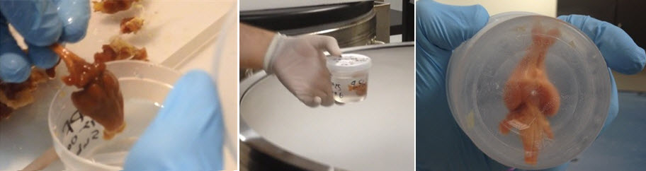