Feeds:
Posts
Comments

Posts Tagged ‘Artificial intelligence’

AI Acquisitions by Big Tech Firms Are Happening at a Blistering Pace: 2019 Recent Data by CBI Insights

Reporter: Stephen J. Williams, Ph.D.

3.4.16

3.4.16   AI Acquisitions by Big Tech Firms Are Happening at a Blistering Pace: 2019 Recent Data by CBI Insights, Volume 2 (Volume Two: Latest in Genomics Methodologies for Therapeutics: Gene Editing, NGS and BioInformatics, Simulations and the Genome Ontology), Part 3: AI in Medicine

Recent report from CBI Insights shows the rapid pace at which the biggest tech firms (Google, Apple, Microsoft, Facebook, and Amazon) are acquiring artificial intelligence (AI) startups, potentially confounding the AI talent shortage that exists.

The link to the report and free download is given here at https://www.cbinsights.com/research/top-acquirers-ai-startups-ma-timeline/

Part of the report:

TECH GIANTS LEAD IN AI ACQUISITIONS

The usual suspects are leading the race for AI: tech giants like Facebook, Amazon, Microsoft, Google, & Apple (FAMGA) have all been aggressively acquiring AI startups in the last decade.

Among the FAMGA companies, Apple leads the way, making 20 total AI acquisitions since 2010. It is followed by Google (the frontrunner from 2012 to 2016) with 14 acquisitions and Microsoft with 10.

Apple’s AI acquisition spree, which has helped it overtake Google in recent years, was essential to the development of new iPhone features. For example, FaceID, the technology that allows users to unlock their iPhone X just by looking at it, stems from Apple’s M&A moves in chips and computer vision, including the acquisition of AI company RealFace.

In fact, many of FAMGA’s prominent products and services came out of acquisitions of AI companies — such as Apple’s Siri, or Google’s contributions to healthcare through DeepMind.

That said, tech giants are far from the only companies snatching up AI startups.

Since 2010, there have been 635 AI acquisitions, as companies aim to build out their AI capabilities and capture sought-after talent (as of 8/31/2019).

The pace of these acquisitions has also been increasing. AI acquisitions saw a more than 6x uptick from 2013 to 2018, including last year’s record of 166 AI acquisitions — up 38% year-over-year.

In 2019, there have already been 140+ acquisitions (as of August), putting the year on track to beat the 2018 record at the current run rate.

Part of this increase in the pace of AI acquisitions can be attributed to a growing diversity in acquirers. Where once AI was the exclusive territory of major tech companies, today, smaller AI startups are becoming acquisition targets for traditional insurance, retail, and healthcare incumbents.

For example, in February 2018, Roche Holding acquired New York-based cancer startup Flatiron Health for $1.9B — one of the largest M&A deals in artificial intelligence. This year, Nike acquired AI-powered inventory management startup Celect, Uber acquired computer vision company Mighty AI, and McDonald’s acquired personalization platform Dynamic Yield.

Despite the increased number of acquirers, however, tech giants are still leading the charge. Acquisitive tech giants have emerged as powerful global corporations with a competitive advantage in artificial intelligence, and startups have played a pivotal role in helping these companies scale their AI initiatives.

Apple, Google, Microsoft, Facebook, Intel, and Amazon are the most active acquirers of AI startups, each acquiring 7+ companies.

To read more on recent Acquisitions in the AI space please see the following articles on this Open Access Online Journal

Diversification and Acquisitions, 2001 – 2015: Trail known as “Google Acquisitions” – Understanding Alphabet’s Acquisitions: A Sector-By-Sector Analysis

Clarivate Analytics expanded IP data leadership by new acquisition of the leading provider of intellectual property case law and analytics Darts-ip

2019 Biotechnology Sector and Artificial Intelligence in Healthcare

Forbes Opinion: 13 Industries Soon To Be Revolutionized By Artificial Intelligence

Artificial Intelligence and Cardiovascular Disease

Multiple Barriers Identified Which May Hamper Use of Artificial Intelligence in the Clinical Setting

Top 12 Artificial Intelligence Innovations Disrupting Healthcare by 2020

The launch of SCAI – Interview with Gérard Biau, director of the Sorbonne Center for Artificial Intelligence (SCAI).

Read Full Post »

Artificial Intelligence and Cardiovascular Disease

Reporter and Curator: Dr. Sudipta Saha, Ph.D.

3.3.18

3.3.18   Artificial Intelligence and Cardiovascular Disease, Volume 2 (Volume Two: Latest in Genomics Methodologies for Therapeutics: Gene Editing, NGS and BioInformatics, Simulations and the Genome Ontology), Part 2: CRISPR for Gene Editing and DNA Repair

Cardiology is a vast field that focuses on a large number of diseases specifically dealing with the heart, the circulatory system, and its functions. As such, similar symptomatologies and diagnostic features may be present in an individual, making it difficult for a doctor to easily isolate the actual heart-related problem. Consequently, the use of artificial intelligence aims to relieve doctors from this hurdle and extend better quality to patients. Results of screening tests such as echocardiograms, MRIs, or CT scans have long been proposed to be analyzed using more advanced techniques in the field of technology. As such, while artificial intelligence is not yet widely-used in clinical practice, it is seen as the future of healthcare.

The continuous development of the technological sector has enabled the industry to merge with medicine in order to create new integrated, reliable, and efficient methods of providing quality health care. One of the ongoing trends in cardiology at present is the proposed utilization of artificial intelligence (AI) in augmenting and extending the effectiveness of the cardiologist. This is because AI or machine-learning would allow for an accurate measure of patient functioning and diagnosis from the beginning up to the end of the therapeutic process. In particular, the use of artificial intelligence in cardiology aims to focus on research and development, clinical practice, and population health. Created to be an all-in-one mechanism in cardiac healthcare, AI technologies incorporate complex algorithms in determining relevant steps needed for a successful diagnosis and treatment. The role of artificial intelligence specifically extends to the identification of novel drug therapies, disease stratification or statistics, continuous remote monitoring and diagnostics, integration of multi-omic data, and extension of physician effectivity and efficiency.

Artificial intelligence – specifically a branch of it called machine learning – is being used in medicine to help with diagnosis. Computers might, for example, be better at interpreting heart scans. Computers can be ‘trained’ to make these predictions. This is done by feeding the computer information from hundreds or thousands of patients, plus instructions (an algorithm) on how to use that information. This information is heart scans, genetic and other test results, and how long each patient survived. These scans are in exquisite detail and the computer may be able to spot differences that are beyond human perception. It can also combine information from many different tests to give as accurate a picture as possible. The computer starts to work out which factors affected the patients’ outlook, so it can make predictions about other patients.

In current medical practice, doctors will use risk scores to make treatment decisions for their cardiac patients. These are based on a series of variables like weight, age and lifestyle. However, they do not always have the desired levels of accuracy. A particular example of the use of artificial examination in cardiology is the experimental study on heart disease patients, published in 2017. The researchers utilized cardiac MRI-based algorithms coupled with a 3D systolic cardiac motion pattern to accurately predict the health outcomes of patients with pulmonary hypertension. The experiment proved to be successful, with the technology being able to pick-up 30,000 points within the heart activity of 250 patients. With the success of the aforementioned study, as well as the promise of other researches on artificial intelligence, cardiology is seemingly moving towards a more technological practice.

One study was conducted in Finland where researchers enrolled 950 patients complaining of chest pain, who underwent the centre’s usual scanning protocol to check for coronary artery disease. Their outcomes were tracked for six years following their initial scans, over the course of which 24 of the patients had heart attacks and 49 died from all causes. The patients first underwent a coronary computed tomography angiography (CCTA) scan, which yielded 58 pieces of data on the presence of coronary plaque, vessel narrowing and calcification. Patients whose scans were suggestive of disease underwent a positron emission tomography (PET) scan which produced 17 variables on blood flow. Ten clinical variables were also obtained from medical records including sex, age, smoking status and diabetes. These 85 variables were then entered into an artificial intelligence (AI) programme called LogitBoost. The AI repeatedly analysed the imaging variables, and was able to learn how the imaging data interacted and identify the patterns which preceded death and heart attack with over 90% accuracy. The predictive performance using the ten clinical variables alone was modest, with an accuracy of 90%. When PET scan data was added, accuracy increased to 92.5%. The predictive performance increased significantly when CCTA scan data was added to clinical and PET data, with accuracy of 95.4%.

Another study findings showed that applying artificial intelligence (AI) to the electrocardiogram (ECG) enables early detection of left ventricular dysfunction and can identify individuals at increased risk for its development in the future. Asymptomatic left ventricular dysfunction (ALVD) is characterised by the presence of a weak heart pump with a risk of overt heart failure. It is present in three to six percent of the general population and is associated with reduced quality of life and longevity. However, it is treatable when found. Currently, there is no inexpensive, noninvasive, painless screening tool for ALVD available for diagnostic use. When tested on an independent set of 52,870 patients, the network model yielded values for the area under the curve, sensitivity, specificity, and accuracy of 0.93, 86.3 percent, 85.7 percent, and 85.7 percent, respectively. Furthermore, in patients without ventricular dysfunction, those with a positive AI screen were at four times the risk of developing future ventricular dysfunction compared with those with a negative screen.

In recent years, the analysis of big data database combined with computer deep learning has gradually played an important role in biomedical technology. For a large number of medical record data analysis, image analysis, single nucleotide polymorphism difference analysis, etc., all relevant research on the development and application of artificial intelligence can be observed extensively. For clinical indication, patients may receive a variety of cardiovascular routine examination and treatments, such as: cardiac ultrasound, multi-path ECG, cardiovascular and peripheral angiography, intravascular ultrasound and optical coherence tomography, electrical physiology, etc. By using artificial intelligence deep learning system, the investigators hope to not only improve the diagnostic rate and also gain more accurately predict the patient’s recovery, improve medical quality in the near future.

The primary issue about using artificial intelligence in cardiology, or in any field of medicine for that matter, is the ethical issues that it brings about. Physicians and healthcare professionals prior to their practice swear to the Hippocratic Oath—a promise to do their best for the welfare and betterment of their patients. Many physicians have argued that the use of artificial intelligence in medicine breaks the Hippocratic Oath since patients are technically left under the care of machines than of doctors. Furthermore, as machines may also malfunction, the safety of patients is also on the line at all times. As such, while medical practitioners see the promise of artificial technology, they are also heavily constricted about its use, safety, and appropriateness in medical practice.

Issues and challenges faced by technological innovations in cardiology are overpowered by current researches aiming to make artificial intelligence easily accessible and available for all. With that in mind, various projects are currently under study. For example, the use of wearable AI technology aims to develop a mechanism by which patients and doctors could easily access and monitor cardiac activity remotely. An ideal instrument for monitoring, wearable AI technology ensures real-time updates, monitoring, and evaluation. Another direction of cardiology in AI technology is the use of technology to record and validate empirical data to further analyze symptomatology, biomarkers, and treatment effectiveness. With AI technology, researchers in cardiology are aiming to simplify and expand the scope of knowledge on the field for better patient care and treatment outcomes.

References:

https://www.news-medical.net/health/Artificial-Intelligence-in-Cardiology.aspx

https://www.bhf.org.uk/informationsupport/heart-matters-magazine/research/artificial-intelligence

https://www.medicaldevice-network.com/news/heart-attack-artificial-intelligence/

https://www.nature.com/articles/s41569-019-0158-5

https://www.ncbi.nlm.nih.gov/pmc/articles/PMC5711980/

www.j-pcs.org/article.asp

http://www.onlinejacc.org/content/71/23/2668

http://www.scielo.br/pdf/ijcs/v30n3/2359-4802-ijcs-30-03-0187.pdf

https://www.escardio.org/The-ESC/Press-Office/Press-releases/How-artificial-intelligence-is-tackling-heart-disease-Find-out-at-ICNC-2019

https://clinicaltrials.gov/ct2/show/NCT03877614

https://www.europeanpharmaceuticalreview.com/news/82870/artificial-intelligence-ai-heart-disease/

https://www.frontiersin.org/research-topics/10067/current-and-future-role-of-artificial-intelligence-in-cardiac-imaging

https://www.news-medical.net/health/Artificial-Intelligence-in-Cardiology.aspx

https://www.sciencedaily.com/releases/2019/05/190513104505.htm

Read Full Post »

Multiple Barriers Identified Which May Hamper Use of Artificial Intelligence in the Clinical Setting

Reporter: Stephen J. Williams, PhD.

From the Journal Science:Science  21 Jun 2019: Vol. 364, Issue 6446, pp. 1119-1120

By Jennifer Couzin-Frankel

3.3.21

3.3.21   Multiple Barriers Identified Which May Hamper Use of Artificial Intelligence in the Clinical Setting, Volume 2 (Volume Two: Latest in Genomics Methodologies for Therapeutics: Gene Editing, NGS and BioInformatics, Simulations and the Genome Ontology), Part 2: CRISPR for Gene Editing and DNA Repair

In a commentary article from Jennifer Couzin-Frankel entitled “Medicine contends with how to use artificial intelligence  the barriers to the efficient and reliable adoption of artificial intelligence and machine learning in the hospital setting are discussed.   In summary these barriers result from lack of reproducibility across hospitals. For instance, a major concern among radiologists is the AI software being developed to read images in order to magnify small changes, such as with cardiac images, is developed within one hospital and may not reflect the equipment or standard practices used in other hospital systems.  To address this issue, lust recently, US scientists and government regulators issued guidance describing how to convert research-based AI into improved medical images and published these guidance in the Journal of the American College of Radiology.  The group suggested greater collaboration among relevant parties in developing of AI practices, including software engineers, scientists, clinicians, radiologists etc. 

As thousands of images are fed into AI algorithms, according to neurosurgeon Eric Oermann at Mount Sinai Hospital, the signals they recognize can have less to do with disease than with other patient characteristics, the brand of MRI machine, or even how a scanner is angled.  For example Oermann and Mount Sinai developed an AI algorithm to detect spots on a lung scan indicative of pneumonia and when tested in a group of new patients the algorithm could detect pneumonia with 93% accuracy.  

However when the group from Sinai tested their algorithm from tens of thousands of scans from other hospitals including NIH success rate fell to 73-80%, indicative of bias within the training set: in other words there was something unique about the way Mt. Sinai does their scans relative to other hospitals.  Indeed, many of the patients Mt. Sinai sees are too sick to get out of bed and radiologists would use portable scanners, which generate different images than stand alone scanners.  

The results were published in Plos Medicine as seen below:

PLoS Med. 2018 Nov 6;15(11):e1002683. doi: 10.1371/journal.pmed.1002683. eCollection 2018 Nov.

Variable generalization performance of a deep learning model to detect pneumonia in chest radiographs: A cross-sectional study.

Zech JR1, Badgeley MA2, Liu M2, Costa AB3, Titano JJ4, Oermann EK3.

Abstract

BACKGROUND:

There is interest in using convolutional neural networks (CNNs) to analyze medical imaging to provide computer-aided diagnosis (CAD). Recent work has suggested that image classification CNNs may not generalize to new data as well as previously believed. We assessed how well CNNs generalized across three hospital systems for a simulated pneumonia screening task.

METHODS AND FINDINGS:

A cross-sectional design with multiple model training cohorts was used to evaluate model generalizability to external sites using split-sample validation. A total of 158,323 chest radiographs were drawn from three institutions: National Institutes of Health Clinical Center (NIH; 112,120 from 30,805 patients), Mount Sinai Hospital (MSH; 42,396 from 12,904 patients), and Indiana University Network for Patient Care (IU; 3,807 from 3,683 patients). These patient populations had an age mean (SD) of 46.9 years (16.6), 63.2 years (16.5), and 49.6 years (17) with a female percentage of 43.5%, 44.8%, and 57.3%, respectively. We assessed individual models using the area under the receiver operating characteristic curve (AUC) for radiographic findings consistent with pneumonia and compared performance on different test sets with DeLong’s test. The prevalence of pneumonia was high enough at MSH (34.2%) relative to NIH and IU (1.2% and 1.0%) that merely sorting by hospital system achieved an AUC of 0.861 (95% CI 0.855-0.866) on the joint MSH-NIH dataset. Models trained on data from either NIH or MSH had equivalent performance on IU (P values 0.580 and 0.273, respectively) and inferior performance on data from each other relative to an internal test set (i.e., new data from within the hospital system used for training data; P values both <0.001). The highest internal performance was achieved by combining training and test data from MSH and NIH (AUC 0.931, 95% CI 0.927-0.936), but this model demonstrated significantly lower external performance at IU (AUC 0.815, 95% CI 0.745-0.885, P = 0.001). To test the effect of pooling data from sites with disparate pneumonia prevalence, we used stratified subsampling to generate MSH-NIH cohorts that only differed in disease prevalence between training data sites. When both training data sites had the same pneumonia prevalence, the model performed consistently on external IU data (P = 0.88). When a 10-fold difference in pneumonia rate was introduced between sites, internal test performance improved compared to the balanced model (10× MSH risk P < 0.001; 10× NIH P = 0.002), but this outperformance failed to generalize to IU (MSH 10× P < 0.001; NIH 10× P = 0.027). CNNs were able to directly detect hospital system of a radiograph for 99.95% NIH (22,050/22,062) and 99.98% MSH (8,386/8,388) radiographs. The primary limitation of our approach and the available public data is that we cannot fully assess what other factors might be contributing to hospital system-specific biases.

CONCLUSION:

Pneumonia-screening CNNs achieved better internal than external performance in 3 out of 5 natural comparisons. When models were trained on pooled data from sites with different pneumonia prevalence, they performed better on new pooled data from these sites but not on external data. CNNs robustly identified hospital system and department within a hospital, which can have large differences in disease burden and may confound predictions.

PMID: 30399157 PMCID: PMC6219764 DOI: 10.1371/journal.pmed.1002683

[Indexed for MEDLINE] Free PMC Article

Images from this publication.See all images (3)Free text


Surprisingly, not many researchers have begun to use data obtained from different hospitals.  The FDA has issued some guidance in the matter but considers “locked” AI software or unchanging software as a medical device.  However they just announced development of a framework for regulating more cutting edge software that continues to learn over time.

Still the key point is that collaboration over multiple health systems in various countries may be necessary for development of AI software which is used in multiple clinical settings.  Otherwise each hospital will need to develop their own software only used on their own system and would provide a regulatory headache for the FDA.

Other articles on Artificial Intelligence in Clinical Medicine on this Open Access Journal include:

Top 12 Artificial Intelligence Innovations Disrupting Healthcare by 2020

The launch of SCAI – Interview with Gérard Biau, director of the Sorbonne Center for Artificial Intelligence (SCAI).

Real Time Coverage @BIOConvention #BIO2019: Machine Learning and Artificial Intelligence #AI: Realizing Precision Medicine One Patient at a Time

50 Contemporary Artificial Intelligence Leading Experts and Researchers

Read Full Post »

Reporter: Gail S. Thornton, M.A.

This article is excerpted from the Harvard Business Review, May 28, 2019

By Moni Miyashita, Michael Brady

3.4.13   The Health Care Benefits of Combining Wearables and AI, Volume 2 (Volume Two: Latest in Genomics Methodologies for Therapeutics: Gene Editing, NGS and BioInformatics, Simulations and the Genome Ontology), Part 3: AI in Medicine

In southeast England, patients discharged from a group of hospitals serving 500,000 people are being fitted with a Wi-Fi-enabled armband that remotely monitors vital signs such as respiratory rate, oxygen levels, pulse, blood pressure, and body temperature.

Under a National Health Service pilot program that now incorporates artificial intelligence to analyze all that patient data in real time, hospital readmission rates are down, and emergency room visits have been reduced. What’s more, the need for costly home visits has dropped by 22%. Longer term, adherence to treatment plans have increased to 96%, compared to the industry average of 50%.

The AI pilot is targeting what Harvard Business School Professor and Innosight co-founder Clay Christensen calls “non-consumption.”  These are opportunity areas where consumers have a job to be done that isn’t currently addressed by an affordable or convenient solution.

Before the U.K. pilot at the Dartford and Gravesham hospitals, for instance, home monitoring had involved dispatching hospital staffers to drive up to 90 minutes round-trip to check in with patients in their homes about once per week. But with algorithms now constantly searching for warning signs in the data and alerting both patients and professionals instantly, a new capability is born: providing healthcare before you knew you even need it.

The biggest promise of artificial intelligence — accurate predictions at near-zero marginal cost — has rightly generated substantial interest in applying AI to nearly every area of healthcare. But not every application of AI in healthcare is equally well-suited to benefit. Moreover, very few applications serve as an appropriate strategic response to the largest problems facing nearly every health system: decentralization and margin pressure.

Take for example, medical imaging AI tools — an area in which hospitals are projected to spend $2 billion annually within four years. Accurately diagnosing diseases from cancers to cataracts is a complex task, with difficult-to-quantify but typically major consequences. However, the task is currently typically part of larger workflows performed by extensively trained, highly specialized physicians who are among some of the world’s best minds. These doctors might need help at the margins, but this is a job already being done. Such factors make disease diagnosis an extraordinarily difficult area for AI to create transformative change. And so the application of AI in such settings  —  even if beneficial  to patient outcomes —  is unlikely to fundamentally improve the way healthcare is delivered or to substantially lower costs in the near-term.

However, leading organizations seeking to decentralize care can deploy AI to do things that have never been done before. For example: There’s a wide array of non-acute health decisions that consumers make daily. These decisions do not warrant the attention of a skilled clinician but ultimately play a large role in determining patient’s health — and ultimately the cost of healthcare.

According to the World Health Organization, 60% of related factors to individual health and quality of life are correlated to lifestyle choices, including taking prescriptions such as blood-pressure medications correctly, getting exercise, and reducing stress. Aided by AI-driven models, it is now possible to provide patients with interventions and reminders throughout this day-to-day process based on changes to the patient’s vital signs.

Home health monitoring itself isn’t new. Active programs and pilot studies are underway through leading institutions ranging from Partners Healthcare, United Healthcare, and the Johns Hopkins School of Medicine, with positive results. But those efforts have yet to harness AI to make better judgements and recommendations in real time. Because of the massive volumes of data involved, machine learning algorithms are particularly well suited to scaling that task for large populations. After all, large sets of data are what power AI by making those algorithms smarter.

By deploying AI, for instance, the NHS program is not only able to scale up in the U.K. but also internationally. Current Health, the venture-capital backed maker of the patient monitoring devices used in the program, recently received FDA clearance to pilot the system in the U.S. and is now testing it with New York’s Mount Sinai Hospital. It’s part of an effort to reduce patient readmissions, which costs U.S. hospitals about $40 billion annually.

The early success of such efforts drives home three lessons in using AI to address non-consumption in the new world of patient-centric healthcare:

1) Focus on impacting critical metrics – for example, reducing costly hospital readmission rates.

Start small to home in on the goal of making an impact on a key metric tied to both patient outcomes and financial sustainability. As in the U.K. pilot, this can be done through a program with select hospitals or provider locations. In another case Grady Hospital, the largest public hospital in Atlanta, points to $4M in saving from reduced readmission rates by 31% over two years thanks to the adoption of an AI tool which identifies ‘at-risk’ patients. The system alerts clinical teams to initiate special patient touch points and interventions.

2) Reduce risk by relying on new kinds of partners.

Don’t try to do everything alone. Instead, form alliances with partners that are aiming to tackle similar problems. Consider the Synaptic Healthcare Alliance, a collaborative pilot program between Aetna, Ascension, Humana, Optum, and others. The alliance is using Blockchain to create a giant dataset across various health care providers, with AI trials on the data getting underway. The aim is to streamline health care provider data management with the goal of reducing the cost of processing claims while also improving access to care. Going it alone can be risky due to data incompatibility issues alone. For instance, the M.D. Anderson Cancer Center had to write off millions in costs for a failed AI project due in part to incompatibility with its electronic health records system. By joining forces, Synaptic’s dataset will be in a standard format that makes records and results transportable.

3) Use AI to collaborate, not compete, with highly-trained professionals.

Clinicians are often looking to augment their knowledge and reasoning, and AI can help. Many medical AI applications do actually compete with doctors. In radiology, for instance, some algorithms have performed image-bases diagnosis as well as or better than human experts. Yet it’s unclear if patients and medical institutions will trust AI to automate that job entirely. A University of California at San Diego pilot in which AI successfully diagnosed childhood diseases more accurately than junior-level pediatricians still required senior doctors to personally review and sign off on the diagnosis. The real aim is always going to be to use AI to collaborate with clinicians seeking higher precision — not try to replace them.

MIT and MGH have developed a deep learning model which identifies patients likely to develop breast cancer in the future. Learning from data on 60,000 prior patients, the AI system allows physicians to personalize their approach to breast cancer screening, essentially creating a detailed risk profile for each patient.

Taken together, these three lessons paired with solutions targeted at non-consumption have the potential to provide a clear path to effectively harnessing a technology that has been subject to rampant over-promising. Longer term, we believe the one of the transformative benefits of AI will be deepening relationships between health providers and patients. The U.K. pilot, for instance, is resulting in more frequent proactive check-ins that never would have happened before. That’s good for both improving health as well as customer loyalty in the emerging consumer-centric healthcare marketplace.

Source:

https://hbr.org/2019/05/the-health-care-benefits-of-combining-wearables-and-ai


Read Full Post »

These twelve artificial intelligence innovations are expected to start impacting clinical care by the end of the decade.

Reporter: Gail S. Thornton, M.A.

This article is excerpted from Health IT Analytics, April 11, 2019.

 By Jennifer Bresnick

3.4.14   These twelve artificial intelligence innovations are expected to start impacting clinical care by the end of the decade, Volume 2 (Volume Two: Latest in Genomics Methodologies for Therapeutics: Gene Editing, NGS and BioInformatics, Simulations and the Genome Ontology), Part 3: AI in Medicine

April 11, 2019 – There’s no question that artificial intelligence is moving quickly in the healthcare industry.  Even just a few months ago, AI was still a dream for the next generation: something that would start to enter regular care delivery in a couple of decades – maybe ten or fifteen years for the most advanced health systems.

Even Partners HealthCare, the Boston-based giant on the very cutting edge of research and reform, set a ten-year timeframe for artificial intelligence during its 2018 World Medical Innovation Forum, identifying a dozen AI technologies that had the potential to revolutionize patient care within the decade.

But over the past twelve months, research has progressed so rapidly that Partners has blown up that timeline. 

Instead of viewing AI as something still lingering on the distant horizon, this year’s Disruptive Dozen panel was tasked with assessing which AI innovations will be ready to fundamentally alter the delivery of care by 2020 – now less than a year away.

Sixty members of the Partners faculty participated in nominating and narrowing down the tools they think will have an almost immediate benefit for patients and providers, explained Erica Shenoy, MD, PhD, an infectious disease specialist at Massachusetts General Hospital (MGH).

“These are innovations that have a strong potential to make significant advancement in the field, and they are also technologies that are pretty close to making it to market,” she said.

The results include everything from mental healthcare and clinical decision support to coding and communication, offering patients and their providers a more efficient, effective, and cost-conscious ecosystem for improving long-term outcomes.

In order from least to greatest potential impact, here are the twelve artificial intelligence innovations poised to become integral components of the next decade’s data-driven care delivery system.

NARROWING THE GAPS IN MENTAL HEALTHCARE

Nearly twenty percent of US patients struggle with a mental health disorder, yet treatment is often difficult to access and expensive to use regularly.  Reducing barriers to access for mental and behavioral healthcare, especially during the opioid abuse crisis, requires a new approach to connecting patients with services.

AI-driven applications and therapy programs will be a significant part of the answer.

“The promise and potential for digital behavioral solutions and apps is enormous to address the gaps in mental healthcare in the US and across the world,” said David Ahern, PhD, a clinical psychologist at Brigham & Women’s Hospital (BWH). 

Smartphone-based cognitive behavioral therapy and integrated group therapy are showing promise for treating conditions such as depression, eating disorders, and substance abuse.

While patients and providers need to be wary of commercially available applications that have not been rigorously validated and tested, more and more researchers are developing AI-based tools that have the backing of randomized clinical trials and are showing good results.

A panel of experts from Partners HealthCare presents the Disruptive Dozen at WMIF19.

Source: Partners HealthCare

STREAMLINING WORKFLOWS WITH VOICE-FIRST TECHNOLOGY

Natural language processing is already a routine part of many behind-the-scenes clinical workflows, but voice-first tools are expected to make their way into the patient-provider encounter in a new way. 

Smart speakers in the clinic are prepping to relieve clinicians of their EHR burdens, capturing free-form conversations and translating the content into structured documentation.  Physicians and nurses will be able to collect and retrieve information more quickly while spending more time looking patients in the eye.

Patients may benefit from similar technologies at home as the consumer market for virtual assistants continues to grow.  With companies like Amazon achieving HIPAA compliance for their consumer-facing products, individuals may soon have more robust options for voice-first chronic disease management and patient engagement.

IDENTIFYING INDIVIDUALS AT HIGH RISK OF DOMESTIC VIOLENCE

Underreporting makes it difficult to know just how many people suffer from intimate partner violence (IPV), says Bharti Khurana, MD, an emergency radiologist at BWH.  But the symptoms are often hiding in plain sight for radiologists.

Using artificial intelligence to flag worrisome injury patterns or mismatches between patient-reported histories and the types of fractures present on x-rays can alert providers to when an exploratory conversation is called for.

“As a radiologist, I’m very excited because this will enable me to provide even more value to the patient instead of simply evaluating their injuries.  It’s a powerful tool for clinicians and social workers that will allow them to approach patients with confidence and with less worry about offending the patient or the spouse,” said Khurana.

REVOLUTIONIZING ACUTE STROKE CARE

Every second counts when a patient experiences a stroke.  In far-flung regions of the United States and in the developing world, access to skilled stroke care can take hours, drastically increasing the likelihood of significant long-term disability or death.

Artificial intelligence has the potential to close the gaps in access to high-quality imaging studies that can identify the type of stroke and the location of the clot or bleed.  Research teams are currently working on AI-driven tools that can automate the detection of stroke and support decision-making around the appropriate treatment for the individual’s needs.  

In rural or low-resource care settings, these algorithms can compensate for the lack of a specialist on-site and ensure that every stroke patient has the best possible chance of treatment and recovery.

AI revolutionizing stroke care

Source: Getty Images

REDUCING ADMINISTRATIVE BURDENS FOR PROVIDERS

The costs of healthcare administration are off the charts.  Recent data from the Center for American progress states that providers spend about $282 billion per year on insurance and medical billing, and the burdens are only going to keep getting bigger.

Medical coding and billing is a perfect use case for natural language processing and machine learning.  NLP is well-suited to translating free-text notes into standardized codes, which can move the task off the plates of physicians and reduce the time and effort spent on complying with convoluted regulations.

“The ultimate goal is to help reduce the complexity of the coding and billing process through automation, thereby reducing the number of mistakes – and, in turn, minimizing the need for such intense regulatory oversight,” Partners says.

NLP is already in relatively wide use for this task, and healthcare organizations are expected to continue adopting this strategy as a way to control costs and speed up their billing cycles.

UNLEASHING HEALTH DATA THROUGH INFORMATION EXCHANGE

AI will combine with another game-changing technology, known as FHIR, to unlock siloes of health data and support broader access to health information.

Patients, providers, and researchers will all benefit from a more fluid health information exchange environment, especially since artificial intelligence models are extremely data-hungry.

Stakeholders will need to pay close attention to maintaining the privacy and security of data as it moves across disparate systems, but the benefits have the potential to outweigh the risks.

“It completely depends on how everyone in the medical community advocates for, builds, and demands open interfaces and open business models,” said Samuel Aronson, Executive Director of IT at Partners Personalized Medicine.

“If we all row in the same direction, there’s a real possibility that we will see fundamental improvements to the healthcare system in 3 to 5 years.”

OFFERING NEW APPROACHES FOR EYE HEALTH AND DISEASE

Image-heavy disciplines have started to see early benefits from artificial intelligence since computers are particularly adept at analyzing patterns in pixels.  Ophthalmology is one area that could see major changes as AI algorithms become more accurate and more robust.

From glaucoma to diabetic retinopathy, millions of patients experience diseases that can lead to irreversible vision loss every year.  Employing AI for clinical decision support can extend access to eye health services in low-resource areas while giving human providers more accurate tools for catching diseases sooner.

REAL-TIME MONITORING OF BRAIN HEALTH

The brain is still the body’s most mysterious organ, but scientists and clinicians are making swift progress unlocking the secrets of cognitive function and neurological disease.  Artificial intelligence is accelerating discovery by helping providers interpret the incredibly complex data that the brain produces.

From predicting seizures by reading EEG tests to identifying the beginnings of dementia earlier than any human, artificial intelligence is allowing providers to access more detailed, continuous measurements – and helping patients improve their quality of life.

Seizures can happen in patients with other serious illnesses, such as kidney or liver failure, explained, Bandon Westover, MD, PhD, executive director of the Clinical Data Animation Center at MGH, but many providers simply don’t know about it.

“Right now, we mostly ignore the brain unless there’s a special need for suspicion,” he said.  “In a year’s time, we’ll be catching a lot more seizures and we’ll be doing it with algorithms that can monitor patients continuously and identify more ambiguous patterns of dysfunction that can damage the brain in a similar manner to seizures.”

AUTOMATING MALARIA DETECTION IN DEVELOPING REGIONS

Malaria is a daily threat for approximately half the world’s population.  Nearly half a million people died from the mosquito-borne disease in 2017, according to the World Health Organization, and the majority of the victims are children under the age of five.

Deep learning tools can automate the process of quantifying malaria parasites in blood samples, a challenging task for providers working without pathologist partners.  One such tool achieved 90 percent accuracy and specificity, putting it on par with pathology experts.

This type of software can be run on a smartphone hooked up to a camera on a microscope, dramatically expanding access to expert-level diagnosis and monitoring.

AI for diagnosing and detecting malaria

Source: Getty Images

AUGMENTING DIAGNOSTICS AND DECISION-MAKING

Artificial intelligence has made especially swift progress in diagnostic specialties, including pathology. AI will continue to speed down the road to maturity in this area, predicts Annette Kim, MD, PhD, associate professor of pathology at BWH and Harvard Medical School.

“Pathology is at the center of diagnosis, and diagnosis underpins a huge percentage of all patient care.  We’re integrating a huge amount of data that funnels through us to come to a diagnosis.  As the number of data points increases, it negatively impacts the time we have to synthesize the information,” she said.

AI can help automate routine, high-volume tasks, prioritize and triage cases to ensure patients are getting speedy access to the right care, and make sure that pathologists don’t miss key information hidden in the enormous volumes of clinical and test data they must comb through every day.

“This is where AI can have a huge impact on practice by allowing us to use our limited time in the most meaningful manner,” Kim stressed.

PREDICTING THE RISK OF SUICIDE AND SELF-HARM

Suicide is the tenth leading cause of death in the United States, claiming 45,000 lives in 2016.  Suicide rates are on the rise due to a number of complex socioeconomic and mental health factors, and identifying patients at the highest risk of self-harm is a difficult and imprecise science.

Natural language processing and other AI methodologies may help providers identify high-risk patients earlier and more reliably.  AI can comb through social media posts, electronic health record notes, and other free-text documents to flag words or concepts associated with the risk of harm.

Researchers also hope to develop AI-driven apps to provide support and therapy to individuals likely to harm themselves, especially teenagers who commit suicide at higher rates than other age groups.

Connecting patients with mental health resources before they reach a time of crisis could save thousands of lives every year.

REIMAGINING THE WORLD OF MEDICAL IMAGING

Radiology is already one of AI’s early beneficiaries, but providers are just at the beginning of what they will be able to accomplish in the next few years as machine learning explodes into the imaging realm.

AI is predicted to bring earlier detection, more accurate assessment of complex images, and less expensive testing for patients across a huge number of clinical areas.

But as leaders in the AI revolution, radiologists also have a significant responsibility to develop and deploy best practices in terms of trustworthiness, workflow, and data protection.

“We certainly feel the onus on the radiology community to make sure we do deliver and translate this into improved care,” said Alexandra Golby, MD, a neurosurgeon and radiologist at BWH and Harvard Medical School.

“Can radiology live up to the expectations?  There are certainly some challenges, including trust and understanding of what the algorithms are delivering.  But we desperately need it, and we want to equalize care across the world.”

Radiologists have been among the first to overcome their trepidation about the role of AI in a changing clinical world, and are eagerly embracing the possibilities of this transformative approach to augmenting human skills.”

“All of the imaging societies have opened their doors to the AI adventure,” Golby said.  “The community very anxious to learn, codevelop, and work with all of the industry partners to turn this technology into truly valuable tools. We’re very optimistic and very excited, and we look forward to learning more about how AI can improve care.”

Source:

https://healthitanalytics.com/news/top-12-artificial-intelligence-innovations-disrupting-healthcare-by-2020


Read Full Post »

AI System Used to Detect Lung Cancer

Reporter: Irina Robu, PhD

3.3.13

3.3.13   AI System Used to Detect Lung Cancer, Volume 2 (Volume Two: Latest in Genomics Methodologies for Therapeutics: Gene Editing, NGS and BioInformatics, Simulations and the Genome Ontology), Part 2: CRISPR for Gene Editing and DNA Repair

Lung cancer is characterized by uncontrolled cell growth in tissues of the lung. The growth spreads beyond the lung by metastasis into nearby tissues. The most common symptoms are coughing (including coughing up blood), weight loss, shortness of breath, and chest pains. The two main types of lung cancer are small-cell lung carcinoma(SCLC) and non-small-cell lung carcinoma (NSCLC). Lung cancer may be seen on chest radiographs and computed tomography(CT) scans. However, computers seem to be as good or better than regular doctors at detecting tiny lung cancers on CT scans according to scientists from Google.

The AI designed by Google was able to interpret images using the same skills as humans to read microscope slides, X-rays, M.R.I.s and other medical scans by feeding huge amounts of data from medical imaging into the systems. It seems that the researchers were able to train computers to recognize patterns linked to a specific condition.

In a new Google study, the scientists applied artificial intelligence to CT scans used to screen people for lung cancer. Current studies have shown that screening can reduce the risk of dying from lung cancer and can also identify spots that might later become malignant.

The researchers created a neural network with multiple layers of processing and trained the AI by giving it many CT scans from patients whose diagnoses were known. This allows radiologists to sort patients into risk groups and decide whether biopsies are needed or follow up to keep track of the suspected regions. Even though the technology seems promising, but it can have pitfalls such as missing tumors, mistaken benign spots for malignancies and push patients into risky procedures.

Yet, the ability to process vast amounts of data may make it imaginable for artificial intelligence to recognize subtle patterns that humans simply cannot see. It is well understood that the systems should be studied extensively before using them for general public use. The lung-screening neural network is not ready for the clinic yet.

SOURCE

https://www.unilad.co.uk/technology/a-i-took-test-to-detect-lung-cancer-and-smashed-it

Read Full Post »

Sepsis Detection using an Algorithm More Efficient than Standard Methods

Reporter : Irina Robu, PhD

3.3.15

3.3.15   Sepsis Detection using an Algorithm More Efficient than Standard Methods, Volume 2 (Volume Two: Latest in Genomics Methodologies for Therapeutics: Gene Editing, NGS and BioInformatics, Simulations and the Genome Ontology), Part 2: CRISPR for Gene Editing and DNA Repair

Sepsis is a complication of severe infection categorized by a systemic inflammatory response with mortality rates between 25% to 30% for severe sepsis and 40% to 70% for septic shock. The most common sites of infection are the respiratory, genitourinary, and gastrointestinal systems, as well as the skin and soft tissue. The first manifestation of sepsis is fever with pneumonia being the most common symptom of sepsis with initial treatment which contains respiratory stabilization shadowed by aggressive fluid resuscitation. When fluid resuscitation fails to restore mean arterial pressure and organ perfusion, vasopressor therapy is indicated.

However, a machine-learning algorithm tested by Christopher Barton, MD from UC-San Francisco has exceeded the four typical methods used for catching sepsis early in hospital patients, giving clinicians up to 48 hours to interfere before the condition has a chance to begin turning dangerous. The four standard methods were Systemic Inflammatory Response Syndrome (SIRS) criteria, Sequential (Sepsis-Related) Organ-Failure Assessment (SOFA) and Modified Early Warning System (MEWS). The purpose of dividing the data sets between two far-flung institutions was to train and test the algorithm on demographically miscellaneous patient populations.

The patients involved in the study were admitted to hospital without sepsis and all had at least one recording of each of six vital signs such as oxygen levels in the blood, heart rate, respiratory rate, temperature, systolic blood pressure and diastolic blood pressure. Even though they were admitted to the hospital without it, some have contracted sepsis during their stay while others did not. Researchers used their algorithm detection versus the standard methods applied at sepsis onset at 24 hours and 48 hours prior.
Even though sepsis affects at least 1.7 million adults mostly outside of the hospital settings, nearly 270,000 die. Researchers are hoping that the algorithm would allow clinicians to interfere prior to the condition becoming deadly.

SOURCE
https://www.aiin.healthcare/topics/diagnostics/sepsis-diagnosis-machine-learning

Read Full Post »

AI App for People with Digestive Disorders

Reporter: Irina Robu, PhD

3.3.14

3.3.14   AI App for People with Digestive Disorders, Volume 2 (Volume Two: Latest in Genomics Methodologies for Therapeutics: Gene Editing, NGS and BioInformatics, Simulations and the Genome Ontology), Part 2: CRISPR for Gene Editing and DNA Repair

Artificial intelligence (AI) constitutes machine learning and deep learning, which allows computers to learn without being clearly programmed every step of the way. The basic principle decrees that AI is machine intelligence leading to the best outcome when given a problem. This sets up AI well for life science applications, which states that AI can be taught to differentiate cells, be used for higher quality imaging techniques, and analysis of genomic data.

Obviously, this type of technology which serves a function and removes the need for explicit programming. It is clear that digital therapeutics will have an essential role in treatment of individuals with gastrointestinal disorders such as IBS. Deep learning is a favorite among the AI facets in biology. The structure of deep learning has its roots in the structure of the human brain which connect to one another through which the data is passed. At each layer, some data is extracted. For example, in cells, one layer may analyze cell membrane, the next some organelle, and so on until the cell can be identified.

A Berlin-based startup,Cara Care uses AI to help people manage their chronic digestive problems and intends to spend the funding raised getting the app in the hands of gastrointestinal patients in the U.S. The company declares its app has already helped up to 400,000 people in Germany and the U.S. manage widespread GI conditions such as reflux, irritable or inflammatory bowel, food intolerances, Crohn’s disease and ulcerative colitis “with a 78.8% treatment success rate.” Cara Care will also use the funding to conduct research and expand collaborations with companies in the pharmaceutical, diagnostics and food-production industries.

SOURCE
https://www.aiin.healthcare/topics/connected-care/ai-app-digestive-disorders-raises-7m?utm_source=newsletter

Read Full Post »

Reported by Dror Nir, PhD

3.3.22

3.3.22   Deep Learning–Assisted Diagnosis of Cerebral Aneurysms, Volume 2 (Volume Two: Latest in Genomics Methodologies for Therapeutics: Gene Editing, NGS and BioInformatics, Simulations and the Genome Ontology), Part 2: CRISPR for Gene Editing and DNA Repair

Deep Learning–Assisted Diagnosis of Cerebral Aneurysms Using the HeadXNet Model

Allison Park, BA1Chris Chute, BS1Pranav Rajpurkar, MS1;  et al, Original Investigation, Health Informatics, June 7, 2019, JAMA Netw Open. 2019;2(6):e195600. doi:10.1001/jamanetworkopen.2019.5600

Key Points

Question  How does augmentation with a deep learning segmentation model influence the performance of clinicians in identifying intracranial aneurysms from computed tomographic angiography examinations?

Findings  In this diagnostic study of intracranial aneurysms, a test set of 115 examinations was reviewed once with model augmentation and once without in a randomized order by 8 clinicians. The clinicians showed significant increases in sensitivity, accuracy, and interrater agreement when augmented with neural network model–generated segmentations.

Meaning  This study suggests that the performance of clinicians in the detection of intracranial aneurysms can be improved by augmentation using deep learning segmentation models.

Abstract

Importance  Deep learning has the potential to augment clinician performance in medical imaging interpretation and reduce time to diagnosis through automated segmentation. Few studies to date have explored this topic.

Objective  To develop and apply a neural network segmentation model (the HeadXNet model) capable of generating precise voxel-by-voxel predictions of intracranial aneurysms on head computed tomographic angiography (CTA) imaging to augment clinicians’ intracranial aneurysm diagnostic performance.

Design, Setting, and Participants  In this diagnostic study, a 3-dimensional convolutional neural network architecture was developed using a training set of 611 head CTA examinations to generate aneurysm segmentations. Segmentation outputs from this support model on a test set of 115 examinations were provided to clinicians. Between August 13, 2018, and October 4, 2018, 8 clinicians diagnosed the presence of aneurysm on the test set, both with and without model augmentation, in a crossover design using randomized order and a 14-day washout period. Head and neck examinations performed between January 3, 2003, and May 31, 2017, at a single academic medical center were used to train, validate, and test the model. Examinations positive for aneurysm had at least 1 clinically significant, nonruptured intracranial aneurysm. Examinations with hemorrhage, ruptured aneurysm, posttraumatic or infectious pseudoaneurysm, arteriovenous malformation, surgical clips, coils, catheters, or other surgical hardware were excluded. All other CTA examinations were considered controls.

Main Outcomes and Measures  Sensitivity, specificity, accuracy, time, and interrater agreement were measured. Metrics for clinician performance with and without model augmentation were compared.

Results  The data set contained 818 examinations from 662 unique patients with 328 CTA examinations (40.1%) containing at least 1 intracranial aneurysm and 490 examinations (59.9%) without intracranial aneurysms. The 8 clinicians reading the test set ranged in experience from 2 to 12 years. Augmenting clinicians with artificial intelligence–produced segmentation predictions resulted in clinicians achieving statistically significant improvements in sensitivity, accuracy, and interrater agreement when compared with no augmentation. The clinicians’ mean sensitivity increased by 0.059 (95% CI, 0.028-0.091; adjusted P = .01), mean accuracy increased by 0.038 (95% CI, 0.014-0.062; adjusted P = .02), and mean interrater agreement (Fleiss κ) increased by 0.060, from 0.799 to 0.859 (adjusted P = .05). There was no statistically significant change in mean specificity (0.016; 95% CI, −0.010 to 0.041; adjusted P = .16) and time to diagnosis (5.71 seconds; 95% CI, 7.22-18.63 seconds; adjusted P = .19).

Conclusions and Relevance  The deep learning model developed successfully detected clinically significant intracranial aneurysms on CTA. This suggests that integration of an artificial intelligence–assisted diagnostic model may augment clinician performance with dependable and accurate predictions and thereby optimize patient care.

Introduction

Diagnosis of unruptured aneurysms is a critically important clinical task: intracranial aneurysms occur in 1% to 3% of the population and account for more than 80% of nontraumatic life-threatening subarachnoid hemorrhages.1 Computed tomographic angiography (CTA) is the primary, minimally invasive imaging modality currently used for diagnosis, surveillance, and presurgical planning of intracranial aneurysms,2,3but interpretation is time consuming even for subspecialty-trained neuroradiologists. Low interrater agreement poses an additional challenge for reliable diagnosis.47

Deep learning has recently shown significant potential in accurately performing diagnostic tasks on medical imaging.8 Specifically, convolutional neural networks (CNNs) have demonstrated excellent performance on a range of visual tasks, including medical image analysis.9 Moreover, the ability of deep learning systems to augment clinician workflow remains relatively unexplored.10 The development of an accurate deep learning model to help clinicians reliably identify clinically significant aneurysms in CTA has the potential to provide radiologists, neurosurgeons, and other clinicians an easily accessible and immediately applicable diagnostic support tool.

In this study, a deep learning model to automatically detect intracranial aneurysms on CTA and produce segmentations specifying regions of interest was developed to assist clinicians in the interpretation of CTA examinations for the diagnosis of intracranial aneurysms. Sensitivity, specificity, accuracy, time to diagnosis, and interrater agreement for clinicians with and without model augmentation were compared.

Methods

The Stanford University institutional review board approved this study. Owing to the retrospective nature of the study, patient consent or assent was waived. The Standards for Reporting of Diagnostic Accuracy (STARD) reporting guideline was used for the reporting of this study.

Data

A total of 9455 consecutive CTA examination reports of the head or head and neck performed between January 3, 2003, and May 31, 2017, at Stanford University Medical Center were retrospectively reviewed. Examinations with parenchymal hemorrhage, subarachnoid hemorrhage, posttraumatic or infectious pseudoaneurysm, arteriovenous malformation, ischemic stroke, nonspecific or chronic vascular findings such as intracranial atherosclerosis or other vasculopathies, surgical clips, coils, catheters, or other surgical hardware were excluded. Examinations of injuries that resulted from trauma or contained images degraded by motion were also excluded on visual review by a board-certified neuroradiologist with 12 years of experience. Examinations with nonruptured clinically significant aneurysms (>3 mm) were included.11

Radiologist Annotations

The reference standard for all examinations in the test set was determined by a board-certified neuroradiologist at a large academic practice with 12 years of experience who determined the presence of aneurysm by review of the original radiology report, double review of the CTA examination, and further confirmation of the aneurysm by diagnostic cerebral angiograms, if available. The neuroradiologist had access to all of the Digital Imaging and Communications in Medicine (DICOM) series, original reports, and clinical histories, as well as previous and follow-up examinations during interpretation to establish the best possible reference standard for the labels. For each of the aneurysm examinations, the radiologist also identified the location of each of the aneurysms. Using the open-source annotation software ITK-SNAP,12 the identified aneurysms were manually segmented on each slice.

Model Development

In this study, we developed a 3-dimensional (3-D) CNN called HeadXNet for segmentation of intracranial aneurysms from CT scans. Neural networks are functions with parameters structured as a sequence of layers to learn different levels of abstraction. Convolutional neural networks are a type of neural network designed to process image data, and 3-D CNNs are particularly well suited to handle sequences of images, or volumes.

HeadXNet is a CNN with an encoder-decoder structure (eFigure 1 in the Supplement), where the encoder maps a volume to an abstract low-resolution encoding, and the decoder expands this encoding to a full-resolution segmentation volume. The segmentation volume is of the same size as the corresponding study and specifies the probability of aneurysm for each voxel, which is the atomic unit of a 3-D volume, analogous to a pixel in a 2-D image. The encoder is adapted from a 50-layer SE-ResNeXt network,1315and the decoder is a sequence of 3 × 3 transposed convolutions. Similar to UNet,16 skip connections are used in 3 layers of the encoder to transmit outputs directly to the decoder. The encoder was pretrained on the Kinetics-600 data set,17 a large collection of YouTube videos labeled with human actions; after pretraining the encoder, the final 3 convolutional blocks and the 600-way softmax output layer were removed. In their place, an atrous spatial pyramid pooling18 layer and the decoder were added.

Training Procedure

Subvolumes of 16 slices were randomly sampled from volumes during training. The data set was preprocessed to find contours of the skull, and each volume was cropped around the skull in the axial plane before resizing each slice to 208 × 208 pixels. The slices were then cropped to 192 × 192 pixels (using random crops during training and centered crops during testing), resulting in a final input of size 16 × 192 × 192 per example; the same transformations were applied to the segmentation label. The segmentation output was trained to optimize a weighted combination of the voxelwise binary cross-entropy and Dice losses.19

Before reaching the model, inputs were clipped to [−300, 700] Hounsfield units, normalized to [−1, 1], and zero-centered. The model was trained on 3 Titan Xp graphical processing units (GPUs) (NVIDIA) using a minibatch of 2 examples per GPU. The parameters of the model were optimized using a stochastic gradient descent optimizer with momentum of 0.9 and a peak learning rate of 0.1 for randomly initialized weights and 0.01 for pretrained weights. The learning rate was scheduled with a linear warm-up from 0 to the peak learning rate for 10 000 iterations, followed by cosine annealing20 over 300 000 iterations. Additionally, the learning rate was fixed at 0 for the first 10 000 iterations for the pretrained encoder. For regularization, L2 weight decay of 0.001 was added to the loss for all trainable parameters and stochastic depth dropout21 was used in the encoder blocks. Standard dropout was not used.

To control for class imbalance, 3 methods were used. First, an auxiliary loss was added after the encoder and focal loss was used to encourage larger parameter updates on misclassified positive examples. Second, abnormal training examples were sampled more frequently than normal examples such that abnormal examples made up 30% of training iterations. Third, parameters of the decoder were not updated on training iterations where the segmentation label consisted of purely background (normal) voxels.

To produce a segmentation prediction for the entire volume, the segmentation outputs for sequential 16-slice subvolumes were simply concatenated. If the number of slices was not divisible by 16, the last input volume was padded with 0s and the corresponding output volume was truncated back to the original size.

Study Design

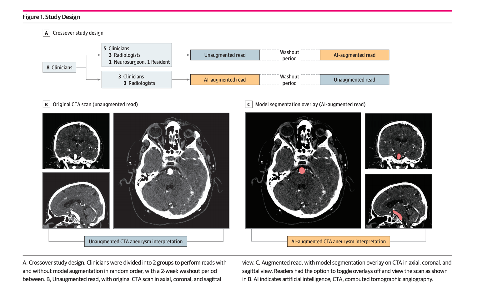
We performed a diagnostic accuracy study comparing performance metrics of clinicians with and without model augmentation. Each of the 8 clinicians participating in the study diagnosed a test set of 115 examinations, once with and once without assistance of the model. The clinicians were blinded to the original reports, clinical histories, and follow-up imaging examinations. Using a crossover design, the clinicians were randomly and equally divided into 2 groups. Within each group, examinations were sorted in a fixed random order for half of the group and sorted in reverse order for the other half. Group 1 first read the examinations without model augmentation, and group 2 first read the examinations with model augmentation. After a washout period of 14 days, the augmentation arrangement was reversed such that group 1 performed reads with model augmentation and group 2 read the examinations without model augmentation (Figure 1A).

Clinicians were instructed to assign a binary label for the presence or absence of at least 1 clinically significant aneurysm, defined as having a diameter greater than 3 mm. Clinicians read alone in a diagnostic reading room, all using the same high-definition monitor (3840 × 2160 pixels) displaying CTA examinations on a standard open-source DICOM viewer (Horos).22 Clinicians entered their labels into a data entry software application that automatically logged the time difference between labeling of the previous examination and the current examination.

When reading with model augmentation, clinicians were provided the model’s predictions in the form of region of interest (ROI) segmentations directly overlaid on top of CTA examinations. To ensure an image display interface that was familiar to all clinicians, the model’s predictions were presented as ROIs in a standard DICOM viewing software. At every voxel where the model predicted a probability greater than 0.5, readers saw a semiopaque red overlay on the axial, sagittal, and coronal series (Figure 1C). Readers had access to the ROIs immediately on loading the examinations, and the ROIs could be toggled off to reveal the unaltered CTA images (Figure 1B). The red overlays were the only indication that was given whether a particular CTA examination had been predicted by the model to contain an aneurysm. Given these model results, readers had the option to take it into consideration or disregard it based on clinical judgment. When readers performed diagnoses without augmentation, no ROIs were present on any of the examinations. Otherwise, the diagnostic tools were identical for augmented and nonaugmented reads.

Statistical Analysis

On the binary task of determining whether an examination contained an aneurysm, sensitivity, specificity, and accuracy were used to assess the performance of clinicians with and without model augmentation. Sensitivity denotes the number of true-positive results over total aneurysm-positive cases, specificity denotes the number of true-negative results over total aneurysm-negative cases, and accuracy denotes the number of true-positive and true-negative results over all test cases. The microaverage of these statistics across all clinicians was also computed by measuring each statistic pertaining to the total number of true-positive, false-negative, and false-positive results. In addition, to convert the models’ segmentation output of the model into a binary prediction, a prediction was considered positive if the model predicted at least 1 voxel as belonging to an aneurysm and negative otherwise. The 95% Wilson score confidence intervals were used to assess the variability in the estimates for sensitivity, specificity, and accuracy.23

To assess whether the clinicians achieved significant increases in performance with model augmentation, a 1-tailed t test was performed on the differences in sensitivity, specificity, and accuracy across all 8 clinicians. To determine the robustness of the findings and whether results were due to inclusion of the resident radiologist and neurosurgeon, we performed a sensitivity analysis: we computed the t test on the differences in sensitivity, specificity, and accuracy across board-certified radiologists only.

The average time to diagnosis for the clinicians with and without augmentation was computed as the difference between the mean entry times into the spreadsheet of consecutive diagnoses; 95% t score confidence intervals were used to assess the variability in the estimates. To account for interruptions in the clinical read or time logging errors, the 5 longest and 5 shortest time to diagnosis for each clinician in each reading were excluded. To assess whether model augmentation significantly decreased the time to diagnosis, a 1-tailed t test was performed on the difference in average time with and without augmentation across all 8 clinicians.

The interrater agreement of clinicians and for the radiologist subset was computed using the exact Fleiss κ.24 To assess whether model augmentation increased interrater agreement, a 1-tailed permutation test was performed on the difference between the interrater agreement of clinicians on the test set with and without augmentation. The permutation procedure consisted of randomly swapping clinician annotations with and without augmentation so that a random subset of the test set that had previously been labeled as read with augmentation was now labeled as being read without augmentation, and vice versa; the exact Fleiss κ values (and the difference) were computed on the test set with permuted labels. This permutation procedure was repeated 10 000 times to generate the null distribution of the Fleiss κ difference (the interrater agreement of clinician annotations with augmentation is not higher than without augmentation) and the unadjusted value calculated as the proportion of Fleiss κ differences that were higher than the observed Fleiss κ difference.

To control the familywise error rate, the Benjamini-Hochberg correction was applied to account for multiple hypothesis testing; a Benjamini-Hochberg–adjusted P ≤ .05 indicated statistical significance. All tests were 1-tailed.25

Results

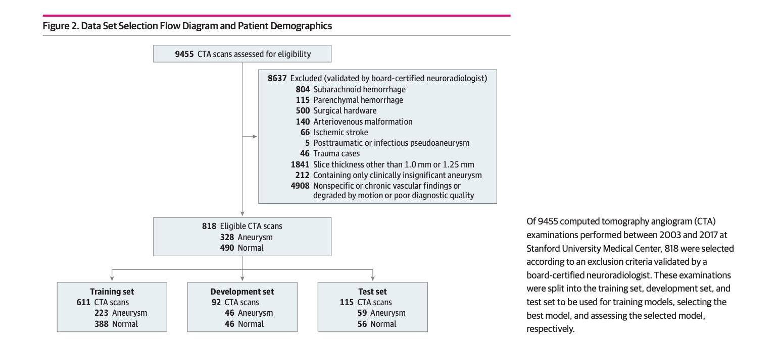
The data set contained 818 examinations from 662 unique patients with 328 CTA examinations (40.1%) containing at least 1 intracranial aneurysm and 490 examinations (59.9%) without intracranial aneurysms (Figure 2). Of the 328 aneurysm cases, 20 cases from 15 unique patients contained 2 or more aneurysms. One hundred forty-eight aneurysm cases contained aneurysms between 3 mm and 7 mm, 108 cases had aneurysms between 7 mm and 12 mm, 61 cases had aneurysms between 12 mm and 24 mm, and 11 cases had aneurysms 24 mm or greater. The location of the aneurysms varied according to the following distribution: 99 were located in the internal carotid artery, 78 were in the middle cerebral artery, 50 were cavernous internal carotid artery aneurysms, 44 were basilar tip aneurysms, 41 were in the anterior communicating artery, 18 were in the posterior communicating artery, 16 were in the vertebrobasilar system, and 12 were in the anterior cerebral artery. All examinations were performed either on a GE Discovery, GE LightSpeed, GE Revolution, Siemens Definition, Siemens Sensation, or a Siemens Force scanner, with slice thicknesses of 1.0 mm or 1.25 mm, using standard clinical protocols for head angiogram or head/neck angiogram. There was no difference between the protocols or slice thicknesses between the aneurysm and nonaneurysm examinations. For this study, axial series were extracted from each examination and a segmentation label was produced on every axial slice containing an aneurysm. The number of images per examination ranged from 113 to 802 (mean [SD], 373 [157]).

The examinations were split into a training set of 611 examinations (494 patients; mean [SD] age, 55.8 [18.1] years; 372 [60.9%] female) used to train the model, a development set of 92 examinations (86 patients; mean [SD] age, 61.6 [16.7] years; 59 [64.1%] female) used for model selection, and a test set of 115 examinations (82 patients; mean [SD] age, 57.8 [18.3] years; 74 [64.4%] female) to evaluate the performance of the clinicians when augmented with the model (Figure 2).

Using stratified random sampling, the development and test sets were formed to include 50% aneurysm examinations and 50% normal examinations; the remaining examinations composed the training set, of which 36.5% were aneurysm examinations. Forty-three patients had multiple examinations in the data set due to examinations performed for follow-up of the aneurysm. To account for these repeat patients, examinations were split so that there was no patient overlap between the different sets. Figure 2 contains pathology and patient demographic characteristics for each set.

A total of 8 clinicians, including 6 board-certified practicing radiologists, 1 practicing neurosurgeon, and 1 radiology resident, participated as readers in the study. The radiologists’ years of experience ranged from 3 to 12 years, the neurosurgeon had 2 years of experience as attending, and the resident was in the second year of training at Stanford University Medical Center. Groups 1 and 2 consisted of 3 radiologists each; the resident and neurosurgeon were both in group 1. None of the clinicians were involved in establishing the reference standard for the examinations.

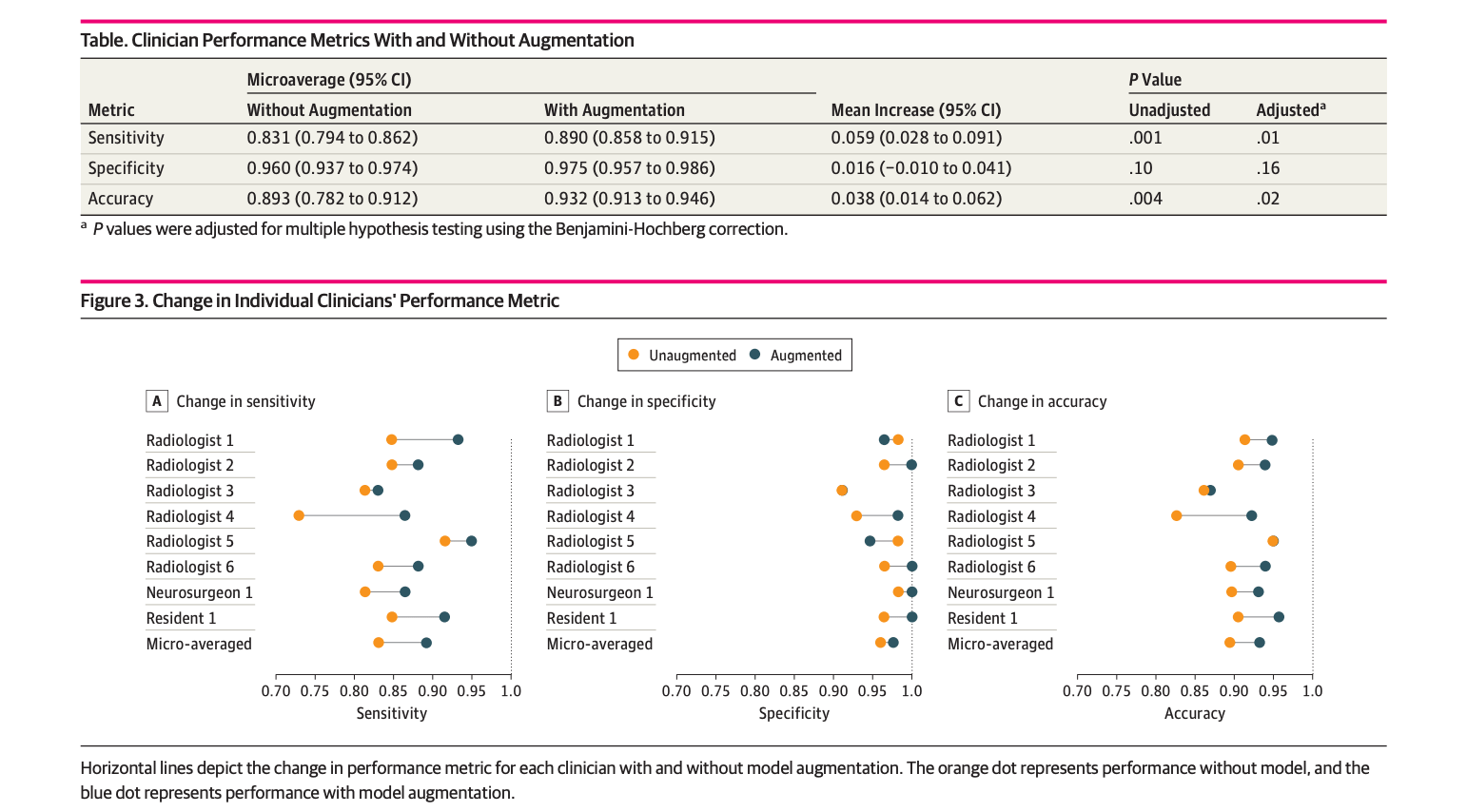
Without augmentation, clinicians achieved a microaveraged sensitivity of 0.831 (95% CI, 0.794-0.862), specificity of 0.960 (95% CI, 0.937-0.974), and an accuracy of 0.893 (95% CI, 0.872-0.912). With augmentation, the clinicians achieved a microaveraged sensitivity of 0.890 (95% CI, 0.858-0.915), specificity of 0.975 (95% CI, 0.957-0.986), and an accuracy of 0.932 (95% CI, 0.913-0.946). The underlying model had a sensitivity of 0.949 (95% CI, 0.861-0.983), specificity of 0.661 (95% CI, 0.530-0.771), and accuracy of 0.809 (95% CI, 0.727-0.870). The performances of the model, individual clinicians, and their microaverages are reported in eTable 1 in the Supplement.

With augmentation, there was a statistically significant increase in the mean sensitivity (0.059; 95% CI, 0.028-0.091; adjusted P = .01) and mean accuracy (0.038; 95% CI, 0.014-0.062; adjusted P = .02) of the clinicians as a group. There was no statistically significant change in mean specificity (0.016; 95% CI, −0.010 to 0.041; adjusted P = .16). Performance improvements across clinicians are detailed in the Table, and individual clinician improvement in Figure 3.

Individual performances with and without model augmentation are shown in eTable 1 in the Supplement. The sensitivity analysis confirmed that even among board-certified radiologists, there was a statistically significant increase in mean sensitivity (0.059; 95% CI, 0.013-0.105; adjusted P = .04) and accuracy (0.036; 95% CI, 0.001-0.072; adjusted P = .05). Performance improvements of board-certified radiologists as a group are shown in eTable 2 in the Supplement.

The mean diagnosis time per examination without augmentation microaveraged across clinicians was 57.04 seconds (95% CI, 54.58-59.50 seconds). The times for individual clinicians are detailed in eTable 3 in the Supplement, and individual time changes are shown in eFigure 2 in the Supplement.

With augmentation, there was no statistically significant decrease in mean diagnosis time (5.71 seconds; 95% CI, −7.22 to 18.63 seconds; adjusted P = .19). The model took a mean of 7.58 seconds (95% CI, 6.92-8.25 seconds) to process an examination and output its segmentation map.Confusion matrices, which are tables reporting true- and false-positive results and true- and false-negative results of each clinician with and without model augmentation, are shown in eTable 4 in the Supplement.

There was a statistically significant increase of 0.060 (adjusted P = .05) in the interrater agreement among the clinicians, with an exact Fleiss κ of 0.799 without augmentation and 0.859 with augmentation. For the board-certified radiologists, there was an increase of 0.063 in their interrater agreement, with an exact Fleiss κ of 0.783 without augmentation and 0.847 with augmentation.

Discussion

In this study, the ability of a deep learning model to augment clinician performance in detecting cerebral aneurysms using CTA was investigated with a crossover study design. With model augmentation, clinicians’ sensitivity, accuracy, and interrater agreement significantly increased. There was no statistical change in specificity and time to diagnosis.Given the potential catastrophic outcome of a missed aneurysm at risk of rupture, an automated detection tool that reliably detects and enhances clinicians’ performance is highly desirable. Aneurysm rupture is fatal in 40% of patients and leads to irreversible neurological disability in two-thirds of those who survive; therefore, an accurate and timely detection is of paramount importance. In addition to significantly improving accuracy across clinicians while interpreting CTA examinations, an automated aneurysm detection tool, such as the one presented in this study, could also be used to prioritize workflow so that those examinations more likely to be positive could receive timely expert review, potentially leading to a shorter time to treatment and more favorable outcomes.The significant variability among clinicians in the diagnosis of aneurysms has been well documented and is typically attributed to lack of experience or subspecialty neuroradiology training, complex neurovascular anatomy, or the labor-intensive nature of identifying aneurysms. Studies have shown that interrater agreement of CTA-based aneurysm detection is highly variable, with interrater reliability metrics ranging from 0.37 to 0.85,6,7,2628 and performance levels that vary depending on aneurysm size and individual radiologist experience.4,6 In addition to significantly increasing sensitivity and accuracy, augmenting clinicians with the model also significantly improved interrater reliability from 0.799 to 0.859. This implies that augmenting clinicians with varying levels of experience and specialties with models could lead to more accurate and more consistent radiological interpretations. Currently, tools to improve clinician aneurysm detection on CTA include bone subtraction,29 as well as 3-D rendering of intracranial vasculature,3032 which rely on application of contrast threshold settings to better delineate cerebral vasculature and create a 3-D–rendered reconstruction to assist aneurysm detection. However, using these tools is labor- and time-intensive for clinicians; in some institutions, this process is outsourced to a 3-D lab at additional costs. The tool developed in this study, integrated directly in a standard DICOM viewer, produces a segmentation map on a new examination in only a few seconds. If integrated into the standard workflow, this diagnostic tool could substantially decrease both cost and time to diagnosis, potentially leading to more efficient treatment and more favorable patient outcomes.Deep learning has recently shown success in various clinical image-based recognition tasks. In particular, studies have shown strong performance of 2-D CNNs in detecting intracranial hemorrhage and other acute brain findings, such as mass effect or skull fractures, on CT head examinations.3336 Recently, one study10 examined the potential role for deep learning in magnetic resonance angiogram–based detection of cerebral aneurysms, and another study37 showed that providing deep learning model predictions to clinicians when interpreting knee magnetic resonance studies increased specificity in detecting anterior cruciate ligament tears. To our knowledge, prior to this study, deep learning had not been applied to CTA, which is the first-line imaging modality for detecting cerebral aneurysms. Our results demonstrate that deep learning segmentation models may produce dependable and interpretable predictions that augment clinicians and improve their diagnostic performance. The model implemented and tested in this study significantly increased sensitivity, accuracy, and interrater reliability of clinicians with varied experience and specialties in detecting cerebral aneurysms using CTA.

Limitations

This study has limitations. First, because the study focused only on nonruptured aneurysms, model performance on aneurysm detection after aneurysm rupture, lesion recurrence after coil or surgical clipping, or aneurysms associated with arteriovenous malformations has not been investigated. Second, since examinations containing surgical hardware or devices were excluded, model performance in their presence is unknown. In a clinical environment, CTA is typically used to evaluate for many types of vascular diseases, not just for aneurysm detection. Therefore, the high prevalence of aneurysm in the test set and the clinician’s binary task could have introduced bias in interpretation. Also, this study was performed on data from a single tertiary care academic institution and may not reflect performance when applied to data from other institutions with different scanners and imaging protocols, such as different slice thicknesses.

Conclusions

A deep learning model was developed to automatically detect clinically significant intracranial aneurysms on CTA. We found that the augmentation significantly improved clinicians’ sensitivity, accuracy, and interrater reliability. Future work should investigate the performance of this model prospectively and in application of data from other institutions and hospitals.

Article Information:

Accepted for Publication: April 23, 2019.

Published: June 7, 2019. doi:10.1001/jamanetworkopen.2019.5600

Open Access: This is an open access article distributed under the terms of the CC-BY License. © 2019 Park A et al. JAMA Network Open.

Corresponding Author: Kristen W. Yeom, MD, School of Medicine, Department of Radiology, Stanford University, 725 Welch Rd, Ste G516, Palo Alto, CA 94304 (kyeom@stanford.edu).

Author Contributions: Ms Park and Dr Yeom had full access to all of the data in the study and take responsibility for the integrity of the data and the accuracy of the data analysis. Ms Park and Messrs Chute and Rajpurkar are co–first authors. Drs Ng and Yeom are co–senior authors.

Concept and design: Park, Chute, Rajpurkar, Lou, Shpanskaya, Ni, Basu, Lungren, Ng, Yeom.

Acquisition, analysis, or interpretation of data: Park, Chute, Rajpurkar, Lou, Ball, Shpanskaya, Jabarkheel, Kim, McKenna, Tseng, Ni, Wishah, Wittber, Hong, Wilson, Halabi, Patel, Lungren, Yeom.

Drafting of the manuscript: Park, Chute, Rajpurkar, Lou, Ball, Jabarkheel, Kim, McKenna, Hong, Halabi, Lungren, Yeom.

Critical revision of the manuscript for important intellectual content: Park, Chute, Rajpurkar, Ball, Shpanskaya, Jabarkheel, Kim, Tseng, Ni, Wishah, Wittber, Wilson, Basu, Patel, Lungren, Ng, Yeom.

Statistical analysis: Park, Chute, Rajpurkar, Lou, Ball, Lungren.

Administrative, technical, or material support: Park, Chute, Shpanskaya, Jabarkheel, Kim, McKenna, Tseng, Wittber, Hong, Wilson, Lungren, Ng, Yeom.

Supervision: Park, Ball, Tseng, Halabi, Basu, Lungren, Ng, Yeom.

Conflict of Interest Disclosures: Drs Wishah and Patel reported grants from GE and Siemens outside the submitted work. Dr Patel reported participation in the speakers bureau for GE. Dr Lungren reported personal fees from Nines Inc outside the submitted work. Dr Yeom reported grants from Philips outside the submitted work. No other disclosures were reported.

Funding/Support: This work was supported by National Institutes of Health National Center for Advancing Translational Science Clinical and Translational Science Award UL1TR001085.

Role of the Funder/Sponsor: The National Institutes of Health had no role in the design and conduct of the study; collection, management, analysis, and interpretation of the data; preparation, review, or approval of the manuscript; and decision to submit the manuscript for publication.

Disclaimer: The content is solely the responsibility of the authors and does not necessarily represent the official views of the National Institutes of Health.

References

1.Jaja  BN, Cusimano  MD, Etminan  N,  et al.  Clinical prediction models for aneurysmal subarachnoid hemorrhage: a systematic review.  Neurocrit Care. 2013;18(1):143-153. doi:10.1007/s12028-012-9792-zPubMedGoogle ScholarCrossref
2.Turan  N, Heider  RA, Roy  AK,  et al.  Current perspectives in imaging modalities for the assessment of unruptured intracranial aneurysms: a comparative analysis and review.  World Neurosurg. 2018;113:280-292. doi:10.1016/j.wneu.2018.01.054PubMedGoogle ScholarCrossref
3.Yoon  NK, McNally  S, Taussky  P, Park  MS.  Imaging of cerebral aneurysms: a clinical perspective.  Neurovasc Imaging. 2016;2(1):6. doi:10.1186/s40809-016-0016-3Google ScholarCrossref
4.Jayaraman  MV, Mayo-Smith  WW, Tung  GA,  et al.  Detection of intracranial aneurysms: multi-detector row CT angiography compared with DSA.  Radiology. 2004;230(2):510-518. doi:10.1148/radiol.2302021465PubMedGoogle ScholarCrossref
5.Bharatha  A, Yeung  R, Durant  D,  et al.  Comparison of computed tomography angiography with digital subtraction angiography in the assessment of clipped intracranial aneurysms.  J Comput Assist Tomogr. 2010;34(3):440-445. doi:10.1097/RCT.0b013e3181d27393PubMedGoogle ScholarCrossref
6.Lubicz  B, Levivier  M, François  O,  et al.  Sixty-four-row multisection CT angiography for detection and evaluation of ruptured intracranial aneurysms: interobserver and intertechnique reproducibility.  AJNR Am J Neuroradiol. 2007;28(10):1949-1955. doi:10.3174/ajnr.A0699PubMedGoogle ScholarCrossref
7.White  PM, Teasdale  EM, Wardlaw  JM, Easton  V.  Intracranial aneurysms: CT angiography and MR angiography for detection prospective blinded comparison in a large patient cohort.  Radiology. 2001;219(3):739-749. doi:10.1148/radiology.219.3.r01ma16739PubMedGoogle ScholarCrossref
8.Suzuki  K.  Overview of deep learning in medical imaging.  Radiol Phys Technol. 2017;10(3):257-273. doi:10.1007/s12194-017-0406-5PubMedGoogle ScholarCrossref
9.Rajpurkar  P, Irvin  J, Ball  RL,  et al.  Deep learning for chest radiograph diagnosis: a retrospective comparison of the CheXNeXt algorithm to practicing radiologists.  PLoS Med. 2018;15(11):e1002686. doi:10.1371/journal.pmed.1002686PubMedGoogle ScholarCrossref
10.Bien  N, Rajpurkar  P, Ball  RL,  et al.  Deep-learning-assisted diagnosis for knee magnetic resonance imaging: development and retrospective validation of MRNet.  PLoS Med. 2018;15(11):e1002699. doi:10.1371/journal.pmed.1002699PubMedGoogle ScholarCrossref
11.Morita  A, Kirino  T, Hashi  K,  et al; UCAS Japan Investigators.  The natural course of unruptured cerebral aneurysms in a Japanese cohort.  N Engl J Med. 2012;366(26):2474-2482. doi:10.1056/NEJMoa1113260PubMedGoogle ScholarCrossref
12.Yushkevich  PA, Piven  J, Hazlett  HC,  et al.  User-guided 3D active contour segmentation of anatomical structures: significantly improved efficiency and reliability.  Neuroimage. 2006;31(3):1116-1128. doi:10.1016/j.neuroimage.2006.01.015PubMedGoogle ScholarCrossref
13.He  K, Zhang  X, Ren  S, Sun  J. Deep residual learning for image recognition. Paper presented at: Proceedings of the IEEE Conference on Computer Vision and Pattern Recognition; June 27, 2016; Las Vegas, NV.
14.Xie  S, Girshick  R, Dollár  P, Tu  Z, He  K. Aggregated residual transformations for deep neural networks. Paper presented at: 2017 IEEE Conference on Computer Vision and Pattern Recognition (CVPR); July 25, 2017; Honolulu, HI.
15.Hu  J, Shen  L, Sun  G. Squeeze-and-excitation networks. Paper presented at: 2018 IEEE Conference on Computer Vision and Pattern Recognition (CVPR); June 21, 2018; Salt Lake City, Utah.
16.Ronneberger  O, Fischer  P, Brox  T. U-net: Convolutional networks for biomedical image segmentation. International Conference on Medical Image Computing and Computer-Assisted Intervention. Basel, Switzerland: Springer International; 2015:234–241.
17.Carreira  J, Zisserman  A. Quo vadis, action recognition? a new model and the kinetics dataset. Paper presented at: 2017 IEEE Conference on Computer Vision and Pattern Recognition (CVPR); July 25, 2017; Honolulu, HI.
18.Chen  L-C, Papandreou  G, Schroff  F, Adam  H. Rethinking atrous convolution for semantic image segmentation. https://arxiv.org/abs/1706.05587. Published June 17, 2017. Accessed May 7, 2019.
19.Milletari  F, Navab  N, Ahmadi  S-A. V-net: Fully convolutional neural networks for volumetric medical image segmentation. Paper presented at: 2016 IEEE Fourth International Conference on 3D Vision (3DV); October 26-28, 2016; Stanford, CA.
20.Loshchilov  I, Hutter  F. Sgdr: Stochastic gradient descent with warm restarts. Paper presented at: 2017 Fifth International Conference on Learning Representations; April 24-26, 2017; Toulon, France.
21.Huang  G, Sun  Y, Liu  Z, Sedra  D, Weinberger  KQ. Deep networks with stochastic depth. European Conference on Computer Vision. Basel, Switzerland: Springer International; 2016:646–661.
22.Horos. https://horosproject.org. Accessed May 1, 2019.
23.Wilson  EB.  Probable inference, the law of succession, and statistical inference.  J Am Stat Assoc. 1927;22(158):209-212. doi:10.1080/01621459.1927.10502953Google ScholarCrossref
24.Fleiss  JL, Cohen  J.  The equivalence of weighted kappa and the intraclass correlation coefficient as measures of reliability.  Educ Psychol Meas. 1973;33(3):613-619. doi:10.1177/001316447303300309Google ScholarCrossref
25.Benjamini  Y, Hochberg  Y.  Controlling the false discovery rate: a practical and powerful approach to multiple testing.  J R Stat Soc Series B Stat Methodol. 1995;57(1):289-300.Google Scholar
26.Maldaner  N, Stienen  MN, Bijlenga  P,  et al.  Interrater agreement in the radiologic characterization of ruptured intracranial aneurysms based on computed tomography angiography.  World Neurosurg. 2017;103:876-882.e1. doi:10.1016/j.wneu.2017.04.131PubMedGoogle ScholarCrossref
27.Wang  Y, Gao  X, Lu  A,  et al.  Residual aneurysm after metal coils treatment detected by spectral CT.  Quant Imaging Med Surg. 2012;2(2):137-138.PubMedGoogle Scholar
28.Yoon  YW, Park  S, Lee  SH,  et al.  Post-traumatic myocardial infarction complicated with left ventricular aneurysm and pericardial effusion.  J Trauma. 2007;63(3):E73-E75. doi:10.1097/01.ta.0000246896.89156.70PubMedGoogle ScholarCrossref
29.Tomandl  BF, Hammen  T, Klotz  E, Ditt  H, Stemper  B, Lell  M.  Bone-subtraction CT angiography for the evaluation of intracranial aneurysms.  AJNR Am J Neuroradiol. 2006;27(1):55-59.PubMedGoogle Scholar
30.Shi  W-Y, Li  Y-D, Li  M-H,  et al.  3D rotational angiography with volume rendering: the utility in the detection of intracranial aneurysms.  Neurol India. 2010;58(6):908-913. doi:10.4103/0028-3886.73743PubMedGoogle ScholarCrossref
31.Lin  N, Ho  A, Gross  BA,  et al.  Differences in simple morphological variables in ruptured and unruptured middle cerebral artery aneurysms.  J Neurosurg. 2012;117(5):913-919. doi:10.3171/2012.7.JNS111766PubMedGoogle ScholarCrossref
32.Villablanca  JP, Jahan  R, Hooshi  P,  et al.  Detection and characterization of very small cerebral aneurysms by using 2D and 3D helical CT angiography.  AJNR Am J Neuroradiol. 2002;23(7):1187-1198.PubMedGoogle Scholar
33.Chang  PD, Kuoy  E, Grinband  J,  et al.  Hybrid 3D/2D convolutional neural network for hemorrhage evaluation on head CT.  AJNR Am J Neuroradiol. 2018;39(9):1609-1616. doi:10.3174/ajnr.A5742PubMedGoogle ScholarCrossref
34.Chilamkurthy  S, Ghosh  R, Tanamala  S,  et al.  Deep learning algorithms for detection of critical findings in head CT scans: a retrospective study.  Lancet. 2018;392(10162):2388-2396. doi:10.1016/S0140-6736(18)31645-3PubMedGoogle ScholarCrossref
35.Jnawali  K, Arbabshirani  MR, Rao  N, Patel  AA. Deep 3D convolution neural network for CT brain hemorrhage classification. Paper presented at: Medical Imaging 2018: Computer-Aided Diagnosis. February 27, 2018; Houston, TX. doi:10.1117/12.2293725
36.Titano  JJ, Badgeley  M, Schefflein  J,  et al.  Automated deep-neural-network surveillance of cranial images for acute neurologic events.  Nat Med. 2018;24(9):1337-1341. doi:10.1038/s41591-018-0147-yPubMedGoogle ScholarCrossref
37.Ueda  D, Yamamoto  A, Nishimori  M,  et al.  Deep learning for MR angiography: automated detection of cerebral aneurysms.  Radiology. 2019;290(1):187-194.PubMedGoogle ScholarCrossref

Read Full Post »

Real Time Coverage @BIOConvention #BIO2019: Machine Learning and Artificial Intelligence: Realizing Precision Medicine One Patient at a Time

Reporter: Stephen J Williams, PhD @StephenJWillia2

The impact of Machine Learning (ML) and Artificial Intelligence (AI) during the last decade has been tremendous. With the rise of infobesity, ML/AI is evolving to an essential capability to help mine the sheer volume of patient genomics, omics, sensor/wearables and real-world data, and unravel the knot of healthcare’s most complex questions.

Despite the advancements in technology, organizations struggle to prioritize and implement ML/AI to achieve the anticipated value, whilst managing the disruption that comes with it. In this session, panelists will discuss ML/AI implementation and adoption strategies that work. Panelists will draw upon their experiences as they share their success stories, discuss how to implement digital diagnostics, track disease progression and treatment, and increase commercial value and ROI compared against traditional approaches.

  • most of trials which are done are still in training AI/ML algorithms with training data sets.  The best results however have been about 80% accuracy in training sets.  Needs to improve
  • All data sets can be biased.  For example a professor was looking at heartrate using a IR detector on a wearable but it wound up that different types of skin would generate a different signal to the detector so training sets maybe population biases (you are getting data from one group)
  • clinical grade equipment actually haven’t been trained on a large set like commercial versions of wearables, Commercial grade is tested on a larger study population.  This can affect the AI/ML algorithms.
  • Regulations:  The regulatory bodies responsible is up to debate.  Whether FDA or FTC is responsible for AI/ML in healtcare and healthcare tech and IT is not fully decided yet.  We don’t have the guidances for these new technologies
  • some rules: never use your own encryption always use industry standards especially when getting personal data from wearables.  One hospital corrupted their system because their computer system was not up to date and could not protect against a virus transmitted by a wearable.
  • pharma companies understand they need to increase value of their products so very interested in how AI/ML can be used.

Please follow LIVE on TWITTER using the following @ handles and # hashtags:

@Handles

@pharma_BI

@AVIVA1950

@BIOConvention

# Hashtags

#BIO2019 (official meeting hashtag)

Read Full Post »

« Newer Posts - Older Posts »