Feeds:
Posts
Comments

Archive for the ‘Electrophysiology’ Category

Ischemia, Infarction, and the Waveforms Q through U, Part 1: How to Read an EKG Curriculum

Reporter: Aviva Lev-Ari, PhD, RN


Watch Video

https://www.youtube.com/v/Ajfk21k3Zmo?fs=1&hl=fr_FR

ECG Interpretation: Ischemia, Infarction, and the Waveforms Q through U, Part 1 Girish L. Kalra, MD Assistant Professor, Department of Medicine, Emory Univer…

Sourced through Scoop.it from: www.youtube.com

See on Scoop.itCardiovascular and vascular imaging

Read Full Post »

UPDATED on 2/25/2019

https://www.medpagetoday.com/cardiology/prevention/78202?xid=nl_mpt_SRCardiology_2019-02-25&eun=g99985d0r&utm_source=Sailthru&utm_medium=email&utm_campaign=CardioUpdate_022519&utm_term=NL_Spec_Cardiology_Update_Active

Medtronic recalled its dual chamber pacemakers (Adapta, Versa, Sensia, Relia, Attesta, Sphera, and Vitatron A, E, G, and Q series) due to a possible software error that can stop pacing.

Steps to minimise replacement of cardiac implantable electronic devices

Reporter: Aviva Lev-Ari, PhD, RN

Pacemaker battery scandal

SOURCE

http://www.bmj.com/content/352/bmj.i228

BMJ 2016; 352 doi: http://dx.doi.org/10.1136/bmj.i228 (Published 04 February 2016)Cite this as: BMJ 2016;352:i228
  1. John Dean, consultant cardiologist 1,
  2. Neil Sulke, consultant cardiologist 2

Author affiliations

  1. Correspondence to: J Dean john.dean2@nhs.net

Much can and should be done to maximise the longevity of existing devices

Imagine spending £3000 on a new watch with a battery embedded in the mechanism that cannot be replaced or recharged. Although the battery is predicted to last 10 years or more, after six years you discover that it is running flat and you’re advised to replace the watch immediately, even though it may keep good time for a year or more.

This mirrors the dilemma faced by all patients with cardiac implantable electronic devices such as pacemakers and implantable cardioverter defibrillators (ICD). But for them the stakes are much higher as replacing the battery exposes them to a risk of serious complications, including life threatening infection.

Over half of all patients with pacemakers require a replacement procedure because the batteries have reached their expected life.1 Some 11-16% need multiple replacements.2 The situation is worse for recipients of an ICD, since the risks of infection at the time of implant and device replacement are higher than with pacemakers and the batteries have a shorter life.3

What is the risk of infection?

With no standard definition or reporting system, infection rates vary widely, and the commonly quoted risk of 0.5% for new implants and 1-5% for replacement procedures may be wrong.4 Infection, even if it seems superficial, usually necessitates extraction of the entire system. Simply treating the infection with antibiotics results in a much poorer outcome.5 The increased risk of infection associated with battery replacement makes it critical that we prolong the life of implantable devices as much as possible. The health economic grounds for minimising the number of replacements are also compelling.6

The current financial model discourages the development of longer life devices. Increasing longevity would reduce profits for manufacturers, implanting physicians, and their institutions. With financial disincentives for both manufacturers and purchasers it is hardly surprising that longer life devices do not exist.

Patients are often assumed to prefer smaller devices, but when offered the choice, over 90% would opt for a larger, longer lasting device over a smaller one that would require more frequent operations to change the battery.7 And given the risks that patients are exposed to during replacement, there is an urgent need to improve longevity by developing longer life batteries and using those in current devices more prudently.

What can be done now?

At present the main drive to improving longevity of pacemakers has been through programming changes aimed at reducing the amount of pacing8 or minimising the drain of current during pacing—for example, using high impedance leads. But devices are usually replaced when there is still substantial life left in the battery. For example, when a pacemaker reaches elective replacement indication, it is usually 3-12 months before it will reach its end of life. And even then, the battery may continue to function for several months. Early replacement may be reasonable for high risk patients (such as those who are entirely dependent on their pacemaker). However, we could delay replacement of the pulse generator until the batteries are virtually depleted in lower risk patients. The increasingly popular innovation of home monitoring of devices would facilitate this.

For ICDs the waste is even more striking; devices reach their elective replacement indication when they are still capable of delivering at least six full energy shocks. Each shock reduces the battery longevity by about 30 days. So for patients who receive no shock therapy we are prematurely discarding a device costing up to £25 000 (€33 000; $36 000), which could last at least another six months (current devices last four to seven years on average). We need to review the timing of replacement of implantable devices in all patients.

CONTINUE READING

http://www.bmj.com/content/352/bmj.i228

REFERENCES

Read Full Post »

What Are Your Medication Options for Heart Arrhythmias?

Reporter: Aviva Lev-Ari, PhD, RN


Heart arrhythmias can be treated with antiarrhythmic drugs, AV nodal blocking drugs, beta blockers, statins, and omega-3 fatty acids.

Sourced through Scoop.it from: heartdisease.about.com

See on Scoop.itCardiovascular Disease: PHARMACO-THERAPY

See our Book

https://www.amazon.com/dp/B07MGSFDWR


Read Full Post »

Hybrid lipid bioelectronic membranes

Larry H. Bernstein, MD, FCAP, Curator

LPBI


Hybrid solid-state chips and biological cells integrated at molecular level

Biological ion channels combine with solid-state transistors to create a new kind of hybrid bioelectronics. Imagine chips with dog-like capability to taste and smell, or even recognize specific molecules.
http://www.kurzweilai.net/hybrid-solid-state-chips-and-biological-cells-integrated-at-molecular-level
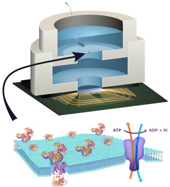
Illustration depicting a biocell attached to a CMOS integrated circuit with a membrane containing sodium-potassium pumps in pores. Energy is stored chemically in ATP molecules. When the energy is released as charged ions (which are then converted to electrons to power the chip at the bottom of the experimental device), the ATP is converted to ADP + inorganic phosphate. (credit: Trevor Finney and Jared Roseman/Columbia Engineering)

Columbia Engineering researchers have combined biological and solid-state components for the first time, opening the door to creating entirely new artificial biosystems.

In this experiment, they used a biological cell to power a conventional solid-state complementary metal-oxide-semiconductor (CMOS) integrated circuit. An artificial lipid bilayer membrane containing adenosine triphosphate (ATP)-powered ion pumps (which provide energy for cells) was used as a source of ions (which were converted to electrons to power the chip).

The study, led by Ken Shepard, Lau Family Professor of Electrical Engineering and professor of biomedical engineering at Columbia Engineering, was published online today (Dec. 7, 2015) in an open-access paper in Nature Communications.

How to build a hybrid biochip

Living systems achieve this functionality with their own version of electronics based on lipid membranes and ion channels and pumps, which act as a kind of “biological transistor.” Charge in the form of ions carry energy and information, and ion channels control the flow of ions across cell membranes.

Solid-state systems, such as those in computers and communication devices, use electrons; their electronic signaling and power are controlled by field-effect transistors.

To build a prototype of their hybrid system, Shepard’s team packaged a CMOS integrated circuit (IC) with an ATP-harvesting “biocell.” In the presence of ATP, the system pumped ions across the membrane, producing an electrical potential (voltage)* that was harvested by the integrated circuit.

“We made a macroscale version of this system, at the scale of several millimeters, to see if it worked,” Shepard notes. “Our results provide new insight into a generalized circuit model, enabling us to determine the conditions to maximize the efficiency of harnessing chemical energy through the action of these ion pumps. We will now be looking at how to scale the system down.”

While other groups have harvested energy from living systems, Shepard and his team are exploring how to do this at the molecular level, isolating just the desired function and interfacing this with electronics. “We don’t need the whole cell,” he explains. “We just grab the component of the cell that’s doing what we want. For this project, we isolated the ATPases because they were the proteins that allowed us to extract energy from ATP.”

The capability of a bomb-sniffing dog, no Alpo required

Next, the researchers plan to go much further, such as recognizing specific molecules and giving chips the potential to taste and smell.

The ability to build a system that combines the power of solid-state electronics with the capabilities of biological components has great promise, they believe. “You need a bomb-sniffing dog now, but if you can take just the part of the dog that is useful — the molecules that are doing the sensing — we wouldn’t need the whole animal,” says Shepard.

The technology could also provide a power source for implanted electronic devices in ATP-rich environments such as inside living cells, the researchers suggest.

*  “In general, integrated circuits, even when operated at the point of minimum energy in subthreshold, consume on the order of 10−2 W mm−2 (or assuming a typical silicon chip thickness of 250 μm, 4 × 10⁻² W mm−3). Typical cells, in contrast, consume on the order of 4 × 10⁻⁶ W mm−3. In the experiment, a typical active power dissipation for the IC circuit was 92.3 nW, and the active average harvesting power was 71.4 fW for the biocell (the discrepancy is managed through duty-cycled operation of the IC).” — Jared M. Roseman et al./Nature Communications


Hybrid integrated biological–solid-state system powered with adenosine triphosphate

Jared M. RosemanJianxun LinSiddharth RamakrishnanJacob K. Rosenstein & Kenneth L. Shepard
Nature Communications 7 Dec 2015; 6(10070)
     http://dx.doi.org:/10.1038/ncomms10070

There is enormous potential in combining the capabilities of the biological and the solid state to create hybrid engineered systems. While there have been recent efforts to harness power from naturally occurring potentials in living systems in plants and animals to power complementary metal-oxide-semiconductor integrated circuits, here we report the first successful effort to isolate the energetics of an electrogenic ion pump in an engineered in vitro environment to power such an artificial system. An integrated circuit is powered by adenosine triphosphate through the action of Na+/K+ adenosine triphosphatases in an integrated in vitro lipid bilayer membrane. The ion pumps (active in the membrane at numbers exceeding 2 × 106mm−2) are able to sustain a short-circuit current of 32.6pAmm−2 and an open-circuit voltage of 78mV, providing for a maximum power transfer of 1.27pWmm−2 from a single bilayer. Two series-stacked bilayers provide a voltage sufficient to operate an integrated circuit with a conversion efficiency of chemical to electrical energy of 14.9%.


Figure 1: Fully hybrid biological–solid-state system.


Fully hybrid biological-solid-state system.

http://www.nature.com/ncomms/2015/151207/ncomms10070/images/ncomms10070-f1.jpg

(a) Illustration depicting biocell attached to CMOS integrated circuit. (b) Illustration of membrane in pore containing sodium–potassium pumps. (c) Circuit model of equivalent stacked membranes, =2.1pA, =98.6G, =575G and =75pF, Ag/AgCl electrode equivalent resistance RWE+RCE<20k, energy-harvesting capacitor CSTOR=100nF combined with switch as an impedance transformation network (only one switch necessary due to small duty cycle), and CMOS IC voltage doubler and resistor representing digital switching load. RL represents the four independent ring oscillator loads. (d) Equivalent circuit detail of stacked biocell. (e) Switched-capacitor voltage doubler circuit schematic.


The energetics of living systems are based on electrochemical membrane potentials that are present in cell plasma membranes, the inner membrane of mitochondria, or the thylakoid membrane of chloroplasts1. In the latter two cases, the specific membrane potential is known as the proton-motive force and is used by proton adenosine triphosphate (ATP) synthases to produce ATP. In the former case, Na+/K+-ATPases hydrolyse ATP to maintain the resting potential in most cells.

While there have been recent efforts to harness power from some naturally occurring potentials in living systems that are the result of ion pump action both in plants2 and animals3, 4 to power complementary metal-oxide semiconductor (CMOS) integrated circuits (ICs), this work is the first successful effort to isolate the energetics of an electrogenic ion pump in an engineered in vitroenvironment to power such an artificial system. Prior efforts to harness power from in vitromembrane systems incorporating ion-pumping ATPases5, 6, 7, 8, 9 and light-activated bacteriorhodopsin9, 10, 11 have been limited by difficulty in incorporating these proteins in sufficient quantity to attain measurable current and in achieving sufficiently large membrane resistances to harness these currents. Both problems are solved in this effort to power an IC from ATP in an in vitro environment. The resulting measurements provide new insight into a generalized circuit model, which allows us to determine the conditions to maximize the efficiency of harnessing chemical energy through the action of electrogenic ion pumps.


ATP-powered IC

Figure 1a shows the complete hybrid integrated system, consisting of a CMOS IC packaged with an ATP-harvesting ‘biocell’. The biocell consists of two series-stacked ATPase bearing suspended lipid bilayers with a fluid chamber directly on top of the IC. Series stacking of two membranes is necessary to provide the required start-up voltage for IC and eliminates the need for an external energy source, which is typically required to start circuits from low-voltage supplies2, 3. As shown inFig. 1c, a matching network in the form of a switched capacitor allows the load resistance of the IC to be matched to that presented by the biocell. In principle, the switch S can be implicit. The biocell charges CSTOR until the self start-up voltage, Vstart, is reached. The chip then operates until the biocell voltage drops below the minimum supply voltage for operation, Vmin. Active current draw from the IC stops at this point, allowing the charge to build up again on CSTOR. In our case, however, the IC leakage current exceeds 13.5nA at Vstart, more than can be provided by the biocell. As a result, an explicit transistor switch and comparator (outside of the IC) are used for this function in the experimental results presented here, which are not powered by the biocell and not included in energy efficiency calculations (see Supplementary Discussion for additional details). The energy from the biocell is used to operate a voltage converter (voltage doubler) and some simple inverter-based ring oscillators in the IC, which receive power from no other sources.

Figure 1: Fully hybrid biological–solid-state system.

http://www.nature.com/ncomms/2015/151207/ncomms10070/images/ncomms10070-f1.jpg


……..   Prior to the addition of ATP, the membrane produces no electrical power and has an Rm of 280G. A 1.7-pA short-circuit (SC) current (Fig. 2b) through the membrane is observed upon the addition of ATP (final concentration 3mM) to the cis chamber where functional, properly oriented enzymes generate a net electrogenic pump current. To perform these measurements, currents through each membrane of the biocell are measured using a voltage-clamp amplifier (inset of Fig. 2b) with a gain of 500G with special efforts taken to compensate amplifier leakage currents. Each ATPase transports three Na+ ions from the cis chamber to the trans chamber and two K+ ions from thetrans chamber to the cis chamber (a net charge movement of one cation) for every molecule of ATP hydrolysed. At a rate of 100 hydrolysis events per second under zero electrical (SC) bias13, this results in an electrogenic current of ~16aA. The observed SC current corresponds to about 105 active ATPases in the membrane or a concentration of about 2 × 106mm−2, about 5% of the density of channels occurring naturally in mammalian nerve fibres14. It is expected that half of the channels inserted are inactive because they are oriented incorrectly.

Figure 2: Single-cell biocell characterization.

http://www.nature.com/ncomms/2015/151207/ncomms10070/images_article/ncomms10070-f2.jpg

(a)…Pre-ATP data linear fit (black line) slope yield Rm=280G. Post ATP data fit to a Boltzmann curve, slope=0.02V (blue line). Post-ATP linear fit (red line) yields Ip=−1.8pA and Rp=61.6G, which corresponds to a per-ATP source resistance of 6.16 × 10¹⁵. The current due to membrane leakage through R_{m} is subtracted in the post-ATP curve…. (b)…


Current–voltage characteristics of the ATPases

Figure 2a shows the complete measured current–voltage (IV) characteristic of a single ATPase-bearing membrane in the presence of ATP. The current due to membrane leakage through Rm is subtracted in the post-ATP curve. The IV characteristic fits a Boltzmann sigmoid curve, consistent with sodium–potassium pump currents measured on membrane patches at similar buffer conditions13, 15, 16. This nonlinear behaviour reflects the fact that the full ATPase transport cycle (three Na+ ions from cis to trans and two K+ ions from trans to cis) time increases (the turn-over rate, kATP, decreases) as the membrane potential increases16. No effect on pump current is expected from any ion concentration gradients produced by the action of the ATPases (seeSupplementary Discussion). Using this Boltzmann fit, we can model the biocell as a nonlinear voltage-controlled current source IATPase (inset Fig. 2a), in which the current produced by this source varies as a function of Vm. In the fourth quadrant, where the cell is producing electrical power, this model can be linearized as a Norton equivalent circuit, consisting of a DC current source (Ip) in parallel with a current-limiting resistor (Rp), which acts to limit the current delivered to the load at increasing bias (IATPase~IpVm/Rp). Figure 2c shows the measured and simulated charging of Cm for a single membrane (open-circuited voltage). A custom amplifier with input resistance Rin>10T was required for this measurement (see Electrical Measurement Methods).


Reconciling operating voltage differences

The electrical characteristics of biological systems and solid-state systems are mismatched in their operating voltages. The minimum operating voltage of solid-state systems is determined by the need for transistors to modulate a Maxwell–Boltzmann (MB) distribution of carriers by several orders of magnitude through the application of a potential that is several multiples of kT/q (where kis Boltzmann’s constant, T is the temperature in degrees Kelvin and q is the elementary charge). Biological systems, while operating under the same MB statistics, have no such constraints for operating ion channels since they are controlled by mechanical (or other conformational) processes rather than through modulation of a potential barrier. To bridge this operating voltage mismatch, the circuit includes a switched-capacitor voltage doubler (Fig. 1d) that is capable of self-startup from voltages as low Vstart=145mV (~5.5kT/q) and can be operated continuously from input voltages from as low as Vmin=110mV (see Supplementary Discussion)…..


Maximizing the efficiency of harvesting energy from ATP

Solid-state systems and biological systems are also mismatched in their operating impedances. In our case, the biocell presents a source impedance, =84.2G, while the load impedance presented by the complete integrated circuit (including both the voltage converter and ring oscillator loads) is approximately RIC=200k. (The load impedance, RL, of the ring oscillators alone is 305k.) This mismatch in source and load impedance is manifest in large differences in power densities. In general, integrated circuits, even when operated at the point of minimum energy in subthreshold, consume on the order of 10−2Wmm−2 (or assuming a typical silicon chip thickness of 250μm, 4 × 10⁻²Wmm−3) (ref. 17). Typical cells, in contrast, consume on the order of 4 × 10⁻⁶Wmm−3 (ref. 18). In our case, a typical active power dissipation for our circuit is 92.3nW, and the active average harvesting power is 71.4fW for the biocell. This discrepancy is managed through duty-cycled operation of the IC in which the circuit is largely disabled for long periods of time (Tcharge), integrating up the power onto a storage capacitor (CSTOR), which is then expended in a very brief period of activity (Trun), as shown in Fig. 3a.

The overall efficiency of the system in converting chemical energy to the energy consumed in the load ring oscillator (η) is given by the product of the conversion efficiency of the voltage doubler (ηconverter) and the conversion efficiency of chemical energy to electrical energy in the biocell (ηbiocell), η=ηconverter × ηbiocell. ηconverter is relatively constant over the range of input voltages at ~59%, as determined by various loading test circuits included in the chip design (Supplementary Figs 1–6). ηbiocell, however, varies with transmembrane potential Vm. η is the efficiency in transferring power to the power ring oscillator loads from the ATP harvested by biocell.

…….

To first order, the energy made available to the Na+/K+-ATPase by the hydrolysis of ATP is independent of the chemical or electric potential of the membrane and is given by |ΔGATP|/(qNA), where ΔGATP is the Gibbs free energy change due to the ATP hydrolysis reaction per mole of ATP at given buffer conditions and NA is Avogadro’s number. Since every charge that passes through IATPase corresponds to a single hydrolysis event, we can use two voltage sources in series with IATPase to independently account for the energy expended by the pumps both in moving charge across the electric potential difference and in moving ions across the chemical potential difference. The dependent voltage source Vloss in this branch fixes the voltage across IATPase, and the total power produced by the pump current source is (|ΔGATP|/NA)(NkATP), which is the product of the energy released per molecule of ATP, the number of active ATPases and the ATP turnover rate. The power dissipated in voltage source Vchem models the work performed by the ATPases in transporting ions against a concentration gradient. In the case of the Na+/K+ ATPase,Vchem is given by . The power dissipated in this source is introduced back into the circuit in the power generated by the Nernst independent voltage sources, and . The power dissipated in the dependent voltage source Vloss models any additional power not used to perform chemical or electrical work. ……


Integration of ATP-harvesting ion pumps could provide a means to power future CMOS microsystems scaled to the level of individual cells22. In molecular diagnostics, the integration of pore-forming proteins such as alpha haemolysin23 or MspA porin24 with CMOS electronics is already finding application in DNA sequencing25. Exploiting the large diversity of function available in transmembrane proteins in these hybrid systems could, for example, lead to highly specific sensing platforms for airborne odorants or soluble molecular entities26, 27. Heavily multiplexed platforms could become high-throughput in vitro drug-screening platforms against this diversity of function. In addition, integration of transmembrane proteins with CMOS may become a convenient alternative to fluorescence for coupling to synthetic biological systems28.


Roseman, J. M. et al. Hybrid integrated biological–solid-state system powered with adenosine triphosphate. Nat. Commun. 6:10070      http://dx.doi.org:/10.1038/ncomms10070 (2015).


  • Rottenberg, H. The measurement of membrane potential and deltapH in cells, organelles, and vesicles. Methods Enzymol. 55, 547569 (1979).
  • Himes, C., Carlson, E., Ricchiuti, R. J., Otis, B. P. & Parviz, B. A. Ultralow voltage nanoelectronics powered directly, and solely, from a tree. IEEE Trans. Nanotechnol. 9, 25(2010).
  • Mercier, P. P., Lysaght, A. C., Bandyopadhyay, S., Chandrakasan, A. P. & Stankovic, K. M.Energy extraction from the biologic battery in the inner ear. Nat. Biotechnol. 30, 12401243(2012).
  • Halámková, L. et al. Implanted Biofuel Cell Operating in a Living Snail. J. Am. Chem. Soc.134, 50405043 (2012).


Read Full Post »

American Society of Physiology Awards

Larry H Bernstein, MD, FCAP, Curator

Leaders in Pharmaceutical Innovation

Series E. 2;


Past Awardees:

2014

Harshita Chodavarapu, Louisiana State Univ. – New Orleans

Jennifer Richards, Saint Louis Univ. – Missouri

2013

Ho-Jin Koh, Joslin Diabetes Center

Danielle Shepherd, West Virginia Univ.

2012

Kavaljit H. Chhabra, LSUHSC – New Orleans

Hariom Yadav,NIDDK – National Institutes of Health

2011

Xuemei Shi, Baylor Col. of Med.

Gina Yosten, St. Louis Univ.

2010

Abid Abdulaziz Kazi, Pennsylvania St. Univ.

Sarah Hoffman Lindsey, Wake Forest Univ

2009

Sharell Monique Bindom, Louisiana St. Univ. Hlth. Sci. Ctr.

Daniele Nunes Ferreira, Univ. of Sao Paulo Sch. of Med.

2008

Michella Soares Coelho, Univ. of Sao Paulo Sch. of Med.

Gordon Ian Smith, Washington Univ. School of Medicine

2007

Andrew Shin, Michigan State Univ.

Carol A. Witczak, Joslin Diabetes Center

2006

Sherry O. Kasper, Lee Univ.

Damian Gaston Romero, Univ. of Mississippi Med. Ctr.

2005

Patrick T. Fueger, Duke Univ. Med. Ctr.

Christos S. Katsanos, Shriners Burns Hosp., Univ. of Texas Med. Br.

2004

Ali Hassan, Georgetown Univ. Med. Ctr.

Pierre Turini, Cent Hospital Univ. – Vaudois

2003

Stephane Cook, CHUV, Lausanne, Switzerland

Edward Wolfgang Lee, Georgetown Univ. Med. Ctr.

2002

Khadijeh Rezaei, Med. Col. of Ohio

Matthew Barber, Michigan St. Univ.


2015  Graham Hardie Univ. of Dundee Col. Life Sci.

2014

Carol F. Elias, Univ. of Michigan

2013

Ellis R. Levin, Univ of California

2012

Michael Schwartz, Univ. of Washington

2011

Christos Mantzoros, Harvard Med. Sch. and VA Boston Healthcare Sys.

2010

Iain CAF Robinson, MRC Natl. Inst. for Med.  Res.-Mill Hill, London

2009

Paul Davis, Ordway Res. Inst., Albany

2008

David Wasserman, Vanderbilt Univ.

2007

Roger D. Cone, Oregon Hlth. Sci. Univ.

2006

Richard N. Bergman, Univ. Southern California

2005

Amira Klip, Hosp. for Sick Children, Toronto

2004

Bert O’Malley, Baylor Col. of Med.

2003

Christopher B. Newgard, Duke Univ. Med. Ctr.

2002

Bruce M. Spiegelman, Dana-Farber Cancer Inst.

2001

Frank Talamantes, Univ. of California, Santa Cruz

2000

Jeffrey S. Flier, Beth Israel Deaconess

1999

Leonard S. Jefferson, Hershey Med. Ctr., Penn. State Univ.

1998

Phyllis M. Wise, Univ. of Kentucky

1997

Ronald Kahn, Harvard Univ.

1996

Robert J. Lefkowitz, Duke Univ.


2015

Karl Deisseroth, M.D., Ph.D., HHMI, Stanford Univ.

2014

Barry E. Levin, M.D., New Jersey Med. Sch. Va Med. Ctr.

2013

Charles Bourque, Ph.D., Research Institute of McGill University Health Centre

2012

Stephen Woods

Univ. of Cincinnati

2011

Larry Swanson

Univ. of Southern California

“Organization of Neural Systems Controlling Eating and Drinking”

2010

Allan Basbaum

Univ. of California, San Francisco

“The Generation and Control of Pain: from Molecules to Circuits to Behavior”

2009

Jeffrey Friedman

The Rockefeller Univ., HHMI Investigator

“Leptin and the Homeostatic Control of Energy Balance”

2008

Eve Marder, Brandeis Univ.

2007

Eric Kandel, Columbia Univ.

2006

Paul Sawchenko, The Salk Inst.

2005

Sten Grillner, Karolinska Inst.

2004

Paul Greengard, Rockefeller Univ.

2003

Fred H. Gage, The Salk Inst.

2002

Celia Sladek, Finch Univ., Chicago Med. Sch.

2001

Gerald D. Fischbach, NINDS, NIH

2000

Catherine Rivier, Salk Inst.

1999

William D. Willis, Jr., Univ. of Texas Med. Br., Galveston

1998

Lawrence B. Cohen, Yale Univ.


2015

Anita Chatarina Aperia Ph.D., M.D., Karolinska Inst.

“Identification of Na,K,ATPase as a signal transducer that regulates mitochondrial functions.”

2014

Raymond A. Frizzel, Ph.D., Univ Pittsburgh Sch. of Med.

“Insulin signal transduction meets vesicle traffic via Rab GTPases and unconventional myosins”

2013

Amira Klip, Ph.D., Univ. of Toronto

“Insulin signal transduction meets vesicle traffic via Rab GTPases and unconventional myosins”

2012

Mark Knepper, NHLBI/NIH

“After the interlude: Cell-level systems biology in the 21st century”

2011

Dennis Brown, Mass. General Hosp.

“Trafficking of proton pumps and aquaporins in urogenital epithelia: a tale of two CTs (cell types)”

2010

Sergio Grinstein, Hosp. for Sick Children, Toronto

2009

Jennifer L. Stow, Univ. of Queensland, Australia

“Control central: the intersection of exocytic and endocytic pathways.”

2008

Douglas C. Eaton, Emory Univ.

2007

David Clapham, Harvard Med. Sch.

2006

Michael J. Welsh, Univ. of Iowa

2005

Randy Schekman, Univ. of California, Berkeley

2004

Peter Agre, Johns Hopkins Univ.

2003

Roger Tsien, Univ. of California, San Diego

2002

Harvey F. Lodish, MIT/Whitehead Inst. for Biomed. Res.

2001

Carolyn W. Slayman, Yale Univ.

2000

Ferid Murad, Univ. of Texas, Houston

1999

Jens Christian Skou, Univ. of Aarhus, Denmark

1998

Sir Andrew Huxley, Trinity Col., UK

1997

Erwin Neher, Max Planck Inst.

1996

Günter Blobel, Rockefeller Univ.

1995

Michael J. Berridge, AFRC Lab. of Molec. Signalling

1994

Hugh E. Huxley, Brandeis Univ.


2015

Jere H. Mitchell, Univ of Texas Southwestern Med. Ctr.

“Abnormal cardiovascular response to exercise in hypertension: contributing neural factors.”

2014
Mohan K. Raizada, Ph.D., University of Florida, Gainesville
“Dysfunctional brain-bone marrow communication in hypertension”

2013
Roger A. L. Dampney, Ph.D.
University of Sydney
“Central mechanisms regulating co-ordinated cardiovascular and respiratory function in stress and arousal”
2011
Allyn Mark, Univ. of Iowa College of Medicine
Lecture: “The Neurobiologic Regulation of Blood Pressure and Activity in Obesity: Insights from Leptin”

2010
Shaun Morrison, Oregon Hlth. & Sci. Univ. Sch. of Med.
“Central Pathways for Thermoregulation”

2009
Murray Esler, Baker Heart Res. Inst., Alfred Hosp., Melbourne
“Autonomic Dysregulation of Blood Pressure: High & Low”

2008
Patrice Guyenet, Univ. of Virginia Hlth. Sys.
“Retrofacial nucleus, central chemoreception and breathing automaticity.”

2007
John Andrew Armour, Univ. of Montreal
“A Little Brain on the Heart”

2006
Gunnar Wallin, Univ. of Stockholm, Goteborg
“Inter-individual differences in sympathetic activity: A key to new insight into cardiovascular regulation.”

2005
Julian F. R. Paton, Univ. of Bristol
“Genes and Proteins in the Blood Brain Barrier Affecting Arterial Pressure Regulation: Implications for the Etiology
of Hypertension”


Pinchas Cohen, M.D.

Dr. Cohen graduated with highest honors in 1986 from the Technion Medical School in Israel, and trained in Pediatrics and Endocrinology at Stanford University until 1992. He was until 1998 an Associate Professor and Pediatric Endocrinology Program Director at the University of Pennsylvania & Children’s Hospital of Philadelphia. He is currently a Professor and Chief of Pediatric Endocrinology at UCLA and the associate director of the UCSD/UCLA Diabetes/Endocrinology Research Center. He was inducted into both the Society of Pediatric Research and the American Pediatric Society (APS). He is the recipient of the American Diabetes Association, Pediatric Endocrine Society, Eli-Lilly & Ross awards, and most recently, the APS Best Science Award. Dr. Cohen published over 250 papers focusing on cancer, aging, growth disorders, diabetes, GH/IGF biology and the emerging science of mitochondrial-derived peptides. He received grants from the National Institutes of Health (NIH), FDA, and various foundations including the Prostate cancer Foundation. He recently received a EUREKA-Award and the NIH-Director-Transformative RO1-Grant. He serves on several NIH study sections and his editorial services include being an associate editor of Pediatric Research and a member of the editorial boards of Journal of Clinical Endocrinology and Metabolism, Endocrinology, and the Journal of GH and IGF Research as well as being an executive officer of the GH Research society, the IGF society and the Endocrine Society Steering Committee.

Publications:

Cohen Pinchas, Rogol Alan D, Weng Wayne, Kappelgaard Anne-Marie, Rosenfeld Ron G, Germak John, Germak John   Efficacy of IGF-based growth hormone (GH) dosing in nonGH-deficient (nonGHD) short stature children with low IGF-I is not related to basal IGF-I levels Clinical endocrinology, 2013; 78(3): 405-14.

Wan JunXiang, Atzmon Gil, Hwang David, Barzlai Nir, Kratzsch Jurgen, Cohen Pinchas   Growth hormone receptor (GHR) exon 3 polymorphism status detection by dual-enzyme-linked immunosorbent assay (ELISA) The Journal of clinical endocrinology and metabolism, 2013; 98(1): E77-81.

Lee Changhan, Yen Kelvin, Cohen Pinchas   Humanin: a harbinger of mitochondrial-derived peptides? Trends in endocrinology and metabolism: TEM, 2013; 24(5): 222-8.

Seligson David B, Yu Hong, Tze Sheila, Said Jonathan, Pantuck Allan J, Cohen Pinchas, Lee Kuk-Wha   IGFBP-3 nuclear localization predicts human prostate cancer recurrence Hormones & cancer, 2013; 4(1): 12-23.

Parrella Edoardo, Maxim Tom, Maialetti Francesca, Zhang Lu, Wan Junxiang, Wei Min, Cohen Pinchas, Fontana Luigi, Longo Valter D   Protein restriction cycles reduce IGF-1 and phosphorylated Tau, and improve behavioral performance in an Alzheimer’s disease mouse model Aging cell, 2013; 12(2): 257-68.

Dean James P, Sprenger Cynthia C, Wan Junxiang, Haugk Kathleen, Ellis William J, Lin Daniel W, Corman John M, Dalkin Bruce L, Mostaghel Elahe, Nelson Peter S, Cohen Pinchas, Montgomery Bruce, Plymate Stephen R  Response of the Insulin-Like Growth Factor (IGF) System to IGF-IR Inhibition and Androgen Deprivation in a Neoadjuvant Prostate Cancer Trial: Effects of Obesity and Androgen Deprivation The Journal of clinical endocrinology and metabolism, 2013; 98(5): E820-8.


CNUP DISTINGUISHED SCIENTIST SEMINAR SERIES

The Distinguished Scientist Seminar Series brings internationally known neuroscientists to Pittsburgh to give lectures of broad interest to the University community. These occasions also allow students and faculty to interact informally with the visitors.

PAST SPEAKERS IN THIS SERIES INCLUDE:

2014

Eric J. Nestler, MD, PhD

Professor and Chair Neuroscience; Director, Friedman Brain Institute,

Professor, Pharmacology & Systems Therapeutics and Psychiatry

Mount Sinai School of Medicine

2011

Rodolfo Llinas, MD, PhD

Thomas and Suzanne Murphy Professor of Neuroscience;

Director, Neuroscience Graduate Program,

Department of Physiology and Neuroscience

NYU Langone Medical Center

2008

Eric I. Knudsen, PhD

Professor of Neurobiology, Stanford eric i. University School of Medicine

2006

Amy Arnsten, PhD

Department of Neurobiology, Yale University School of Medicine

2005

Gina G. Turrigiano, PhD

Associate Professor of Biology, Brandeis University

“Homeostatic Plasticity in the Developing Visual Cortex”

2004

Chris J. McBain, PhD

Branch Chief, Laboratory of Cellular and Synaptic Neurophysiology, NICHD

“Do Lilliputian-Sized Mossy Fiber-Interneuron Synapses Hold the Balance of Power?”


2002–03

Carla J. Shatz, PhD

Chair, Department of Neurobiology, Harvard Medical School

“Brain Waves and Immune Genes in Synaptic Remodeling During Development”

Alan F. Sved, PhD

Professor and Chair, Department of Neuroscience; Co-Director, Center for Neuroscience, University of Pittsburgh

“The Neurobiology of Hypertension: Studies on the Central Neural Control of Blood Pressure”

2002

Paul M. Plotsky, PhD

Director, Stress Neurobiology Laboratory and SmithKline Beecham Professor of Psychiatry and Behavioral Sciences, Emory University School of Medicine

“Altering the Developmental Trajectory of the Brain: Short and Long Term Consequences of Early Experience in Animal Models”


1999–2000

Tobias Bonhoeffer, PhD

Director, Max-Planck Institute of Neurobiology, Martinsreid

“Activity Dependent Plasticity: New Insights into Functional and Morphological Changes on the Synaptic Level”

Judy L. Cameron, PhD

Associate Professor of Psychiatry, University of Pittsburgh; Associate Scientist, Oregon Regional Primate Research Center, and Department of Physiology and Pharmacology, Oregon Health Sciences University

“Neural Mechanisms Underlying the Development of Anxiety and Depression”


1998–99

Linda Buck, PhD

Associate Professor of Neurobiology, Department of Neurobiology, Harvard Medical School, and Associate Investigator, Howard Hughes Medical Institute

“Reconstructing Smell”

Steven T. DeKosky, MD

Professor of Psychiatry, Neurology, and Neurobiology, Western Psychiatric Institute and Clinic and University of Pittsburgh

“Brain Injury and Self Repair: Modeling Human Therapies in Experimental Models”

Patricia Goldman-Rakic, PhD

Professor of Neuroscience, Yale University School of Medicine

“Functional and Neurochemical Architecture of Prefrontal Cortex”

Corey S. Goodman, PhD

Professor of Neurobiology, and Investigator, Howard Hughes Medical Institute; Department of Molecular and Cell Biology, University of California, Berkeley

“Wiring up the Brain: Mechanisms and Molecules that Control Axon Guidance”


1997–98

Robert Desimone, PhD

Chief, Laboratory of Neuropsychology and Scientific Director, National Institute of Mental Health

“Neuronal Mechanisms of Attention”

James L. McClelland, PhD

Professor of Psychology and Computer Science, Carnegie Mellon University; Co-Director, Center for the Neural Basis of Cognition

“Reopening the Critical Period: A Hebbian Account of Successes and Failures in Adult Learning and Memory”


1996–97

Eric Frank, PhD

Professor, Department of Neurobiology, University of Pittsburgh

“Strategies for the Formation of Specific Synaptic Connections in the Developing Spinal Cord”


Michael E. Greenberg, PhD

Professor, Department of Neurology and Neurobiology, Harvard Medical School; Director, Division of Neuroscience, Children’s Hospital, Boston

“Neurotrophin and Neurotransmitter Regulation of Gene Expression and Neuronal Adaptive Responses”


Ronald M. Lindsay, PhD

Vice President, Neurobiology, Regeneron Pharmaceuticals, Inc.

“Neurotrophic Factors: Biology, Trafficking and Therapeutic Potential of the Neurotrophins and CNTF in PNS and CNS Disorders”


Nicholas C. Spitzer, PhD

Professor of Biology, University of California at San Diego

“Breaking the Code: Regulating Neuronal Differentiation by Patterns of Calcium Transients”


2015 Distinguished Career Contributions Award.

Marta Kutas, PhD

“45 years of Cognitive Electrophysiology: neither just psychology nor just the brain but the visible electrical interface between the twain”

Marta Kutas, MD
Distinguished Professor and Chair, Cognitive Science and Distinguished Adjunct Professor of Neurosciences, and Director of the Center for Research in Language, University of California, San Diego.

I’ve spent my scientific life demonstrating that event related brain potentials (ERPs) – warts and all – are temporally exquisite instruments for investigating what the brain does – loosely, the mind. ERPs are effective instruments because they are continuous and instantaneous reflections of brain activity (neuronal communication) which have been proven systematically sensitive to sensory, motor, and psychological variables. Moreover, after careful study in their own right, ERPs in known paradigms, can offer opportunities for looking at what the brain considers qualitatively similar or just quantitatively different and by when, at brain activity that may or may not lead to overt behavior, as well as at hypothetical psychological processes that may not otherwise be readily accessible. I was smitten with ERPs from the beginning; others have warmed up more slowly, if at all. I plan to share aspects of my scientific journey: P3 latency and mental chronometry, RP and specific movement preparation, N400, meaning and modularity, the nogo N200 and seriality of language production, and what ERP data say about the functional role of the visual system in accessing knowledge about an object from its name.

A scientific refrain

Brain brain please don’t go away

And do come again each and every day

Please help me find the right connection

That missing link to my mind to help instruct me

On how I think (for I think I do), upon reflection.

Nu? How it is my neural and body cells construct

What I see, what I hear

What I think, and what I fear

but dare not or care not to reveal in utterances aloud.

yet have routinely allowed to be read

from sensors bound to my head

Electrical and magnetic

— empirically prophetic.

About

Marta Kutas is Distinguished Professor and Chair, Cognitive Science and Distinguished Adjunct Professor of Neurosciences, and Director of the Center for Research in Language, University of California, San Diego. Born behind the Iron Curtain, Kutas immigrated to the United States with her family after the Hungarian Revolution. She received her B.A. in Psychology from Oberlin College in 1971, and her M.A. and Ph.D. in Biological Psychology with Professor Emanuel Donchin (and Michael G. Coles) from the University of Illinois, Urbana-Champaign in 1977. She then packed up all her stuff, arrived in San Diego, January 1, 1978, and has yet to leave except for a two year gap as a visitor at the psychology department at Hebrew University, Jerusalem, Israel. Kutas went to the UCSD Department of Neurosciences as a postdoctoral fellow to work with Professors Steven A. Hillyard (and Robert Galambos). Two years later, Kutas was fortunate to receive two Research Scientist Development Awards from NIMH back to back and ten years whizzed by. She next joined the (first!) Department of Cognitive Science as a Professor soon after it opened its doors. Kutas holds Honorary Degrees from Oberlin College and Radboud Universiteit Nijmegen. She is interested in the relationships between mind, body, brain, and behavior, which she investigates as part of a scientific village with lots of head scratching, elbow grease, with behavioral and cognitive electrophysiological measures and paradigms.

Previous Winner:

2014 Marsel Mesulam, M.D., Northwestern University

2013 Robert T. Knight, M.D., University of California, Berkeley

2012 Morris Moscovitch, Ph.D., University of Toronto

Read Full Post »

Cardiac Resynchronization Therapy (CRT) Improves Symptoms and Reduces Mortality and Readmission among Selected Patients with Heart Failure and Left Ventricular Systolic Dysfunction

Reporter: Aviva Lev-Ari, PhD, RN

UPDATED on 1/17/2022

Heart Failure Management – Why CRT? | Medtronic

https://www.medtronic.com/us-en/healthcare-professionals/therapies-procedures/cardiac-rhythm/heart-failure-management/why-crt.html

QRS Duration, Bundle-Branch Block Morphology, and Outcomes Among Older Patients With Heart Failure Receiving Cardiac Resynchronization Therapy

Pamela N. Peterson, MD, MSPH1,2,3; Melissa A. Greiner, MS4; Laura G. Qualls, MS4; Sana M. Al-Khatib, MD, MHS4,5; Jeptha P. Curtis, MD6; Gregg C. Fonarow, MD7; Stephen C. Hammill, MD8; Paul A. Heidenreich, MD9; Bradley G. Hammill, MS4; Jonathan P. Piccini, MD, MHS4,5; Adrian F. Hernandez, MD, MHS4,5; Lesley H. Curtis, PhD4,5; Frederick A. Masoudi, MD, MSPH2,3

Importance  The benefits of cardiac resynchronization therapy (CRT) in clinical trials were greater among patients with left bundle-branch block (LBBB) or longer QRS duration.

Objective  To measure associations between QRS duration and morphology and outcomes among patients receiving a CRT defibrillator (CRT-D) in clinical practice.

Design, Setting, and Participants  Retrospective cohort study of Medicare beneficiaries in the National Cardiovascular Data Registry’s ICD Registry between 2006 and 2009 who underwent CRT-D implantation. Patients were stratified according to whether they were admitted for CRT-D implantation or for another reason, then categorized as having either LBBB or no LBBB and QRS duration of either 150 ms or greater or 120 to 149 ms.

Main Outcomes and Measures  All-cause mortality; all-cause, cardiovascular, and heart failure readmission; and complications. Patients underwent follow-up for up to 3 years, with follow-up through December 2011.

Results  Among 24 169 patients admitted for CRT-D implantation, 1-year and 3-year mortality rates were 9.2% and 25.9%, respectively. All-cause readmission rates were 10.2% at 30 days and 43.3% at 1 year. Both the unadjusted rate and adjusted risk of 3-year mortality were lowest among patients with LBBB and QRS duration of 150 ms or greater (20.9%), compared with LBBB and QRS duration of 120 to 149 ms (26.5%; adjusted hazard ratio [HR], 1.30 [99% CI, 1.18-1.42]), no LBBB and QRS duration of 150 ms or greater (30.7%; HR, 1.34 [99% CI, 1.20-1.49]), and no LBBB and QRS duration of 120 to 149 ms (32.3%; HR, 1.52 [99% CI, 1.38-1.67]). The unadjusted rate and adjusted risk of 1-year all-cause readmission were also lowest among patients with LBBB and QRS duration of 150 ms or greater (38.6%), compared with LBBB and QRS duration of 120 to 149 ms (44.8%; adjusted HR, 1.18 [99% CI, 1.10-1.26]), no LBBB and QRS duration of 150 ms or greater (45.7%; HR, 1.16 [99% CI, 1.08-1.26]), and no LBBB and QRS duration of 120 to 149 ms (49.6%; HR, 1.31 [99% CI, 1.23-1.40]). There were no observed associations with complications.

Conclusions and Relevance  Among fee-for-service Medicare beneficiaries undergoing CRT-D implantation in clinical practice, LBBB and QRS duration of 150 ms or greater, compared with LBBB and QRS duration less than 150 ms or no LBBB regardless of QRS duration, was associated with lower risk of all-cause mortality and of all-cause, cardiovascular, and heart failure readmissions.

Clinical trials have shown that cardiac resynchronization therapy (CRT) improves symptoms and reduces mortality and readmission among selected patients with heart failure and left ventricular systolic dysfunction. Following broad implementation of CRT, it was recognized that one-third to one-half of patients receiving the therapy for heart failure do not improve.1 Identification of patients likely to benefit from CRT is particularly important, because CRT defibrillator (CRT-D) implantation is expensive, invasive, and associated with important procedural risks.

A primary question regarding optimal patient selection for CRT is whether patients with longer QRS duration or left bundle-branch block (LBBB) morphology derive greater benefit than others. Current guidelines recommend selection of patients primarily on the basis of QRS duration and morphology based predominantly on meta-analyses and subgroup analyses of clinical trials evaluating either QRS duration or morphology. Only 1 study specifically evaluated the combination of QRS duration and morphology but did not assess meaningful patient outcomes.2 Thus, the role of QRS duration and morphology in the selection of patients for CRT in contemporary clinical practice remains unclear.

The objectives of this study were to determine the long-term outcomes of unselected patients undergoing CRT-D implantation in real-world settings and associations between combinations of QRS duration and presence of LBBB and longitudinal outcomes, including mortality, readmission, and complications following CRT-D implantation in a large population of Medicare beneficiaries who received CRT-Ds.

SOURCE

http://jama.jamanetwork.com/article.aspx?articleid=1728715&utm_content=sidebar-related&utm_term=alsolike&utm_source=Silverchair%20Information%20Systems&utm_medium=email&utm_campaign=JAMA:OnlineFirst08/30/2015

Read Full Post »


7 Risks of Beta-Blockers Your Doctor Doesn’t Tell You

Reporter: Aviva Lev-Ari, PhD, RN


By Jerry Shaw    |   Monday, 06 April 2015 11:59 AM EDT

According to Patient, beta-blockers are drugs prescribed to many patients to lower blood pressure, treat angina, control abnormal heart rhythms and prevent heart attack. The medication is effective and powerful but there are still beta-blocker risks you should discuss with your doctor.

Beta-blockers slow the heart by blocking adrenaline your body produces naturally. According to WebMD, although many people who take beta-blockers will not have side effects, others may experience fatigue, dizziness or shortness of breath, as well as headache, upset stomach, constipation or diarrhea, or other minor discomforts.

Here are seven risks of beta-blockers your doctor doesn’t tell you:

1. People with asthma or COPD should not take beta-blockers, which may trigger severe asthma attacks or otherwise worsen symptoms, according to the Mayo Clinic. Doctors don’t normally prescribe them for those conditions.

2. If you have diabetes, beta-blockers could prevent warning signs of low blood sugar like a rapid heartbeat. Be sure to monitor your blood sugar as directed if you take beta-blockers, reports the Mayo Clinic.

3. Beta-blockers may trigger a modest increase in triglycerides, fats in the blood, while slightly decreasing high-density lipoprotein, the “good” cholesterol that cleans the arteries of unhealthy cholesterol, according to the Mayo Clinic. Although these effects are generally temporary, be sure you have regular cholesterol checks.

4 Hidden Symptoms Could Cause a Heart Attack; Take This Test to Reveal Them — Click Here Now

4. Beta blockers are occasionally prescribed for other conditions not related to blood pressure. Doctors don’t usually prescribe them for low blood pressure or a slow pulse, which can lower the heart rate, causing dizziness and lightheadedness, according to WebMD. Patients should record their pulse regularly and contact their doctor if the pulse is slower than normal.

5. Beta blockers stimulate the muscles that surround the air passages so they contract and lead to difficulty in breathing, according to MedicineNet.com.

6. Talk to your doctor about all other medications you take, including those sold over the counter. Aspirin, for example, may interact with your prescribed beta-blockers and reduce the effects.

7. Suddenly stopping the medication could increase beta-blocker risks such as heart troubles or even a heart attack. A doctor will advise you on stopping the medication by slowly decreasing the dosage.

SOURCE

https://www.newsmax.com/FastFeatures/beta-blocker-risks/2015/04/06/id/636670/

Sourced through Scoop.it from: www.newsmax.com

See on Scoop.itCardiovascular Disease: PHARMACO-THERAPY

Read Full Post »

Premature Ventricular Contraction percentage predicts new Systolic Dysfunction and clinically diagnosed CHF and overall Mortality

Reporter: Aviva Lev-Ari, PhD, RN


Cardiovascular Health Study (CHS)

This study has been completed. ClinicalTrials.gov processed this record on July 13, 2015
Sponsor:
Information provided by:
National Heart, Lung, and Blood Institute (NHLBI)
ClinicalTrials.gov Identifier:
NCT00005133
First received: May 25, 2000
Last updated: May 1, 2009
Last verified: May 2009
  Purpose

To determine the extent to which known risk factors predict coronary heart disease and stroke in the elderly, to assess the precipitants of coronary heart disease and stroke in the elderly, and to identify the predictors of mortality and functional impairments in clinical coronary disease or stroke.

SOURCE

https://clinicaltrials.gov/ct2/show/NCT00005133?term=Cardiovascular+Health+Study&rank=2

Although links between frequent PVCs and ongoing heart failure have been observed, the current analysis, based on a cohort from the Cardiovascular Health Study (CHS), provides “the first evidence that PVC percentage predicts new systolic dysfunction, as well as clinically diagnosed CHF and overall mortality,” say the authors in their report, published in the July 14, 2015 issue of the Journal of the American College of Cardiology. It also raises the issue of whether PVCs might sometimes be an appropriate target for treatments aimed at preventing heart failure.

The observational study can’t demonstrate causality, note the authors, led by Dr Jonathan W Dukes (University of California, San Francisco). But overall, the findings “suggest that PVCs might be an important cause of occult or ‘idiopathic’ cardiomyopathy and might be an important determinant of incident CHF among those with other established CHF risk factors.”

Ablate PVCs in HF, LVEF Can Improve

“There’s this general notion that PVCs are very benign, which is certainly what I was taught, even in my general cardiology fellowship, before the more recent data that came out of the electrophysiology labs,” senior author Dr Gregory M Marcus (UCSF) said in an interview with heartwire from Medscape.

In recent years, he said, it’s been appreciated that ablation of PVCs in patients with lots of them can improve quality of life by alleviating symptoms such as syncope. And there are series of patients with PVCs and primarily nonischemic cardiomyopathy in the EP literature suggesting that “if you ablate those PVCs, their heart failure improves and often their reduced ejection fraction normalizes,” according to Marcus. “Many of us have seen that and witnessed it firsthand in many of our own patients.”

Although the analysis tried to control for such factors, she said, the question remains “whether PVCs are causing deterioration in EF and HF or if they are simply a marker of underlying disease. If the former is true, then treating PVCs would help. But if the latter is true, then treating PVCs may not make a difference.”

Marcus acknowledges that PVCs may be simply a risk marker in people with sick hearts. “But even if that’s the case, I think it’s potentially a very useful marker.” He said he hopes the report will help “motivate future research in potentially two different directions. One, might ablation be an effective therapy to prevent heart failure in the right patients? Alternatively, could this be used to help predict heart failure and implement other strategies, such as beta-blockers, to prevent heart failure in those patients?”

CASTing a New Light on Treatment of PVCs

The Cardiac Arrhythmia Suppression Trial (CAST), Marcus noted, “taught us a lot of important lessons. More generally, it was a great example of the need to look at hard outcomes rather than secondary or surrogate outcomes.”

As cardiology textbooks have since noted, CAST randomized about 2300 patients who had asymptomatic or only mildly symptomatic PVCs after acute MI to receive one of three antiarrhythmic agents or placebo. The drugs, which included the class Ic agents encainide and flecainide, were mostly effective at suppressing PVCs. But over a mean 10 months of follow-up, patients who had received those drugs showed steep rise in rate of arrhythmic death (the primary end point) as well as nonfatal cardiac arrest, almost certainly due to proarrhythmic effects.

The widely learned lesson: post-MI suppression of PVCs, a surrogate for the pathology behind sudden cardiac death in ischemic heart disease, doesn’t lower its risk; in fact, treatment of surrogate markers can make things a lot worse. (Importantly, CAST was conducted in the early days of arrhythmia ablation and implantable defibrillators, which were not options for its patients.)

As a result, according to Marcus, class Ic agents are generally avoided in patients with structural heart disease. “I think that while the proarrhythmic effects of those drugs were known, they weren’t fully appreciated, and CAST taught us to be wary of them.”


The CHS is sponsored by the National Heart, Lung, and Blood Institute. Dukes and Marcus report that they have no relevant financial relationships; disclosures for the other authors are in the report. Santangeli and Marchlinski report that they have no relevant financial relationships. Al-Khatib says she has no relevant financial relationships with industry.


SOURCE

http://www.medscape.com/viewarticle/847859?nlid=84244_2562&src=wnl_edit_medp_card&uac=93761AJ&spon=2&impID=760872&faf=1

Read Full Post »

Optogenetics: The Promise for development of Biological Alternatives to the Electronic Pacemaker: Pacing and Resynchronizing Heartbeat by Activating Light-sensitive Proteins: ion-channel ChR2, overexpressed in Excitable cells in Heart Muscle Cells to modulate their Electrical Activity

Reporter: Aviva Lev-Ari, PhD, RN

Optogenetics for in vivo cardiac pacing and resynchronization therapies

Nature Biotechnology 33, 750–754 (2015) doi:10.1038/nbt.3268
Received
28 February 2014
Accepted
22 May 2015
Published online
22 June 2015

Abnormalities in the specialized cardiac conduction system may result in slow heart rate or mechanical dyssynchrony. Here we apply optogenetics, widely used to modulate neuronal excitability1, 2, 3, 4, for cardiac pacing and resynchronization. We used adeno-associated virus (AAV) 9 to express the Channelrhodopsin-2 (ChR2) transgene at one or more ventricular sites in rats. This allowed optogenetic pacing of the hearts at different beating frequencies with blue-light illumination both in vivo and in isolated perfused hearts. Optical mapping confirmed that the source of the new pacemaker activity was the site of ChR2 transgene delivery. Notably, diffuse illumination of hearts where the ChR2 transgene was delivered to several ventricular sites resulted in electrical synchronization and significant shortening of ventricular activation times. These findings highlight the unique potential of optogenetics for cardiac pacing and resynchronization therapies.

The study was conducted by Dr. Udi Nussinovitch as part of his PhD work in Professor Gepstein’s laboratory at the Technion. Dr. Nussinovitch is currently an intern at the Department of Internal Medicine at Rambam.

The optogenetic technology employed allowed researchers to selectively activate light-sensitive proteins (such as the ion-channel ChR2, first identified in algae), which were overexpressed in excitable cells (such as nerve or muscle cells), in an attempt to modulate (either augment or suppress) their electrical activity. Optogenetics has become an important tool in brain research and the current study is the first to translate this important innovation to pace and resynchronize the heartbeat.

In the study, conducted in rats, the researchers first directed a beam of blue light at an area in the heart where the light-sensitive genes were delivered. This resulted in effective pacing of the heart at different rates as dictated by the frequency of the blue light flashes applied. Subsequently, a more advanced experiment was conducted, in which various locations in the rat hearts expressing ChR2 were activated simultaneously by light, resulting in improved synchronization of the contractions of the ventricles.

Professor Gepstein stresses that this is a preliminary study, and that “in order to translate the aforementioned approach to the clinical arena, we must overcome some significant hurdles. We must

  • improve the penetration of light through the tissues,
  • ensure continuous expression of the protein in the heart for many years, and
  • develop a unique pacing device that will provide the necessary illumination.

But despite all of this, the results of the study demonstrate the unique potential of optogenetics for both

  • cardiac pacing (as an alternative to electronic pacemakers) and
  • resynchronization (for the treatment of heart failure with ventricular dys-synchrony) therapies.”

SOURCES

Nature Biotechnology 33, 750–754 (2015) doi:10.1038/nbt.3268

http://pard.technion.ac.il/2015/06/22/the-illuminated-heart/

Other related articles in this Open Access Online Scientific Journal include the following: 

All Articles in the Electrophysiology Research Category in the Journal

https://pharmaceuticalintelligence.com/wp-admin/edit.php?category_name=electrophysiology

Atrioventricular (AV) Conduction Disease (block): Human Mutations affecting the Voltage Clock

Aviva Lev-Ari, PhD, RN

http://pharmaceuticalintelligence.com/2013/12/18/atrioventricular-av-conduction-disease-block-human-mutations-affecting-the-voltage-clock/

Selective Ion Conduction

Larry H Bernstein, MD, FCAP

http://pharmaceuticalintelligence.com/2013/10/07/selective-ion-conduction/

Genetics of Conduction Disease: Atrioventricular (AV) Conduction Disease (block): Gene Mutations – Transcription, Excitability, and Energy Homeostasis

Aviva Lev-Ari, PhD, RN

http://pharmaceuticalintelligence.com/2013/04/28/genetics-of-conduction-disease-atrioventricular-av-conduction-disease-block-gene-mutations-transcription-excitability-and-energy-homeostasis/

Obesity associated with reduced posterior LA endocardial voltage and infiltration of contiguous posterior LA muscle by epicardial fat, representing a unique substrate for atrial fibrillation (AF)

Aviva Lev-Ari, PhD, RN

http://pharmaceuticalintelligence.com/2015/07/07/obesity-associated-with-reduced-posterior-la-endocardial-voltage-and-infiltration-of-contiguous-posterior-la-muscle-by-epicardial-fat-representing-a-unique-substrate-for-atrial-fibrillation-af/

Diagnostics Industry and Drug Development in the Genomics Era: Mid 80s to Present

Larry H Bernstein, MD, FCAP

http://pharmaceuticalintelligence.com/2014/11/21/diagnostics-industry-and-drug-development-in-the-genomics-era-mid-80s-to-present/

Cardiovascular Biology  – A Bibliography of Research @Technion

Aviva Lev-Ari, PhD, RN

http://pharmaceuticalintelligence.com/2014/05/27/cardiovascular-biology-a-bibliography-of-research-technion/

Summary of Translational Medicine – e-Series A: Cardiovascular Diseases, Volume Four – Part 1

Larry H Bernstein, MD, FCAP and Aviva Lev-Ari, PhD, RN

http://pharmaceuticalintelligence.com/2014/04/28/summary-of-translational-medicine-cardiovascular-diseases-part-1/

Read Full Post »

Obesity associated with reduced posterior LA endocardial voltage and infiltration of contiguous posterior LA muscle by epicardial fat, representing a unique substrate for atrial fibrillation (AF)

Reporter: Aviva Lev-Ari, PhD, RN

Original Investigation |

Electrophysiological, Electroanatomical, and Structural Remodeling of the Atria as Consequences of Sustained Obesity

Listen to the Audio Commentary:

00:00|00:00

Other Options:
Download MP3 | Subscribe to Podcast

Commentary by Dr. Valentin Fuster

Rajiv Mahajan, MD, PhD; Dennis H. Lau, MBBS, PhD; Anthony G. Brooks, PhD; Nicholas J. Shipp, PhD; Jim Manavis, PhD; John P.M. Wood, D Phil; John W. Finnie, BVSc, PhD§; Chrishan S. Samuel, PhD; Simon G. Royce, PhD; Darragh J. Twomey, MBBS; Shivshanker Thanigaimani, PhD; Jonathan M. Kalman, MBBS, PhD; Prashanthan Sanders, MBBS, PhD
J Am Coll Cardiol. 2015;66(1):1-11. doi:10.1016/j.jacc.2015.04.058

Abstract

Background  Obesity and atrial fibrillation (AF) are public health issues with significant consequences.

Objectives  This study sought to delineate the development of global electrophysiological and structural substrate for AF in sustained obesity.

Methods  Ten sheep fed ad libitum calorie-dense diet to induce obesity over 36 weeks were maintained in this state for another 36 weeks; 10 lean sheep with carefully controlled weight served as controls. All sheep underwent electrophysiological and electroanatomic mapping; hemodynamic and imaging assessment (echocardiography and dual-energy x-ray absorptiometry); and histology and molecular evaluation. Evaluation included atrial voltage, conduction velocity (CV), and refractoriness (7 sites, 2 cycle lengths), vulnerability for AF, fatty infiltration, atrial fibrosis, and atrial transforming growth factor (TGF)-β1 expression.

Results  Compared with age-matched controls, chronically obese sheep demonstrated greater total body fat (p < 0.001); LA volume (p < 0.001); LA pressure (p < 0.001), and PA pressures (p < 0.001); reduced atrial CV (LA p < 0.001) with increased conduction heterogeneity (p < 0.001); increased fractionated electrograms (p < 0.001); decreased posterior LA voltage (p < 0.001) and increased voltage heterogeneity (p < 0.001); no change in the effective refractory period (ERP) (p > 0.8) or ERP heterogeneity (p > 0.3). Obesity was associated with more episodes (p = 0.02), prolongation (p = 0.01), and greater cumulative duration (p = 0.02) of AF. Epicardial fat infiltrated the posterior LA in the obese group (p < 0.001), consistent with reduced endocardial voltage in this region. Atrial fibrosis (p = 0.03) and TGF-β1 protein (p = 0.002) were increased in the obese group.

Conclusions  Sustained obesity results in global biatrial endocardial remodeling characterized by LA enlargement, conduction abnormalities, fractionated electrograms, increased profibrotic TGF-β1 expression, interstitial atrial fibrosis, and increased propensity for AF. Obesity was associated with reduced posterior LA endocardial voltage and infiltration of contiguous posterior LA muscle by epicardial fat, representing a unique substrate for AF.

SOURCE

http://content.onlinejacc.org/article.aspx?articleID=2375086

Read Full Post »

« Newer Posts - Older Posts »