Feeds:
Posts
Comments

Posts Tagged ‘CRISPR/Cas9’

Demet Sag, PhD, CRA, GCP


Gene engineering and editing specifically are becoming more attractive. There are many applications derived from microbial origins to correct genomes in many organisms including human to find solutions in health.

There are four customizable DNA specific binding protein applications to edit the gene expression in translational genomics. The targeted DNA double-strand breaks (DSBs) could greatly stimulate genome editing through HR-mediated recombination events.  We can mainly name these site-specific DNA DSBs:


  1. meganucleases derived from microbial mobile genetic elements (Smith et al., 2006),
  2. zinc finger (ZF) nucleases based on eukaryotic transcription factors (Urnov et al., 2005;Miller et al., 2007),
  3. transcription activator-like effectors (TALEs) from Xanthomonasbacteria (Christian et al., 2010Miller et al., 2011Boch et al., 2009; Moscou and Bogdanove, 2009), and
  4. most recently the RNA-guided DNA endonuclease Cas9 from the type II bacterial adaptive immune system CRISPR (Cong et al., 2013;Mali et al., 2013a).

There is a new ground breaking study published in Science by Valentino Gantz and Ethan Bier of the University of California, San Diego, described an approach called mutagenic chain reaction (MCR).

This group developed a new technology for editing genes that can be transferable change to the next generation by combining microbial immune defense mechanism, CRISPR/Cas9 that is the latest ground breaking technology for translational genomics with gene therapy-like approach.

  • In short, this so-called “mutagenic chain reaction” (MCR) introduces a recessive mutation defined by CRISPR/Cas9 that lead into a high rate of transferable information to the next generation. They reported that when they crossed the female MCR offspring to wild type flies, the yellow phenotype observed more than 95 percent efficiency.


Development and Applications of CRISPR-Cas9 for Genome Engineeri

Structural and Metagenomic Diversity of Cas9 Orthologs

(A) Crystal structure of Streptococcus pyogenes Cas9 in complex with guide RNA and target DNA.

(B) Canonical CRISPR locus organization from type II CRISPR systems, which can be classified into IIA-IIC based on their cas gene clusters. Whereas type IIC CRISPR loci contain the minimal set of cas9, cas1, andcas2, IIA and IIB retain their signature csn2 and cas4 genes, respectively.

(C) Histogram displaying length distribution of known Cas9 orthologs as described in UniProt, HAMAP protein family profile MF_01480.

(D) Phylogenetic tree displaying the microbial origin of Cas9 nucleases from the type II CRISPR immune system. Taxonomic information was derived from greengenes 16S rRNA gene sequence alignment, and the tree was visualized using the Interactive Tree of Life tool (iTol).

(E) Four Cas9 orthologs from families IIA, IIB, and IIC were aligned by ClustalW (BLOSUM). Domain alignment is based on the Streptococcus pyogenes Cas9, whereas residues highlighted in red indicate highly conserved catalytic residues within the RuvC I and HNH nuclease domains.

(Cell. Author manuscript; available in PMC 2015 Feb 27.Published in final edited form as:

Cell. 2014 Jun 5; 157(6): 1262–1278.doi:  10.1016/j.cell.2014.05.010)


The uniqueness of this study comes from:


  • There is a big difference between the new type of mutation and traditional mutation is expressivity of the character since previously mutations were passive and non-transferable at 100% rate. However,  in classical Mendelian Genetics, only one fourth f the recessive traits can be presented in new generation. Yet, in this case this can be achieve about 97% plus transferred to new generation.


  • MCR alterations is active that is they convert matching sequences at the same target site so mutated sites took over the wild type character without degenerating by wild type alleles segregating independently during the breeding process


  • Therefore, the altered sequences routinely replace the wild type (original) sequences at that site. The data demonstrated that among 92 flies, only one female became wild type but remaining 41 females had yellow eyes yet all 50 males showed wild type eye coloring at the second generation.


  • The genetic engineering of the genome occurred in a single generation with high efficiency.


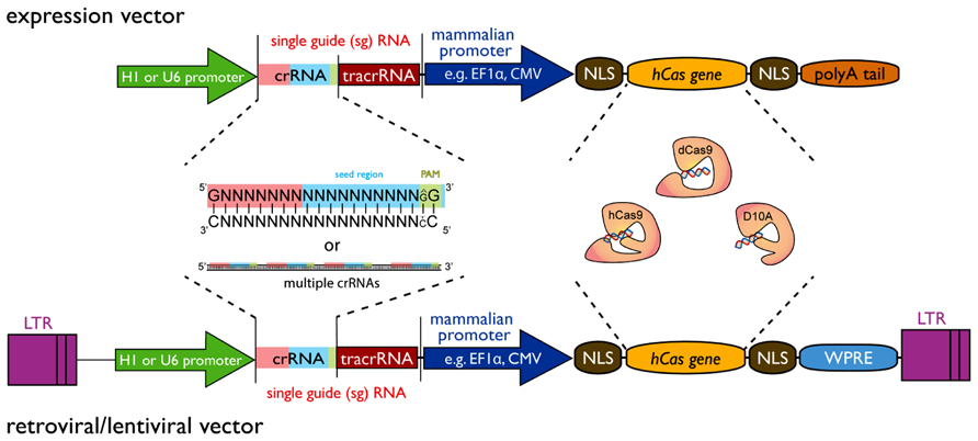
Their technique developed by Gantz and Bier had three basic parts:


  1. Both somatic and germline cells expressed a Cas9 gene,


  1. A guide RNA (gRNA) targeted to a genomic sequence of interest,


  1. The Cas9/gRNA cassettes have the flanking homolog arms that matches the two genomic sequences immediately adjacent to either side of the target cut site


There are many applications in translational genomics that requires multiple steps to make it perfect for complicated organisms, such as plants, mosquitoes and human diseases.

Short Walk from Past to the Future of CRISPR/Cas9

Development and Applications of CRISPR-Cas9 for Genome Engineeri

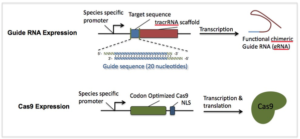
The RNA-guided Cas9 nuclease from the microbial clustered regularly interspaced short palindromic repeats (CRISPR) adaptive immune system can be used to facilitate efficient genome engineering in eukaryotic cells by simply specifying a 20-nt targeting sequence within its guide RNA.

CRISPR/Cas systems are part of the adaptive immune system of bacteria and archaea, protecting them against invading nucleic acids such as viruses by cleaving the foreign DNA in a sequence-dependent manner.

The latest ground-breaking technology for genome editing is based on RNA-guided engineered nucleases, which already hold great promise due to their:

  • simplicity,
  • efficiency and
  • versality

Although CRISPR arrays were first identified in the Escherichia coli genome in 1987 (Ishino et al., 1987),

their biological function was not understood until 2005, when it was shown that the spacers were homologous to viral and plasmid sequences suggesting a role in adaptive immunity (Bolotin et al., 2005; Mojica et al., 2005; Pourcel et al., 2005).

Two years later, CRISPR arrays were confirmed to provide protection against invading viruses when combined with Cas genes (Barrangou et al., 2007).

The mechanism of this immune system based on RNA-mediated DNA targeting was demonstrated shortly thereafter (Brouns et al., 2008; Deltcheva et al., 2011; Garneau et al., 2010; Marraffini and Sontheimer, 2008).


The most widely used system is the type II clustered regularly interspaced short palindromic repeat (CRISPR)/Cas9 (CRISPR-associated) system from Streptococcus pyogenes (Jinek et al., 2012).

Then, five independent groups demonstrated that the two-component system was functional in eukaryotes (human, mouse and zebrafish), indicating that the other functions of the CRISPR locus genes were supported by endogenous eukaryotic enzymes (Cho et al., 2013Cong et al., 2013Hwang et al., 2013Jinek et al., 2013 and Mali et al., 2013).

Beginning with target design, gene modifications can be achieved within as little as 1-2 weeks, and modified colonial cell lines can be derived within 2-3 weeks


Development and Applications of CRISPR-Cas9 for Genome Engineeri

Genome editing with site-specific nucleases.

Double-strand breaks induced by a nuclease at a specific site can be repaired either by non-homologous end joining (NHEJ) or homologous recombination (HR).  In most cases, NHEJ causes random insertions or deletions (indels), which can result in frameshift mutations if they occur in the coding region of a gene, effectively creating a gene knockout.

Alternatively, when the DSB generates overhangs, NHEJ can mediate the targeted introduction of a double-stranded DNA template with compatible overhangs

Even though the generation of breaks in both DNA strands induces recombination at specific genomic loci, NHEJ is by far the most common DSB repair mechanism in most organisms, including higher plants, and the frequency of targeted integration by HR remains much lower than random integration.

  • Unlike its predecessors, the CRISPR/Cas9 system does not require any protein engineering steps, making it much more straightforward to test multiple gRNAs for each target gene


  • Unlike ZFNs and TALENs, the CRISPR/Cas9 system can cleave methylated DNA in human cells (Hsu et al., 2013), allowing genomic modifications that are beyond the reach of the other nucleases (Ding et al., 2013).


  • The main practical advantage of CRISPR/Cas9 compared to ZFNs and TALENs is the ease of multiplexing. The simultaneous introduction of DSBs at multiple sites can be used to edit several genes at the same time (Li et al., 2013; Mao et al., 2013) and can be particularly useful to knock out redundant genes or parallel pathways.


  • Finally, the open access policy of the CRISPR research community has promoted the widespread uptake and use of this technology in contrast, for example, to the proprietary nature of the ZFN platform.

The community provides access to plasmids (e.g., via the non-profit repository Addgene), web tools for selecting gRNA sequences and predicting specificity:

Downside:

One area that will likely need to be addressed when moving to more complex genomes, for instance, is off-target CRISPR/Cas9 activity since fruit fly has only four chromosomes and less likely to have off-target effects. However, this study provided proof of principle.

  • Yet, this critics is not new since one of the few criticisms of the CRISPR/Cas9 technology is the relatively high frequency of off-target mutations reported in some of the earlier studies (Cong et al., 2013; Fu et al., 2013; Hsu et al., 2013; Jiang et al., 2013a; Mali et al., 2013; Pattanayak et al., 2013).


Several strategies have been developed to reduce off-target genome editing, the most important of which is the considered design of the gRNA.


  • fusions of catalytically inactive Cas9 and FokI nuclease have been generated, and these show comparable efficiency to the nickases but substantially higher (N140-fold) specificity than the wild-type enzyme (Guilinger et al., 2014; Tsai et al., 2014)


  • Altering the length of the gRNA can also minimize non-target modifications. Guide RNAs with two additional guanidine residues at the 5′ end were able to avoid off-target sites more efficiently than normal gRNAs but were also slightly less active at on-target sites (Cho et al., 2014)

Development and Applications of CRISPR-Cas9 for Genome Engineeri

What more:

The CRISPR/Cas9 system can be used for several purposes in addition to genome editing:

  • The ectopic regulation of gene expression, which can provide useful information about gene functions and can also be used to engineer novel genetic regulatory circuits for synthetic biology applications.


  • The external control of gene expression typically relies on the use of inducible or repressible promoters, requiring the introduction of a new promoter and a particular treatment (physical or chemical) for promoter activation or repression.


  • Disabled nucleases can be used to regulate gene expression because they can still bind to their target DNA sequence. This is the case with the catalytically inactive version of Cas9 which is known as dead Cas9 (dCas9).


  • Preparing the host for an immunotherapy is possible if it is combined with TLR mechanism:

On the other hand, the host mechanism needs to be review carefully for the design of an effective outcome.

The mechanism of microbial response and infectious tolerance are complex.


During microbial responses, Toll-like receptors (TLRs) play a role to differentiate and determine the microbial structures as a ligand to initiate production of cytokines and pro-inflammatory agents to activate specific T helper cells.


Uniqueness of TLR comes from four major characteristics of each individual TLR :


  1. ligand specificity,
  2. signal transduction pathways,
  3. expression profiles and
  4. cellular localization.


Thus, TLRs are important part of the immune response signaling mechanism to initiate and design adoptive responses from innate (naïve) immune system to defend the host.


TLRs are expressed cell type specific patterns and present themselves on APCs (DCs, MQs, monocytes) with a rich expression  levels Specific TLR stimulat ion links innate and acquired responses through simple recognition of pathogen-associated molecular patterns (PAMPs) or co-stimulation of PAMPs with other TLR or non-TLR receptors, or even better with proinflammatory cytokines.


Some examples of ligand – TLR specificity shown in Table1, which are bacterial lipopeptides, Pam3Cys through TLR2, double stranded (ds) RNAs through TLR3, lipopolysaccharide (LPS) through TLR4, bacterial flagellin through TLR5, single stranded RNAs through TLR7/8, synthetic anti-viral compounds imiquinod through TLR 7 and resiquimod through TLR8, unmethylated CpG DNA motifs through TLR9.


The specificity is established by correct pairing of a TLR with its proinflammatory cytokine(s), so that these permutations influence creation and maintenance of cell differentiat ion.

Development and Applications of CRISPR-Cas9 for Genome Engineeri


  • Immunotherapy: The immune cells can be used as a sensor to scavenger the circulating malformed cells in vivo diagnostics or attack and remember them, for instance, relapse of cancer, re-infection with a same or similar agent (bacteria or virus) etc.

Not only using unique microbial and other model organism properties but also using the human host defense mechanism during innate immune responses may bring a new combat to create a new method of precision medicine. This can be a new type of immunotherapy, immune cell mediated gene therapy or vaccine even a step for an in vivo diagnostics.


Molecular Genetics took a long road from discovery of restriction enzymes, developing PCR assays, cloning were the beginning. Now, having technology to sequence and compare the sequences between organisms also help to design more sophisticated methods.

Generating mutant lines in Drosophila with the classical genetics methods relies on P elements, a type of transposon and balancers after crossing selected flies with specific markers. This fly pushing is a very tedious work but powerful to identify primary pathways, mechanisms and gene interactions in system and translational  genomics.

 Thus, Microbial Immunomodulation is an important factor not only using the microorganisms or their mechanisms but also modulating the immune cells based on the host interaction may generate new types of diagnostics and targeted therapy tools.


Microbial immunomodulation. Microbes from the environment, and from the various microbiota, modulate the immune system. Some of this is due to direct effects of defined microbial products on elements of the immune system. But modulation of the immune system also secondarily alters the host–microbiota relationship and leads to changes in the composition of the microbiota, and so to further changes in immunoregulation (shown as indirect pathways). At the end of the day balance is the key for survival.

microbial immunomodulationGrahamnihms199923f2 A. W. Rook,*,1 Christopher A. Lowry,2 and Charles L. Raison3  Microbial ‘Old Friends’, immunoregulation and stress resilience  Evol Med Public Health. 2013; 2013(1): 46–64. Published online 2013 Apr 9. doi:  10.1093/emph/eot004 PMCID: PMC3868387

http://www.ncbi.nlm.nih.gov/pmc/articles/PMC2881665/bin/nihms199923f2.jpg


CRISPR-Cas9 mediated NHEJ in transient transfection experiments.

Table 1.
Species Transformation method Cas9 codon optimization Promoters (Cas9,  gRNA) Target Mutation frequency Detection method Off-target (no. of sites analyzed) Detection method Multiplex (deletion) Reference
Arabidopsis thaliana PEG-protoplast transfection Arabidopsis (with intron) CaMV35SPDK, AtU6 PDS3<comma> FLS2 1.1–5.6% PCR + sequencing Li et al. (2013)
A. thaliana Leaf agroinfiltration Arabidopsis (with intron) CaMV35SPDK, AtU6 PDS3 2.70% PCR + sequencing Yes (48 bp) Li et al. (2013)
A. thaliana PEG-protoplast transfection Arabidopsis (with intron) CaMV35SPDK,  AtU6 RACK1b<comma> RACK1c 2.5–2.7% PCR + sequencing No (1 site) PCR + sequencing Li et al. (2013)
A. thaliana Leaf agroinfiltration C. reinhardtii CaMV35S, AtU6 Co-transfected GFP n.a. Pre-digested PCR + RE Jiang et al. 2013a and Jiang et al. 2013b
Nicotiana benthamiana PEG-protoplast transfection Arabidopsis (with intron) CaMV35SPDK, AtU6 PDS3 37.7–38.5% PCR + sequencing Li et al. (2013)
N. benthamiana Leaf agroinfiltration Arabidopsis (with intron) CaMV35SPDK,  AtU6 PDS3 4.80% PCR + sequencing Li et al. (2013)
N. benthamiana Leaf agroinfiltration Human CaMV35S,  AtU6 PDS 1.8–2.4% PCR + RE No (18 sites) PCR + RE Nekrasov et al. (2013)
N. benthamiana Leaf agroinfiltration C. reinhardtii CaMV35S, AtU6 Co-transfected GFP n.a. pre-digested PCR + RE Jiang et al. 2013a and Jiang et al. 2013b
N. benthamiana Leaf agroinfiltration Human CaMV35S, CaMV35S PDS 12.7–13.8% Upadhyay et al. (2013)
Nicotiana tabacum PEG-protoplast transfection Tobacco 2xCaMV35S, AtU6 PDS<comma> PDR6 16.27–20.3% PCR + RE Yes (1.8 kb) Gao et al. (2014)
Oryza sativa PEG-protoplast transfection Rice 2xCaMV35S, OsU3 PDS<comma> BADH2<comma> MPK2<comma> Os02g23823 14.5–38.0% PCR + RE Noa (3 sites) PCR + RE Shan et al. (2013)
O. sativa PEG-protoplast transfection Human CaMV35S,  OsU3 or OsU6 MPK5 3–8% RE + qPCR and T7E1 assay No (2 sites) Yes (1 site with a mismatch at position 12) RE + PCR Xie and Yang (2013)
O. sativa PEG-protoplast transfection Rice CaMV35S,  OsU6 SWEET14 n.a. pre-digested PCR + RE Jiang et al. 2013a and Jiang et al. 2013b
O. sativa PEG-protoplast transfection Rice ZmUbi,  OsU6 KO1 KOL5; CPS4 CYP99A2; CYP76M5 CYP76M6 n.a. PCR + sequencing Yes (115<comma> 170<comma> 245 kb) Zhou et al. (2014)
Triticum aestivum PEG-protoplast transfection Rice 2xCaMV35S, TaU6 MLO 28.50% PCR + RE Shan et al. (2013)
T. aestivum PEG-protoplast transfection Plant ZmUbi, TaU6 MLO-A1 36% T7E1 Wang et al. 2014a and Wang et al. 2014b
T. aestivum Agrotransfection of cells from immature embryos Human CaMV35S,  CaMV35S PDS<comma> INOX 18–22% PCR + sequencing Upadhyay et al. (2013)
T. aestivum Agrotransfection of cells from immature embryos Human CaMV35S,  CaMV35S INOX PCR + sequencing No* PCR + RE Yes (53 bp) Upadhyay et al. (2013)
Zea mays PEG-protoplast transfection Rice 2xCaMV35S,  ZmU3 IPK 16.4–19.1% PCR + RE Liang et al. (2014)
Citrus sinensis Leaf agroinfiltration Human CaMv35S,  CaMV35S PDS 3.2–3.9% PCR + RE No (8 sites) PCR + RE Jia et al. (2014)


References:

A brief overview of CRISPR-mediated immunity and explain how the emerging new properties of this defense system are being repurposed for genome engineering in bacteria, yeast, human cells, insects, fish, worms, plants, frogs, pigs, and rodents.

Also look at F1000Prime Rep. 2014; 6: 3. For the list of microorganisms use in CRISPR applications.

Bikard D, Jiang W, Samai P, Hochschild A, Zhang F, Marraffini LA. Programmable repression and activation of bacterial gene expression using an engineered CRISPR-Cas system. Nucleic Acids Res. 2013;41:7429–37. doi: 10.1093/nar/gkt520.

Jiang W, Bikard D, Cox D, Zhang F, Marraffini LA. RNA-guided editing of bacterial genomes using CRISPR-Cas systems. Nat Biotechnol. 2013;31:233–9. doi: 10.1038/nbt.2508.

Qi LS, Larson MH, Gilbert LA, Doudna JA, Weissman JS, Arkin AP, Lim WA. Repurposing CRISPR as an RNA-guided platform for sequence-specific control of gene expression. Cell. 2013;152:1173–83. doi: 10.1016/j.cell.2013.02.022

Esvelt KM, Mali P, Braff JL, Moosburner M, Yaung SJ, Church GM. Orthogonal Cas9 proteins for RNA-guided gene regulation and editing. Nat Methods.2013;10:1116–21. doi: 10.1038/nmeth.2681.

Gilbert LA, Larson MH, Morsut L, Liu Z, Brar GA, Torres SE, Stern-Ginossar N, Brandman O, Whitehead EH, Doudna JA, Lim WA, Weissman JS, Qi LS. CRISPR-mediated modular RNA-guided regulation of transcription in eukaryotes.Cell. 2013;154:442–51. doi: 10.1016/j.cell.2013.06.044.

DiCarlo JE, Norville JE, Mali P, Rios X, Aach J, Church GM. Genome engineering in Saccharomyces cerevisiae using CRISPR-Cas systems. Nucleic Acids Res. 2013;41:4336–43. doi: 10.1093/nar/gkt135.

Cheng AW, Wang H, Yang H, Shi L, Katz Y, Theunissen TW, Rangarajan S, Shivalila CS, Dadon DB, Jaenisch R. Multiplexed activation of endogenous genes by CRISPR-on, an RNA-guided transcriptional activator system. Cell Res.2013;23:1163–71. doi: 10.1038/cr.2013.122.

 Hou Z, Zhang Y, Propson NE, Howden SE, Chu L, Sontheimer EJ, Thomson JA. Efficient genome engineering in human pluripotent stem cells using Cas9 from Neisseria meningitidis. Proc Natl Acad Sci USA. 2013;110:15644–9. doi: 10.1073/pnas.1313587110.

Ran FA, Hsu PD, Lin C, Gootenberg JS, Konermann S, Trevino AE, Scott DA, Inoue A, Matoba S, Zhang Y, Zhang F. Double nicking by RNA-guided CRISPR Cas9 for enhanced genome editing specificity. Cell. 2013;154:1380–9. doi: 10.1016/j.cell.2013.08.021.

Cho SW, Kim S, Kim JM, Kim J. Targeted genome engineering in human cells with the Cas9 RNA-guided endonuclease. Nat Biotechnol. 2013;31:230–2. doi: 10.1038/nbt.2507.

Le Cong, Ran FA, Cox D, Lin S, Barretto R, Habib N, Hsu PD, Wu X, Jiang W, Marraffini LA, Zhang F. Multiplex genome engineering using CRISPR/Cas systems.Science. 2013;339:819–23. doi: 10.1126/science.1231143.

Cradick TJ, Fine EJ, Antico CJ, Bao G. CRISPR/Cas9 systems targeting β-globin and CCR5 genes have substantial off-target activity. Nucleic Acids Res.2013;41:9584–92. doi: 10.1093/nar/gkt714.

Ding Q, Regan SN, Xia Y, Oostrom LA, Cowan CA, Musunuru K. Enhanced efficiency of human pluripotent stem cell genome editing through replacing TALENs with CRISPRs. Cell Stem Cell. 2013;12:393–4. doi: 10.1016/j.stem.2013.03.006.

Ebina H, Misawa N, Kanemura Y, Koyanagi Y. Harnessing the CRISPR/Cas9 system to disrupt latent HIV-1 provirus. Sci Rep. 2013;3:2510. doi: 10.1038/srep02510.

Fu Y, Foden JA, Khayter C, Maeder ML, Reyon D, Joung JK, Sander JD. High-frequency off-target mutagenesis induced by CRISPR-Cas nucleases in human cells.Nat Biotechnol. 2013;31:822–6. doi: 10.1038/nbt.2623.

Hsu PD, Scott DA, Weinstein JA, Ran FA, Konermann S, Agarwala V, Li Y, Fine EJ, Wu X, Shalem O, Cradick TJ, Marraffini LA, Bao G, Zhang F. DNA targeting specificity of RNA-guided Cas9 nucleases. Nat Biotechnol. 2013;31:827–32. doi: 10.1038/nbt.2647.

Jinek M, East A, Cheng A, Lin S, Ma E, Doudna J. RNA-programmed genome editing in human cells. Elife. 2013;2:e00471. doi: 10.7554/eLife.00471.

Maeder ML, Linder SJ, Cascio VM, Fu Y, Ho QH, Joung JK. CRISPR RNA-guided activation of endogenous human genes. Nat Methods. 2013;10:977–9. doi: 10.1038/nmeth.2598.

Mali P, Aach J, Stranges PB, Esvelt KM, Moosburner M, Kosuri S, Yang L, Church GM. CAS9 transcriptional activators for target specificity screening and paired nickases for cooperative genome engineering. Nat Biotechnol. 2013;31:833–8. doi: 10.1038/nbt.2675.

Mali P, Yang L, Esvelt KM, Aach J, Guell M, DiCarlo JE, Norville JE, Church GM. RNA-guided human genome engineering via Cas9. Science. 2013;339:823–6. doi: 10.1126/science.1232033.

Pattanayak V, Lin S, Guilinger JP, Ma E, Doudna JA, Liu DR. High-throughput profiling of off-target DNA cleavage reveals RNA-programmed Cas9 nuclease specificity. Nat Biotechnol. 2013;31:839–43. doi: 10.1038/nbt.2673.

Perez-Pinera P, Kocak DD, Vockley CM, Adler AF, Kabadi AM, Polstein LR, Thakore PI, Glass KA, Ousterout DG, Leong KW, Guilak F, Crawford GE, Reddy TE, Gersbach CA. RNA-guided gene activation by CRISPR-Cas9-based transcription factors. Nat Methods. 2013;10:973–6. doi: 10.1038/nmeth.2600.

Yang L, Guell M, Byrne S, Yang JL, Los Angeles A de, Mali P, Aach J, Kim-Kiselak C, Briggs AW, Rios X, Huang P, Daley G, Church G. Optimization of scarless human stem cell genome editing. Nucleic Acids Res. 2013;41:9049–61. doi: 10.1093/nar/gkt555.

Bassett AR, Tibbit C, Ponting CP, Liu J. Highly efficient targeted mutagenesis of Drosophila with the CRISPR/Cas9 system. Cell Rep. 2013;4:220–8. doi: 10.1016/j.celrep.2013.06.020.

Gratz SJ, Cummings AM, Nguyen JN, Hamm DC, Donohue LK, Harrison MM, Wildonger J, O’Connor-Giles KM. Genome engineering of Drosophila with the CRISPR RNA-guided Cas9 nuclease. Genetics. 2013;194:1029–35. doi: 10.1534/genetics.113.152710.

Yu Z, Ren M, Wang Z, Zhang B, Rong YS, Jiao R, Gao G. Highly efficient genome modifications mediated by CRISPR/Cas9 in Drosophila. Genetics.2013;195:289–91. doi: 10.1534/genetics.113.153825.

Kondo S, Ueda R. Highly improved gene targeting by germline-specific cas9 expression in Drosophila. Genetics. 2013;195:715–21. doi: 10.1534/genetics.113.156737.

Chang N, Sun C, Gao L, Zhu D, Xu X, Zhu X, Xiong J, Xi JJ. Genome editing with RNA-guided Cas9 nuclease in zebrafish embryos. Cell Res. 2013;23:465–72. doi: 10.1038/cr.2013.45.

Hwang WY, Fu Y, Reyon D, Maeder ML, Kaini P, Sander JD, Joung JK, Peterson RT, Yeh JJ. Heritable and precise zebrafish genome editing using a CRISPR-Cas system. PLoS ONE. 2013;8:e68708. doi: 10.1371/journal.pone.0068708.

Hwang WY, Fu Y, Reyon D, Maeder ML, Tsai SQ, Sander JD, Peterson RT, Yeh JJ, Joung JK. Efficient genome editing in zebrafish using a CRISPR-Cas system.Nat Biotechnol. 2013;31:227–9. doi: 10.1038/nbt.2501.

Jao L, Wente SR, Chen W. Efficient multiplex biallelic zebrafish genome editing using a CRISPR nuclease system. Proc Natl Acad Sci USA. 2013;110:13904–9. doi: 10.1073/pnas.1308335110.

Xiao A, Wang Z, Hu Y, Wu Y, Luo Z, Yang Z, Zu Y, Li W, Huang P, Tong X, Zhu Z, Lin S, Zhang B. Chromosomal deletions and inversions mediated by TALENs and CRISPR/Cas in zebrafish. Nucleic Acids Res. 2013;41:e141. doi: 10.1093/nar/gkt464.

Chen C, Fenk LA, Bono M de. Efficient genome editing in Caenorhabditis elegans by CRISPR-targeted homologous recombination. Nucleic Acids Res.2013;41:e193. doi: 10.1093/nar/gkt805.

Chiu H, Schwartz HT, Antoshechkin I, Sternberg PW. Transgene-Free Genome Editing in Caenorhabditis elegans Using CRISPR-Cas. Genetics. 2013;195:1167–71. doi: 10.1534/genetics.113.155879.

Cho SW, Lee J, Carroll D, Kim J, Lee J. Heritable Gene Knockout in Caenorhabditis elegans by Direct Injection of Cas9-sgRNA Ribonucleoproteins.Genetics. 2013;195:1177–80. doi: 10.1534/genetics.113.155853.

Friedland AE, Tzur YB, Esvelt KM, Colaiácovo MP, Church GM, Calarco JA. Heritable genome editing in C. elegans via a CRISPR-Cas9 system. Nat Methods.2013;10:741–3. doi: 10.1038/nmeth.2532.

Katic I, Großhans H. Targeted Heritable Mutation and Gene Conversion by Cas9-CRISPR in Caenorhabditis elegans. Genetics. 2013;195:1173–6. doi: 10.1534/genetics.113.155754.

Lo T, Pickle CS, Lin S, Ralston EJ, Gurling M, Schartner CM, Bian Q, Doudna JA, Meyer BJ. Precise and heritable genome editing in evolutionarily diverse nematodes using TALENs and CRISPR/Cas9 to engineer insertions and deletions.Genetics. 2013;195:331–48. doi: 10.1534/genetics.113.155382.

Tzur YB, Friedland AE, Nadarajan S, Church GM, Calarco JA, Colaiácovo MP. Heritable Custom Genomic Modifications in Caenorhabditis elegans via a CRISPR-Cas9 System. Genetics. 2013;195:1181–5. doi: 10.1534/genetics.113.156075.

Waaijers S, Portegijs V, Kerver J, Lemmens BBLG, Tijsterman M, van den Heuvel S, Boxem M. CRISPR/Cas9-Targeted Mutagenesis in Caenorhabditis elegans. Genetics. 2013;195:1187–91. doi: 10.1534/genetics.113.156299.

Dickinson DJ, Ward JD, Reiner DJ, Goldstein B. Engineering the Caenorhabditis elegans genome using Cas9-triggered homologous recombination.Nat Methods. 2013;10:1028–34. doi: 10.1038/nmeth.2641.

Feng Z, Zhang B, Ding W, Liu X, Yang D, Wei P, Cao F, Zhu S, Zhang F, Mao Y, Zhu J. Efficient genome editing in plants using a CRISPR/Cas system. Cell Res.2013;23:1229–32. doi: 10.1038/cr.2013.114.

Li J, Norville JE, Aach J, McCormack M, Zhang D, Bush J, Church GM, Sheen J. Multiplex and homologous recombination-mediated genome editing in Arabidopsis and Nicotiana benthamiana using guide RNA and Cas9. Nat Biotechnol. 2013;31:688–91. doi: 10.1038/nbt.2654.

Nekrasov V, Staskawicz B, Weigel D, Jones JDG, Kamoun S. Targeted mutagenesis in the model plant Nicotiana benthamiana using Cas9 RNA-guided endonuclease. Nat Biotechnol. 2013;31:691–3. doi: 10.1038/nbt.2655.

Shan Q, Wang Y, Li J, Zhang Y, Chen K, Liang Z, Zhang K, Liu J, Xi JJ, Qiu J, Gao C. Targeted genome modification of crop plants using a CRISPR-Cas system.Nat Biotechnol. 2013;31:686–8. doi: 10.1038/nbt.2650.

 Xie K, Yang Y. RNA-Guided Genome Editing in Plants Using a CRISPR-Cas System. Mol Plant. 2013;6:1975–83. doi: 10.1093/mp/sst119.

Miao J, Guo D, Zhang J, Huang Q, Qin G, Zhang X, Wan J, Gu H, Qu L. Targeted mutagenesis in rice using CRISPR-Cas system. Cell Res. 2013;23:1233–6. doi: 10.1038/cr.2013.123.

Jiang W, Zhou H, Bi H, Fromm M, Yang B, Weeks DP. Demonstration of CRISPR/Cas9/sgRNA-mediated targeted gene modification in Arabidopsis, tobacco, sorghum and rice. Nucleic Acids Res. 2013;41:e188. doi: 10.1093/nar/gkt780.

Upadhyay SK, Kumar J, Alok A, Tuli R. RNA Guided Genome Editing for Target Gene Mutations in Wheat. G3 (Bethesda) 2013

Nakayama T, Fish MB, Fisher M, Oomen-Hajagos J, Thomsen GH, Grainger RM. Simple and efficient CRISPR/Cas9-mediated targeted mutagenesis in Xenopus tropicalis. Genesis. 2013 doi: 10.1002/dvg.22720.

Tan W, Carlson DF, Lancto CA, Garbe JR, Webster DA, Hackett PB, Fahrenkrug SC. Efficient nonmeiotic allele introgression in livestock using custom endonucleases. Proc Natl Acad Sci USA. 2013;110:16526–31. doi: 10.1073/pnas.1310478110.

Li D, Qiu Z, Shao Y, Chen Y, Guan Y, Liu M, Li Y, Gao N, Wang L, Lu X, Zhao Y, Liu M. Heritable gene targeting in the mouse and rat using a CRISPR-Cas system.Nat Biotechnol. 2013;31:681–3. doi: 10.1038/nbt.2661.

Li W, Teng F, Li T, Zhou Q. Simultaneous generation and germline transmission of multiple gene mutations in rat using CRISPR-Cas systems. Nat Biotechnol.2013;31:684–6. doi: 10.1038/nbt.2652.

Shen B, Zhang J, Wu H, Wang J, Ma K, Li Z, Zhang X, Zhang P, Huang X. Generation of gene-modified mice via Cas9/RNA-mediated gene targeting. Cell Res. 2013;23:720–3. doi: 10.1038/cr.2013.46.

Wang H, Yang H, Shivalila CS, Dawlaty MM, Cheng AW, Zhang F, Jaenisch R. One-step generation of mice carrying mutations in multiple genes by CRISPR/Cas-mediated genome engineering. Cell. 2013;153:910–8. doi: 10.1016/j.cell.2013.04.025.


Previously Published at Leaders in Pharmaceutical Intelligence:


CRISPR/Cas9: Contributions on Endoribonuclease Structure and Function, Role in Immunity and Applications in Genome Engineering larryhbern 2015/03/27
Published
CRISPR-CAS editing brings cloning of woolly mammoth one step closer to reality 2012pharmaceutical 2015/03/26
Published
GUIDE-seq: First genome-wide method of detecting off-target DNA breaks induced by CRISPR-Cas nucleases 2012pharmaceutical 2014/12/22
Published
The Patents for CRISPR, the DNA editing technology as the Biggest Biotech Discovery of the Century 2012pharmaceutical 2014/12/05
Published
CRISPR: Applications for Autoimmune Diseases @UCSF 2012pharmaceutical 2014/11/04
Published
“Gene Editing at CRISPR Speed”: Services and Tools 2012pharmaceutical 2014/10/29
Published
Licensing CRISPR-Cas9 Technology from Broad Institute: Clontech, Horizon Discovery, Sage Labs 2012pharmaceutical 2014/10/28
Published
CRISPR-Cas9 Discovery and Development of Programmable Genome Engineering – Gabbay Award Lectures in Biotechnology and Medicine – Hosted by Rosenstiel Basic Medical Sciences Research Center, 10/27/14 3:30PM Brandeis University, Gerstenzang 121 2012pharmaceutical 2014/10/26
Published
Inactivation of the human papillomavirus E6 or E7 gene in cervical carcinoma cells using a bacterial CRISPR/Cas 2012pharmaceutical 2014/10/24
Published
CRISPR-Cas9 Foundational Technology originated at UC, Berkeley & UCSF, Broad Institute is developing Biotech Applications — Intellectual Property emerging as Legal Potential Dispute 2012pharmaceutical 2014/06/18
Published
2:15 – 2:45, 6/13/2014, Jennifer Doudna “The biology of CRISPRs: from genome defense to genetic engineering” 2012pharmaceutical 2014/06/13
Published
CRISPR @MIT – Genome Surgery 2012pharmaceutical 2014/04/21
Published
Gene Therapy and the Genetic Study of Disease: @Berkeley and @UCSF – New DNA-editing technology spawns bold UC initiative as Crispr Goes Global 2012pharmaceutical 2014/03/27
Published
Evaluate your Cas9 Gene Editing Vectors: CRISPR/Cas Mediated Genome Engineering – Is your CRISPR gRNA optimized for your cell lines? 2012pharmaceutical 2014/03/25
Published
CRISPR-Cas: A powerful new tool for precise genetic engineering 2012pharmaceutical 2013/11/29
Published
A NEW ERA OF GENETIC MANIPULATION   Demet Sag, Ph.D., CRA, GCP 31 mins ago
Published
Manipulate Signaling Pathways [7.6] larryhbern 2015/04/08
Published
RNAi – On Transcription and Metabolic Control larryhbern 2015/03/26
Published
Real Time Conference Coverage for Scientific and Business Media: Unique Twitter Hashtags and Handles per Conference Presentation/Session 2012pharmaceutical 2015/03/24
Published
Advances in Gene Editing Technology: New Gene Therapy Options in Personalized Medicine 2012pharmaceutical 2015/03/16
Published
Annual Margaret Pittman Lecture, honors the NIH’s first female lab chief, March 11, 2015, 3:00:00 PM by Jennifer Doudna, Ph.D., University of California, Berkeley 2012pharmaceutical 2015/03/11
Published
Protecting Your Biotech IP and Market Strategy: Notes from Life Sciences Collaborative 2015 Meeting sjwilliamspa 2015/03/11
Published
Genomics Diagnostics Companies attractive to Institutional Investors 2012pharmaceutical 2015/03/10
Published
attn #1: Investors in HealthCare — Platforms in the Ecosystem of Regulatory & Reimbursement – Integrated Informational Platforms in Medical Devices, Global Oncology Drugs Market and Peer-Reviewed Curations: Cancer, Genomics and Cardiovascular – Draft 2012pharmaceutical 2015/02/24
Last Modified
attn #2: Investors in HealthCare — Cardiovascular Medical Devices: Platforms in the Ecosystem of Regulatory & Reimbursement with Integrated Informational Platforms of Peer-Reviewed Global Scientific Curations on Medical Devices and Cardiac Surgery, Interventional Cardiology and Cardiovascular Imaging – Draft 2012pharmaceutical 2015/02/24
Last Modified
attn #3: Investors in HealthCare — Platforms in the Ecosystem of Regulatory & Reimbursement – Integrated Informational Platforms in Orthopedic Medical Devices, and Global Peer-Reviewed Scientific Curations: Bone Disease and Orthopedic Medicine – Draft 2012pharmaceutical 2015/02/23
Last Modified
attn #7: Investors in HealthCare — Platforms in the Ecosystem of Regulatory & Reimbursement – Integrated Informational Platforms in Medical Devices, Global Oncology Drugs Market and Peer-Reviewed Curations: Cancer, Genomics and Cardiovascular – Draft 2012pharmaceutical 2015/02/22
Last Modified
attn #6: Investors in HealthCare — Platforms in the Ecosystem of Regulatory & Reimbursement – Integrated Informational Platforms in Medical Devices, Global Oncology Drugs Market and Peer-Reviewed Curations: Cancer, Genomics and Cardiovascular – Draft 2012pharmaceutical 2015/02/22
Last Modified
attn #5: Investors in HealthCare — Platforms in the Ecosystem of Regulatory & Reimbursement – Integrated Informational Platforms in Medical Devices, Global Oncology Drugs Market and Peer-Reviewed Curations: Cancer, Genomics and Cardiovascular – Draft 2012pharmaceutical 2015/02/22
Last Modified
attn #4: Investors in HealthCare — Platforms in the Ecosystem of Regulatory & Reimbursement – Integrated Informational Platforms in Medical Devices, Global Oncology Drugs Market and Peer-Reviewed Curations: Cancer, Genomics and Cardiovascular – Draft 2012pharmaceutical 2015/02/22
Last Modified
7:55AM – 9AM, January 26, 2015 – Introduction and Overview – LIVE @Silicon Valley 2015 Personalized Medicine World Conference, Mountain View, CA 2012pharmaceutical 2015/01/26
Published
Litigation on the Way: Broad Institute Gets Patent on Revolutionary Gene-Editing Method 2012pharmaceutical 2014/12/05
Published
3:15PM 11/12/2014 – Discussion Complex Disorders @10th Annual Personalized Medicine Conference at the Harvard Medical School, Boston 2012pharmaceutical 2014/11/12
Published
Twitter is Becoming a Powerful Tool in Science and Medicine sjwilliamspa 2014/11/06
Published
New Frontiers in Gene Editing: Transitioning From the Lab to the Clinic, February 19-20, 2015 | The InterContinental San Francisco | San Francisco, CA 2012pharmaceutical 2014/10/29
Published
Geneticist George Church: A Future Without Limits 2012pharmaceutical 2014/10/24
Published
Metabolomics is about Metabolic Systems Integration larryhbern 2014/10/13
Published
Using RNA-seq and targeted nucleases to identify mechanisms of drug resistance in acute myeloid leukemia 2012pharmaceutical 2014/09/26
Published
New Frontiers in Gene Editing — Cambridge Healthtech Institute’s Inaugural, February 19-20, 2015 | The Inter Continental San Francisco | San Francisco, CA 2012pharmaceutical 2014/08/27
Published
The role and importance of transcription factors larryhbern 2014/08/06
Published
  Pathology Emergence in the 21st Century larryhbern 2014/08/03
Published
  Regulation of somatic stem cell Function larryhbern 2014/07/29
Published
  Prediction of the Winner RNA Technology, the FRONTIER of SCIENCE on RNA Biology, Cancer and Therapeutics & The Start Up Landscape in Boston 2012pharmaceutical 2014/06/16
Published
  Lecture Contents delivered at Koch Institute for Integrative Cancer Research, Summer Symposium 2014: RNA Biology, Cancer and Therapeutic Implications, June 13, 2014 @MIT 2012pharmaceutical 2014/06/16
Published
  3:45 – 4:15, 2014, Scott Lowe “Tumor suppressor and tumor maintenance genes” 2012pharmaceutical 2014/06/13
Published
  11:30 – 12:00, 6/13/2014, Daniel Anderson “Intracellular RNA delivery” 2012pharmaceutical 2014/06/13
Published
  9:10 – 9:30, 6/13/2014, Phillip Sharp “Why RNA Biology?” Phillip Sharp, PhD Institute Professor, Koch Institute for Integrative Cancer Research, Massachusetts Institute of Technology 2012pharmaceutical 2014/06/13
Published
  The SCID Pig II: Researchers Develop Another SCID Pig, And Another Great Model For Cancer Research sjwilliamspa 2014/06/11
Published
  Koch Institute for Integrative Cancer Research @MIT – Summer Symposium 2014: RNA Biology, Cancer and Therapeutic Implications, June 13, 2014 8:30AM – 4:30PM, Kresge Auditorium @MIT 2012pharmaceutical 2014/06/04
Published
  An expanded-DNA Biology from Scripps Research Institute: Beyond A-T and C-G: Applications for new Medicines and Nanotechnology 2012pharmaceutical 2014/05/11
Published
  Foundation Medicine reported 4,702 Clinical Tests in Q1, 715 were the FoundationOne Heme Cancer Test, average Reimbursement of $3,400 per Test 2012pharmaceutical 2014/05/08
Published
  The Cancer Research Concentration @ Leaders in Pharmaceutical Business Intelligence sjwilliamspa 2014/05/06
Published
  Aviva’s Perspective on New Oncology Database Asset Positioning by Business Scenario – Password protected 2012pharmaceutical 2014/05/05
Published
  Cancer Research: Curations and Reporting: Aviva Lev-Ari, PhD, RN 2012pharmaceutical 2014/04/20
Published
  Predictions on Biotech Sector’s Two-year Boom 2012pharmaceutical 2014/03/27
Published
  DNA: One man’s trash is another man’s treasure, but there is no JUNK after all Demet Sag, Ph.D., CRA, GCP 2013/06/24
Published
  Ribozymes and RNA Machines – Work of Jennifer A. Doudna 2012pharmaceutical 2013/04/15
Published
  Zebrafish—Susceptible to Cancer larryhbern 2013/04/02
Published
  Diagnosing Diseases & Gene Therapy: Precision Genome Editing and Cost-effective microRNA Profiling 2012pharmaceutical 2013/03/28
Published
  Directions for Genomics in Personalized Medicine larryhbern 2013/01/27
Published


About the author:

Dr Sag has a Bachelor’s degree in Basic and Industrial Microbiology as a Sum cum Laude among 450 graduating class of Science faculty,  an MSc in Microbial Engineering and Biotechnology (Bioprocessing improvement) and PhD in Molecular and Developmental Genetics (Functional Genome and Stem Cell Biology).

She is an translational functional genomic scientist to develop diagnostics and targeted therapies by non-invasive methods for personalized medicine from bench to bedside and engineering tools through clinical trials and regulatory affairs.

You may contact with her at 858-729-4942 or by demet.sag@gmail.com if you have questions.


Read Full Post »

CRISPR/Cas9: Contributions on Endoribonuclease Structure and Function, Role in Immunity and Applications in Genome Engineering

Writer and Curator:Larry H Bernstein, MD, FCAP 

2.2.25

2.2.25   CRISPR/Cas9: Contributions on Endoribonuclease Structure and Function, Role in Immunity and Applications in Genome Engineering, Volume 2 (Volume Two: Latest in Genomics Methodologies for Therapeutics: Gene Editing, NGS and BioInformatics, Simulations and the Genome Ontology), Part 2: CRISPR for Gene Editing and DNA Repair

This is the fourth contribution to a series on transcriptional control and cellular remodeling. The previous dealt with RNAs – mRNA, miRNA, RNAi, siRNA, shRNA, small RNAs, lncRNAs, DICER, SLICER, RISC, recombination, and related processes.  It is clear that the classical model was limited, is history, and could not predict a large universe encompassing DNA, RNA, transcription, translation, signaling, proteins, protein conformation, mRNA-miRNA interactions, protein-protein interactions, inter- and intracellular interactions, and cellular remodeling.

Cutting it close: CRISPR-associated endoribonuclease structure and function
Hochstrasser ML and Doudna JA
Trends in Biochemical Sciences, Jan 2015; 40(1):58-66
http://dx.doi.org/10.1016/j.tibs.2014.10.007

RNAi pathways in eukaryotes, as in archaea, possess an adaptive immune system consisting of repetitive genetic elements known as clustered regularly clustered interspersed short palindromic repeats (CRISPERS) and CRISPR-associated (cas) proteins. CRISPR-cas systems require small RNAs for sequence-specific detection and degradation of complex nucleic acids. Cas 5 and cas 6 enzymes have evolved to specifically recognize and process CRISPR-derived transcripts to function as small RNAs used as guides by interference complexes.

Figure 1. Overview of CRISPR RNA (crRNA) processing and comparison between CRISPR–Cas interference systems. There are three main pathways of CRISPR adaptive immunity (Types I–III) and several subtypes, each typified by a different set of Cas proteins. The first stage of the CRISPR–Cas system is acquisition, in which a foreign DNA sequence is incorporated into the host CRISPR locus. Next, the entire repeat-spacer array is transcribed into a long precursor crRNA (pre-crRNA). A single cleavage within each repeat sequence generates shorter, mature crRNAs. Some crRNAs undergo an additional trimming step. The enzymes responsible for catalysis and exact mode of crRNA processing differ in each system. The crRNA is loaded into an interference complex where it serves as a guide for targeting invasive DNA, or in Type III-B systems, RNA.

Figure 2. Fundamental structural features of CRISPR endoRNases. (A) Topology diagram of a typical Cas6 C-terminal RRM fold with key structural features labeled. (B) Two views of Thermus thermophilus Cas6e (PDB: 2Y8W) colored as in (A). For clarity, the N-terminal RRM fold has been omitted in the left panel. (C) Comparison of structures of Cas6 and Cas5c enzymes associated with different CRISPR subtypes (in parentheses), highlighting shared structural elements, colored as in (A) and (B), with the Cas5 ‘thumb’ in black (PDB: 4ILL, 2XLK, 3UFC, 4F3M). Note that no active site residues are shown for Pyrococcus furiosus Cas6-3nc because this protein is non-catalytic.

Figure 3. Structure and sequence-specific RNA binding by Cas6 enzymes. (A) First two images: Thermus thermophilus Cas6A in the apo form and bound to its product CRISPR RNA (crRNA) (PDB: apo – 4C97, product-bound – 4C8Z). Second two images: electrostatic surface potential rendering of the same enzyme in two views with the first eight nucleotides of the Pyrococcus furiosus crRNA 30 handle (PDB: 3PKM) modeled onto the structure based on alignment of the two proteins, as in Niewoehner et al. [30]. For simplicity, only one subunit of the non-crystallographic dimer is shown. (B) Pseudomonas aeruginosa Cas6f bound to its cognate RNA (PDB: 2XLK). Close-up views highlight the active site and sequence-specific interactions by the groove-binding element. (C) Sulfolobus solfataricus Cas6-1A bound to its pre-crRNA substrate (PDB: 4ILL). The active site and sequence-specific contacts made by the glycine-rich loop are shown in detail. For simplicity, only one subunit of the SsoCas6-1A dimer is shown.

CRISPRs (clustered regularly interspaced short palindromic repeats) are DNA loci containing short repetitions of base sequences. Each repetition is followed by short segments of “spacer DNA” from previous exposures to a virus.[2]

CRISPRs are found in approximately 40% of sequenced bacteria genomes and 90% of sequenced archaea.[3][4]

CRISPRs are often associated with cas genes that code for proteins related to CRISPRs. The CRISPR/Cas system is a prokaryotic immune system that confers resistance to foreign genetic elements such as plasmids and phages[5][6] and provides a form of acquired immunity. CRISPR spacers recognize and cut these exogenous genetic elements in a manner analogous to RNAi in eukaryotic organisms.[2]

Since 2013, the CRISPR/Cas system has been used for gene editing (adding, disrupting or changing the sequence of specific genes) and gene regulation in species throughout the tree of life.[7] By delivering the Cas9 protein and appropriate guide RNAs into a cell, the organism’s genome can be cut at any desired location.

It may be possible to use CRISPR to build RNA-guided gene drives capable of altering the genomes of entire populations.

Gene-editing predecessors

In the early 2000s, researchers developed zinc finger nucleases, synthetic proteins whose DNA-binding domains enable them to cut DNA at specific spots. Later, synthetic nucleases called TALENs provided an easier way to target specific DNA and were predicted to surpass zinc fingers. They both depend on making custom proteins for each DNA target, a more cumbersome procedure than guide RNAs. CRISPRs are more efficient and can target more genes than these earlier techniques.

Repeats and spacers

CRISPR loci range in size from 24 to 48 base pairs. They usually show some dyad symmetry, implying the formation of a secondary structure such as a hairpin, but are not truly palindromic. Repeats are separated by spacers of similar length. Some CRISPR spacer sequences exactly match sequences from plasmids and phages, although some spacers match the prokaryote’s genome (self-targeting spacers). New spacers can be added rapidly in response to phage infection.

http://en.wikipedia.org/wiki/CRISPR

http://upload.wikimedia.org/wikipedia/commons/thumb/5/5f/Crispr.png/1024px-Crispr.png

http://www.frontiersin.org/files/Articles/58953/fgene-04-00193-r2/image_m/fgene-04-00193-g001.jpg

http://2013.igem.org/wiki/images/thumb/c/c0/CRISPR_Cooperativity_2.png/720px-CRISPR_Cooperativity_2.png

http://img.scoop.it/2Y0f1M2hXSr35d9-xn4WVTl72eJkfbmt4t8yenImKBVvK0kTmF0xjctABnaLJIm9

A CRISPR CASe for high-throughput silencing

A CRISPR CASe for high-throughput silencing

dual gRNA vector

dual gRNA vector

Genome editing with RNA-guided Cas9 nuclease in Zebrafish embryos

Genome editing with RNA-guided Cas9 nuclease in Zebrafish embryos

The role of CRISPR–Cas systems in adaptive immunity and beyond

Barrangue R
Current Opinion in Immunology 2015; 32:36–41
http://dx.doi.org/10.1016/j.coi.2014.12.008

CRISPR–Cas immune systems. CRISPR-encoded immunization and interference. In the adaptation stage, exogenous DNA is sampled and a novel spacer is integrated into the CRISPR locus; in the expression stage, the CRISPR array is transcribed and processed into small interfering CRISPR RNAs (crRNAs) that guide Cas endonucleases towards target complementary DNA in the interference stage.

Cas-mediated DNA targeting and cleavage. The Cas9 endonuclease forms a ribonucleoprotein complex in combination with the dual guide RNA (crRNA and tracrRNA), and the target dsDNA. First, the Cas9:guide RNA complex binds to proto-spacer adjacent motif (PAM) and drives the formation of an R-loop in the target DNA for genesis of a double stranded break using the RuvC and HNH nickase domains. The former primarily involves the recognition (REC) Cas9 lobe (top), and the latter is primarily driven by the nuclease (NUC) lobe (bottom). Cas-mediated targeting can aim at phage DNA for antiviral resistance (cleaved viral DNA cannot replicate), plasmid DNA to preclude the uptake and dissemination of plasmids (cleaved plasmid DNA cannot replicate), and chromosomal DNA for genome editing (insertion of mutations using endogenous DNA repair systems at the site of cleavage) or transcriptional control (dCas9 binding blocks RNA polymerase).

CRISPR-Cas9 Knockin Mice for Genome Editing and Cancer Modeling
Cell Oct 9, 2014; 159:440–455
http://dx.doi.org/10.1016/j.cell.2014.09.014

Figure2. Ex Vivo Genome Editing of Primary Immune Cells Derived from Constitutive Cas9-Expressing Mice (A) Schematic of ex vivo genome editing experimental flow. (B)Flow cytometry histogram of bone marrow cells from constitutive Cas9-expressing (green) and wild-type (blue) mice, showing Cas9-P2A-EGFP expressiononlyinCas9mice.Dataareplottedasa percentage of the total number of cells. (C) sgRNA design for targeting the mouse Myd88 locus. (D) sgRNA design for targeting the mouse A20 locus. (E) Myd88 indel analysis of constitutive Cas9expressing DCs transduced with either a Myd88targeting sgRNA (sgMyd88-1 and sgMyd88-2) or controls (CTR, average of four control sgRNAs), showing indel formation only in Myd88-targeted cells. Data are plotted as the percent of Illumina sequencing reads containing indels at the target site. Mutations are categorized as frameshift (fs, yellow bar) or non-frameshift (nfs, orange bar). (F) A20 indel analysis of constitutive Cas9-expressing DCs transduced with either an A20-targeting sgRNA (sgA20-1) or controls (CTR, average of four control sgRNAs), showing indel formation only in A20-targeted cells. Data are plotted as the percent of Illumina sequencing reads containing indels at the target site. Mutations are categorized as frameshift (fs, yellow bar) or non-frameshift (nfs, orange bar). (G) Myd88 mRNA quantification of constitutive Cas9-expressing DCs transduced with either Myd88-targeting sgRNA (sgMyd88-1 or sgMyd882) or controls (CTR, average of six control sgRNAs), showing reduced expression only in Myd88-targeted cells. Data are plotted as Myd88 mRNA levels from Nanostring nCounter analysis. (H) Immunoblot of constitutive Cas9-expressing DCs transduced with either Myd88-targeting sgRNA (sgMyd88-1 or sgMyd88-2) or controls (four control sgRNAs), showing depletion of MyD88 protein only in Myd88-targeted cells. b-actin was used as a loading control. (*) Overexposed, repeated-measurement. (I) Nanostring nCounter analysis of constitutive Cas9-expressing DCs transduced with either Myd88-targeting sgRNA (sgMyd88-1 or sgMyd882) or shRNA (shMyd88), A20-targeting sgRNA (sgA20-1 or sgA20-2), or shRNA (shA20), showing analtered LPS response.(Inset)Theclustershowing the highest difference between Myd88- and A20 targeting sgRNAs, including key inflammatory genes(IL1a,IL1b,Cxcl1,Tnf,etc.).(Red)High;(blue) low; (white) unchanged; based on fold change relative to measurements with six control sgRNAs. See also Figure S2.

Figure 5. In Vivo Tumor Formation in AAV9-KPL-Injected Mice (A) Lung mCT images of Cre-dependent Cas9 mice injected with either AAV9-KPL or AAV9-sgLacZ 2 months posttransduction, showing tumor formation (indicated by the arrowhead) only in AAV9-KPL injected mice. (B)LungmCT3DrenderingofCre-dependent Cas9 mice injected with AAV9-KPL 2 months posttransduction, showing tumor formation (indicated by a yellow oval). (C) Major tumor burden quantification of Cre-dependent Cas9 mice injected with either AAV9-KPL or AAV9-sgLacZ, showing significant tumor burden in AAV9KPL-injected mice. Data are plotted as mean ± SEM. **p < 0.005. (D) Representative lung H&E images of Cre-dependent Cas9 mice injected with either AAV9-KPL or AAV9-sgLacZ 9 weeks posttransduction, showing heterogeneous tumor formation in AAV9-KPL-injected mice. Arrowheads highlight a representative subset of tumors within the lungs of AAV9-KPL injected mice.

Development and Applications of CRISPR-Cas9 for Genome Engineering
Zhu PD, Lander ES, Zhang F
Cell Jun 5, 2014; 157:1262-1278
http://dx.doi.org/10.1016/j.cell.2014.05.010

Figure 1. Applications of Genome Engineering Genetic and epigenetic control of cells with genome engineering technologies is enabling a broad range of applications from basic biology to biotechnology and medicine. (Clockwise from top) Causal genetic mutations or epigenetic variants associated with altered biological function or disease phenotypes can nowberapidlyandefficientlyrecapitulated inanimalorcellularmodels (Animal models, Genetic variation). Manipulating biological circuits couldalso facilitate the generation of useful synthetic materials, such as algae-derived, silicabased diatoms for oral drug delivery (Materials). Additionally, precise genetic engineering of important agricultural crops could confer resistance to environmental deprivation or pathogenic infection, improving food security while avoiding the introduction of foreign DNA (Food). Sustainable and cost-effective biofuels are attractive sources for renewable energy, which could be achieved by creating efficient metabolic pathways for ethanol production in algae or corn (Fuel). Direct in vivo correction of genetic or epigenetic defects in somatic tissue would be permanent genetic solutions that address the root cause of genetically encoded disorders (Gene surgery). Finally, engineering cells to optimize high yield generation of drug precursors in bacterial factories could significantly reduce the cost and accessibility of useful therapeutics (Drug development).

Figure 2. Genome Editing Technologies Exploit Endogenous DNA Repair Machinery (A) DNA double-strand breaks (DSBs) are typically repaired by nonhomologous end-joining (NHEJ) or homology-directed repair (HDR). In the errorprone NHEJ pathway, Ku heterodimers bind to DSB ends and serve as a molecular scaffold for associated repair proteins. Indels are introduced when the complementary strands undergo end resection and misaligned repair due to microhomology, eventually leading to frameshift mutations and gene knockout. Alternatively, Rad51 proteins may bind DSB ends during the initial phase of HDR, recruiting accessory factors that direct genomic recombination with homology arms on an exogenous repair template. Bypassing the matching sister chromatid facilitates the introduction of precise gene modifications. (B) Zinc finger (ZF) proteins and transcription activator-like effectors (TALEs) are naturally occurring DNA-binding domains that can be modularly assembled to target specific sequences. ZF and TALE domains each recognize 3 and 1 bp of DNA, respectively. Such DNA-binding proteins can be fused to the FokI endonuclease to generate programmable site-specific nucleases. (C) The Cas9 nuclease from the microbial CRISPR adaptive immune system is localized to specific DNA sequences via the guide sequence on its guide RNA (red), directly base-pairing with the DNA target. Binding of a protospacer-adjacent motif (PAM, blue) downstream of the target locus helps to direct Cas9-mediated DSBs.

Figure 3. Key Studies Characterizing and Engineering CRISPR Systems Cas9 has also been referred to as Cas5, Csx12, and Csn1 in literature prior to 2012. For clarity, we exclusively adopt the Cas9 nomenclature throughout this Review. CRISPR, clustered regularly interspaced short palindromic repeats; Cas, CRISPR-associated; crRNA, CRISPR RNA; DSB, double-strand break; tracrRNA, trans-activating CRISPR RNA.

Figure 4. Natural Mechanisms of Microbial CRISPR Systems in Adaptive Immunity Following invasion of the cell by foreign genetic elements from bacteriophages or plasmids (step 1: phage infection), certain CRISPR-associated (Cas) enzymes acquire spacers from the exogenous protospacer sequences and install them into the CRISPR locus within the prokaryotic genome (step 2: spacer acquisition). These spacers are segregated between direct repeats that allow the CRISPR system to mediate self and nonself recognition. The CRISPR array is a noncoding RNA transcript that is enzymatically maturated through distinct pathways that are unique to each type of CRISPR system (step 3: crRNA biogenesis and processing). In types I and III CRISPR, the pre-crRNA transcript is cleaved within the repeats by CRISPR-associated ribonucleases, releasing multiple small crRNAs. Type III crRNA intermediates are further processed at the 30 end by yet-to-be-identified RNases to produce the fully mature transcript. In type II CRISPR, an associated trans-activating CRISPR RNA (tracrRNA) hybridizes with the direct repeats, forming an RNA duplex that is cleaved and processed by endogenous RNase III and other unknown nucleases. Maturated crRNAs from type I and III CRISPR systems are then loaded onto effector protein complexes for target recognition and degradation. In type II systems, crRNA-tracrRNA hybrids complex with Cas9 to mediate interference. Both type I and III CRISPR systems use multiprotein interference modules to facilitate target recognition. In type I CRISPR, the Cascade complex is loaded with a crRNA molecule, constituting a catalytically inert surveillance complex that recognizes target DNA. The Cas3 nuclease is then recruited to the Cascade-bound R loop, mediating target degradation. In type III CRISPR, crRNAs associate either with Csm or Cmr complexes that bind and cleave DNA and RNA substrates, respectively. In contrast, the type II system requires only the Cas9 nuclease to degrade DNA matching its dual guide RNA consisting of a crRNA-tracrRNA hybrid.

Figure 5. Structural and Metagenomic Diversity of Cas9 Orthologs (A) Crystal structure of Streptococcus pyogenes Cas9 in complex with guide RNA and target DNA. (B) Canonical CRISPR locus organization from type II CRISPR systems, which can be classified into IIA-IIC based on their cas gene clusters. Whereas type IIC CRISPR loci contain the minimal set of cas9, cas1, and cas2, IIA and IIB retain their signature csn2 and cas4 genes, respectively. (C) Histogram displaying length distribution of known Cas9 orthologs as described in UniProt, HAMAP protein family profile MF_01480. (D) Phylogenetic tree displaying the microbial origin of Cas9 nucleases from the type II CRISPR immune system. Taxonomic information was derived from greengenes 16S rRNA gene sequence alignment, and the tree was visualized using the Interactive Tree of Life tool (iTol). (E) Four Cas9 orthologs from families IIA, IIB, and IIC were aligned by ClustalW (BLOSUM). Domain alignment is based on the Streptococcus pyogenes Cas9, whereas residues highlighted in red indicate highly conserved catalytic residues within the RuvC I and HNH nuclease domains.

Figure 6. Applications ofCas9 as aGenome Engineering Platform (A) The Cas9 nuclease cleaves DNA via its RuvC and HNHnucleasedomains,eachofwhichnicks a DNA strand to generate blunt-end DSBs. Either catalytic domain can be inactivated to generate nickase mutants that cause single-strand DNA breaks. (B) Two Cas9 nickase complexes with appropriatelyspacedtargetsitescanmimictargetedDSBs viacooperative nicks, doubling thelengthof target recognition without sacrificing cleavage efficiency. (C) Expression plasmids encoding the Cas9 gene and a short sgRNA cassette driven by the U6 RNA polymerase III promoter can be directly transfected into cell lines of interest. (D) Purified Cas9 protein and in vitro transcribed sgRNA can be microinjected into fertilized zygotes for rapid generation of transgenic animal models. (E) For somatic genetic modification, high-titer viral vectors encoding CRISPR reagents can be transduced into tissues or cells of interest. (F) Genome-scale functional screening can be facilitated by mass synthesis and delivery of guide RNA libraries. (G) Catalytically dead Cas9 (dCas9) can be converted into a general DNA-binding domain and fused to functional effectors such as transcriptional activators or epigenetic enzymes. The modularity of targeting and flexible choice of functional domains enable rapid expansion of the Cas9 toolbox. (H) Cas9 coupled to fluorescent reporters facilitates live imaging of DNA loci for illuminating the dynamics of genome architecture. (I) Reconstituting split fragments of Cas9 via chemical or optical induction of heterodimer domains, such as the cib1/cry2 system from Arabidopsis, confers temporal control of dynamic cellular processes.

Characterization and Optimization of the CRISPR/Cas System for Applications in Genome Engineering
http://nrs.harvard.edu/urn-3:HUL.InstRepos:12407619

Two important advances in the last several decades have propelled our understanding of molecular processes far beyond descriptions of biology at macroscopic levels and fundamentally altered the way that we comprehend organisms, tissues, and cells. First, growing hand in hand with the exponential expansion of computing power, the development of genome sequencing technology, enabling high resolution mapping of DNA sequences, has allowed us to define, down to the nucleotide level, differences between multiple species, members of a species, and within an individual, between classes of cells, as well as diseased and malignant cells. At this point, our ability to make sense of this wealth of genomic information is only limited by our ability to make ever-more precise cellular and genomic alterations to which we may ascribe a phenotypic change. To achieve this, we have concurrently created tools that have allowed us to query the functions of genes and genetic variations from scales large to small by means of first random and then targeted mutagenesis, followed by increasingly refined means of manipulating either the genome directly or the activity of the genes themselves at the level of RNA or protein.

The ability to precisely manipulate the genome in a targeted manner is fundamental to driving both basic science research and development of medical therapeutics. Until recently, this has been primarily achieved through coupling of a nuclease domain with customizable protein modules that recognize DNA in a sequence-specific manner such as zinc finger or transcription activator-like effector domains. Though these approaches have allowed unprecedented precision in manipulating the genome, in practice they have been limited by the reproducibility, predictability, and specificity of targeted cleavage, all of which are partially attributable to the nature of protein-mediated DNA sequence recognition. It has been recently shown that the microbial CRISPR-Cas system can be adapted for eukaryotic genome editing. Cas9, an RNA guided DNA endonuclease, is directed by a 20-nt guide sequence via Watson-Crick base-pairing to its genomic target. Cas9 subsequently induces a double-stranded DNA break that results in targeted gene disruption through non-homologous end-joining repair or gene replacement via homologous recombination. Finally, the RNA guide and protein nuclease dual component system allows simultaneous delivery of multiple guide RNAs (sgRNA) to achieve multiplex genome editing with ease and efficiency.

The potential effects of off-target genomic modification represent a significant caveat to genome editing approaches in both research and therapeutic applications. Prior work from our lab and others has shown that Cas9 can tolerate some degree of mismatch with the guide RNA to target DNA base pairing. To increase substrate specificity, we devised a technique that uses a Cas9 nickase mutant with appropriately paired guide RNAs to efficiently inducing double-stranded breaks via simultaneous nicks on both strands of target DNA. As single-stranded nicks are repaired with high fidelity, targeted genome modification only occurs when the two opposite-strand nicks are closely spaced. This double nickase approach allows for marked reduction of off-target genome modification while maintaining robust on-target cleavage efficiency, making a significant step towards addressing one of the primary concerns regarding the use of genome editing technologies.

The ability to multiplex genome engineering by simply co-delivering multiple sgRNAs is a versatile property unique to the CRISPR-Cas system. While co-transfection of multiple guides is readily feasible in tissue culture, many in vivo and therapeutic applications would benefit from a compact, single vector system that would allow robust and reproducible multiplex editing. To achieve this, we first generated and functionally validated alternate sgRNA architectures to characterize the structure-function relationship of the Cas9 protein with the sgRNA in DNA recognition and cleavage. We then applied this knowledge towards the development and optimization of a tandem synthetic guide RNA (tsgRNA) scaffold that allows for a single promoter to drive expression of a single RNA transcript encoding two sgRNAs, which are subsequently processed into individual active sgRNAs.

A programmable genome editing tool fundamentally consists of two key elements: a DNA recognition domain conferring target specificity and a nuclease domain, ideally without any sequence specificity on its own. A key breakthrough came with the observation that the restriction enzyme FokI has molecularly distinct binding and cleavage domains, and that swapping of recognition domains could alter FokI targeting specificity. Prior to this realization, zinc fingers were discovered as a class of protein motifs in X. laevi, and found to be frequently occurring in mammalian cells as transcription factors where bind DNA in a modular, sequence specific manner. Each individual module of a Cys2-His2 zinc finger domain, the most commonly used ZF-type domain in genome engineering applications, contains approximately 30 amino acids that fold to interact with 3-bp of DNA.

With the creation of custom zinc-finger arrays capable of targeting any DNA sequence, either through stringing together of pre-defined modules with known, predicted 3bp-binding affinity or selection-based protocols with randomized ZF array libraries to account and optimize for inter-modular interactions, the pairing of the DNA-targeting ZF and FokI nuclease components created a new class of zinc finger nucleases (ZFNs) that quickly proved to be an adaptable and efficient method for targeting specific genomic loci in a variety of model organisms. While zinc finger technology can in theory target any specific genomic sequence, the difficulty of accurately predicting protein conformational folding and DNA-protein interactions prior to array assembly can make ZFN construction a somewhat tedious and costly process involving a substantial validation phase prior to practical use.

More recently, an analogous, simpler alternative was developed following the deciphering of the DNA recognition patterns of another class of proteins: the transcription activator-like effector proteins (TALEs). First observed in the rice pathogen Xanthomonas, these proteins consisted of naturally occurring modular arrays of 33-35 amino acid domains, each interacting with a single base pair. Although the single base discrimination of TALE modules compared to 3bp recognition in ZF domains provides greater ease and flexibility in designing TALE arrays to genomic targets, the inherently repetitive nature of TALE repeats posed a technical challenge that required the development of new assembly methodologies. Even so, given the modular separation of DNA recognition activity from nuclease or other effector domains, TALE-derived proteins have been able to quickly co-opt existing technology generated by the studies involving ZF proteins to similarly demonstrate effective genome editing capabilities in a wide variety of model organisms and systems.

One of the major limitations of the aforementioned genome-engineering technologies is their intrinsic dependence on protein-DNA interactions to drive specificity. As such, even after following rational design or thorough in vitro selection processes, it is necessary to perform extensive in vitro validation as protein activity and affinity may vary depending on the specific context in unpredictable ways. Practically, these factors necessitate the construction of multiple sets of TALENs or ZFNs for each locus targeted and, as a consequence, make high-throughput screening applications less tractable.

Although not directly manipulating the genome, the use of small-interfering RNAs (siRNA) to modulate gene expression represents a powerful alternative technology that is not bound by many of the short-comings of these existing genome editing technologies and revolutionized our ability to functionally interrogate the genome. The foundational observation was first made in C. elegans, that the introduction of double-stranded RNA into a cell results in potent post- transcriptional silencing of gene or genes carrying sequences complementary to the exogenous sequence. There are a number of key features that made the RNAi approach particularly tractable and drove its widespread and rapid adoption in basic science research.

  1. RNAi is an extremely efficient method of gene silencing. It is not uncommon to achieve greater than 85% gene knockdown, which, while not complete, is often more than sufficient for inducing a phenotype by which to assess gene function.
  2. siRNA targeting is mediated by predictable Watson-Crick base-pairing. This has allowed the elucidation of design parameters to both maximize on-target silencing and minimize off-target effects. Additionally, the relative ease of designing and creating siRNA constructs allows for rapid prototyping and validation of new targets.
  3. the mechanism of siRNA action takes advantage of a highly-conserved endogenous pathway for processing small RNAs, which minimizes the amount of material that needs to be delivered for adequate effect.

This has had a number of key impacts including but not limited to the possibility of multiplexed delivery to silence more than a single gene at a time or to target a single gene with multiple siRNAs to maximize knock-down, as well as the generation of large siRNA libraries allowing the development of high-throughput screening methodologies for rapid phenotyping in different contexts. The efficacy, predictability, and generalizability of RNAi technologies provided it with enough compelling qualities to become a truly disruptive technology in the field of genome engineering.

Re-purposing the bacterial CRISPR/Cas system for genome editing

The RNA-guided CRISPR (clustered regularly interspaced short palindromic repeats) endonuclease system was first observed in E. coli in 1987 by its striking eponymous genomic structure evolved as an adaptive immune system, bacteria and archaea use a set of CRISPR- associated (Cas) genes to incorporate exogenous material into the CRISPR locus, and subsequently transcribe them as RNA templates for targeted destruction of the mobile elements at either DNA or RNA level.

Three types of CRISPR systems have been identified to date, differing in their targets as well as mechanisms of action. Type I and III CRISPR systems employ an ensemble of Cas gene to carryout RNA processing, recognition of target, and cleavage33,34. By contrast, the type II CRISPRCas system makes use of a single endonuclease, Cas9, to locate and cleave target DNA. Cas9 is guided by a pair non-coding RNAs, a guide-bearing and variable crRNA and a required auxiliary transactivating crRNA (tracrRNA). The crRNA contains a 20-nt guide sequence, also known as a spacer, that determines target specificity by via Watson-Crick base-pairing with target DNA, followed by the invariant “direct repeat” portion that base-pairs with the “antirepeat” portion of the tracrRNA to form an RNA duplex. In the native bacterial system, multiple crRNAs are co-transcribed as a pre-crRNA array before being processed down to individual units for directing Cas9 against various targets. In the CRISPR-Cas system derived from Streptococcus pyogenes, the target DNA sequence always precedes a 5’-NGG protospacer adjacent motif (PAM), which can differ depending on the CRISPR system.

The S. pyogenes CRISPR-Cas system was the first to be reconstituted in mammalian cells through the heterologous expression of human codon-optimized Cas9 and the two RNA components. By altering the the 20-nt guide sequence within the sgRNA, Cas9 can be redirected toward any target bearing an appropriate PAM. Furthermore, elements from the crRNA and tracrRNA can be artificially linked to create a chimeric, single guide RNA (sgRNA), further simplifying the system for eukaryotic gene targeting.

At an overall structural level, Cas9 contains two nuclease domains, HNH and RuvC, each of which cleaves one strand of the target DNA. A mutation in either one of its catalytic domains converts Cas9 nuclease into a nickase, which has shown to induce single-stranded breaks for high-fidelity HDR applications, potentially ameliorating unwanted indel mutations from off target DSBs. Finally, a catalytically inactive or dead Cas9 (dCas9) with mutations in both DNA-cleaving catalytic residues can serve as an RNA-guided DNA-binding scaffold for localizing target effector domains that gene expression at the transcriptional level.

Engineering synthetic TALE and CRISPR-Cas9 transcription factors for regulating gene expression
Methods 2014; 69:188-197
http://dx.doi.org/10.1016/j.ymeth.2014.06.014

Fig.1. TheTAL effectorDNA-binding domain.(A) Through a DNA–protein interaction, each TALE repeatbinds one bp of DNA.TheTALE repeat is shown in blue, and the repeat variable di residue (RVD) at the 12th and 13th position are shown in green and red, respectively. (B) TALEs can be linked in tandem to recognize virtually any DNA sequence. The desired string of TALEs is then fused to an effector domain to induce a specific action at a predetermined DNA sequence. Crystal structure adapted from [60].

Fig. 2. The CRISPR/Cas9 DNA-binding domain. The Cas9 protein forms a complex with the gRNA, which recognizes a specific 20 bp DNA target sequence, known as the protospacer. A short sequence directly downstream from the protospacer, the protospacer adjacent motif (PAM),is requiredfor Cas9-mediated cleavage. ThePAM sequence is highly variable between different organisms (Table 2). With only two amino acid substitutions (D10A and H840A), Cas9 endonuclease activity can been eliminated while maintaining its RNA-guided DNA-binding activity. This deactivated Cas9 (dCas9) functions as a modular DNA-binding domain, similar to TALEs. RNA-guided transcriptional activators and repressors have been created by fusing dCas9 with different effector domains.

Fig.3. Golden gate assembly ofTALEs.Golden Gate assembly makesuse of type IIS restriction enzymes, including BsaI, BsmBI,and Esp3I, that cleave outside their recognition sequence to create unique overhangs. Therefore it is possible to digest and ligate multiple inserts into a destination plasmid with a single restriction enzyme in a single reaction. In step 1, single RVDs are excised from module plasmids and ligated into the desired array plasmid (sample overhangs are shown). This platform allows for construction ofup to 10RVDsinto each array plasmid. Importantly,the array plasmids confer spectinomycin resistance (SpecR) rather than tetracycline resistance (TetR). This ensures that only successfully assembled array plasmids are propagated. In step 2, the array plasmids and the last repeat (LR) plasmid are assembled in a second Golden Gate reaction to obtain the final desired TALE construct. Similar to step 1, in step 2 the final backbone vector confers ampicillin resistance (AmpR), rather than spectinomycin or tetracycline resistance, to ensure that only successfully assembled vectors are propagated. Replacement of the b-galactosidase expression cassette (LacZ) in the final step allows for blue-white screening of successful ligations. Figure adapted from [37].

Fig. 4. Custom gRNA cloning. The most common gRNA cloning methods make use of the BbsI type IIS restriction enzyme that cleaves outside its recognition sequence to create unique overhangs. Single stranded oligonucleotides containing each protospacer are annealed to create overhangs that are compatible with the BbsI sites in the destination vector. Upon ligation, the protospacer is inserted directly following the human U6 promoter and in front of the remainder of the chimeric gRNA sequence. The underlined G indicates the transcriptional start site.

The CRISPR/Cas9 gRNA Targeting System

The recent discovery of the CRISPR/Cas9 sysem has provided researchers an invaluable tool to target and modify any genomic sequence with high levels of efficacy and specificity. The system, consisting of a nuclease (Cas9) and a DNA-directed guide RNA (gRNA), allows for sequence-specific cleavage of target sequence containing a protospacer adaptor motif “NGG”. By changing the gRNA target sequence, virtually any gene sequence upstream of a PAM motif can be targeted by the CRISPR/Cas9 system, enabling the possibility of systematic targeting of sequences on a genomic scale. The most successful gene targeting using the CRISPR/Cas9 system is through expression of multiple gRNAs to guide the enzyme complex to several locations within the target gene to be cut or nicked.

The scalability of the Multiplex gRNA Cloning Kit allows for simultaneous cloning of two or more gRNAs at once into a single vector. This enables researchers to perform more advanced CRISPR/Cas9 techniques such as tandem double-nicking (4 gRNAs total) to remove defined genomic segments using Cas9 Nickase with significantly decreased chances for off-target effects.

The cloning of four gRNAs will require the researcher to perform three separate PCR reactions with separate primer pairs and blocks. Once the correct size amplicons are generated and gel-purified, they can be mixed at equimolar ratios (1:1:1) based on their concentrations and used as inserts in the subsequent fusion reaction with a suitable linearized destination vector.

https://www.systembio.com/downloads/Multiplex-gRNA-Cas9-system_ver5.pdf

https://www.systembio.com/images/How-quad-plex-cloning-works.jpg

https://www.addgene.org/static/data/easy-thumbnails/filer_public/cms/filer_public/7a/b2/7ab294b8-7f7a-4c30-8650-dbb520e2beb4/grna-and-cas9_1.jpg__600x277_q85_subsampling-2_upscale.png

Generation and utility of genetically humanized mouse models
Scheer N, Snaith M, Wolf CR, and Seibler J
Drug Discov Today 2013; 18(23/24):1200-1210
http://dx.doi.org/10.1016/j.drudis.2013.07.007

Applications of genetically humanized mouse models
Type of humanized mouse model Applications
Proteins involved in drug metabolismand disposition Drug–drug interaction studies
Identification and safety assessmentof human metabolites

Assessment of drug bioavailability

and clearance
PKPD modelling

Proteins of the immune and hematopoietic system Studying infectious diseases
Vaccine development and testingStudying autoimmune disorders, ..

involving the immune system

Discovery and testing of antibodies

for therapeutic use

Supported engraftment of human

cells in mice

Proteins involved in pathogeninfection Studying human infectiousdiseases
Aneuploidies or chromosomalre-arrangements Studying human hereditarydiseases
Drug targets Efficacy testing
Human regulatory elements Studying human gene expressionand regulation
Human proto-oncogenes ortumor suppressor genes Cancerogenicity testing

Components of the major pathway for drug metabolism and disposition.

Ligand-dependent activation of the xenobiotic receptors PXR, CAR, PPARa and AHR leads to a translocation to the nucleus and, together with their respective heterodimerization partners retinoic X receptor (RXR) and aryl hydrocarbon receptor nuclear translocator (ARNT), to binding of corresponding response elements and an induction of target genes.

Identifying Drug-Target Selectivity of Small-Molecule CRM1/XPO1 Inhibitors by CRISPR-Cas9 Genome Editing
JE Neggers, et al.
Chemistry & Biology , Jan 22, 2015; 22:107–116
http://dx.doi.org/10.1016/j.chembiol.2014.11.015

Figure 1. Generation of a Mutant XPO1C528S Cell Line Using CRISPR/Cas9 Genome Editing and Homologous Recombination (A) A schematic presentation of the two SINE compounds KPT-185 and KPT-330. (B) Schematic overview of the CRISPR/Cas9induced homologous recombination of human XPO1. Exons are represented by open thick arrows. The blue arrow indicates the sgRNA target site, and small arrowheads beneath the exons indicate forward and reverse PCR A or sequencing primersB.Thesiteofrecombinationis enlarged, and the location of the double strand break (scissors and arrow) is shown. Both the WT XPO1 and donor mutant template sequences are shown at the bottom (magenta, PAM motif; bold, cysteine 528 codon; red, template mutations; underlined, sgRNA sequence). (C) Sequencing chromatogram of genomic DNA of the XPO1 region around the targeted cysteine codon (in bold) from XPO1C528S cells (clone 6).See also Figure S1 and Table S1. (D) Partial protein sequence of XPO1 in WT and mutant XPO1C528S cells (clone 6). Residue 528 of XPO1 is shown in bold. (E) Sequencing chromatogram of the mRNA from XPO1C528S cells (clone 6) in the XPO1 region around the targeted cysteine codon. (F) Visualization of XPO1 protein expression in WT and mutant XPO1C528S cells (clone 6) by immunoblot with b-tubulin as loading control. (G) Relative comparison of XPO1 mRNA expression levels quantified with a probe specific to exon 2 of XPO1 (unpaired student’s t test p value, <0.0001). GAPDH and b-actin were used as internal controls. (H) Relative comparison of mean XPO1 protein expression in WT and XPO1C528S cells (clone 6) as measured by immunofluorescence staining and quantified by confocal fluorescence microscopy (unpaired student’s t test p value, <0.0001). Error bars indicate the 95% confidence interval.

Repurposing CRISPR as an RNA-Guided platform for sequence-specific control of gene expression
LS Qi. MH Larson, LA Gilbert, JA Duoda, et al.
Cell Feb 28, 2013; 152:1173–1183
http://dx.doi.org/10.1016/j.cell.2013.02.022

Figure 1. Design of the CRISPR Interference System (A)Theminimalinterferencesystemconsistsofasingleproteinandadesigned sgRNA chimera. The sgRNA chimera consists of three domains (boxed region): a 20 nt complementary region for specific DNA binding, a 42 nt hairpin for Cas9 binding (Cas9 handle), and a 40 nt transcription terminator derived from S. pyogenes. The wild-type Cas9 protein contains the nuclease activity. The dCas9 protein is defective in nuclease activity. (B) The wild-type Cas9 protein binds to the sgRNA and forms a protein-RNA complex. The complex binds to specific DNA targets by Watson-Crick base pairing between the sgRNA and the DNA target. In the case of wild-type Cas9, the DNA will be cleaved due to the nuclease activity of the Cas9 protein. We hypothesize that the dCas9 is still able to form a complex with the sgRNA and bind to specific DNA target. When the targeting occurs on the protein-coding region, it could block RNA polymerase and transcript elongation. See also Figure S1

Figure 2. CRISPRi Effectively Silences Transcription Elongation and Initiation (A) The CRISPRi system consists of an inducible Cas9 protein and a designed sgRNA chimera. The dCas9 contains mutations of the RuvC1 and HNH nuclease domains. The sgRNA chimera contains three functional domains, as described in Figure 1. (B) Sequence of designed sgRNA (NT1) and the DNA target. NT1 targets the nontemplate DNA strand of the mRFP-coding region. Only the region surrounding the base-pairing motif (20 nt) is shown. Base-pairing nucleotides are shown in orange, and the dCas9-binding hairpin is in blue. The PAM sequence is shown in red. (C) CRISPRi blocks transcription elongation in a strand-specific manner. A synthetic fluorescence-based reporter system containing an mRFP-coding gene is inserted into the E.coli MG1655 genome (then sfA locus). Six sgRNAs that bind to either the template DNA strand or the nontemplate DNA strand are coexpressed with the dCas9 protein, with their effects on the target mRFP measured by in vivo fluorescence assay. Only sgRNAs that bind to the nontemplate DNA strand showed silencing (10- to 300-fold). The control shows fluorescence of the cells with dCas9 protein but without the sgRNA. (D) CRISPRi blocks transcription initiation. Five sgRNAs are designed to bind to different regions around an E.coli promoter (J23119). The transcription start site is labeled as +1. The dotted oval shows the initial RNAP complex that covers a 75 bp region from 55 to +20. Only sgRNAs targeting regions inside of the initial RNAP complex show repression (P1–P4). Unlike transcription elongation block, silencing is independent of the targeted DNA strand. (E) CRISPRi regulation is reversible. Both dCas9 and sgRNA (NT1) are under the control of an aTc-inducible promoter. Cell culture was maintained during exponential phase. At timeT=0, 1mM of a Tc was supplemented to cells with OD=0.001. Repression of target mRFP starts within 10min.The fluorescence signal decays in a way that is consistent with cell growth, suggesting that the decay is due to cell division. In 240 min, the fluorescence reaches the fully repressed level. At T= 370 min, a T cis washed away from the growth media, and cells are diluted back to OD = 0.001. Fluorescence starts to increase after 50 min and takes about 300 min to rise to the same level as the positive control. Positive control: always without the inducer; negative control: always with 1 mM aTc inducer. Fluorescence results in (C)–(E) represent average and SEM of at least three biological replicates. See also Figures S2 and S3.

Figure 3. CRISPRi Functions by Blocking Transcription Elongation (A) FLAG-tagged RNAP molecules were immunoprecipitated, and the associated nascent mRNA transcripts were sequenced. (Top) Sequencing results of the nascent
mRFP transcript in cells without sgRNA. (Bottom) Results in cells with sgRNA. In the presence of sgRNA, a strong transcriptional pause is observed 19 bp upstream of the target site, after which the number of sequencing reads drops precipitously. (B) A proposed CRISPRi mechanism based on physical collision between RNAP and dCas9-sgRNA. The distance from the center of RNAP to its front edge is ~19 bp, which matches well with our measured distance between the transcription pause site and 30 of sgRNA base-pairing region. The paused RNAP aborts transcription elongation upon encountering the dCas9-sgRNA roadblock.

Figure 4. Targeting Specificity of the CRISPRi System (A) Genome-scale mRNA sequencing (RNA-seq) confirms that CRISPRi targeting has no off-target effects. The sgRNA NT1 that bindsto the mRFP coding region is used. The dCas9, mRFP, and sfGFP genes are highlighted. (B)Multiple sgRNAs can independently silence two fluorescent protein reporters in the same cell. Each sgRNA specifically represses its cognate gene,but not the other gene. When both sgRNAs are present, both genes are silenced. Error bars represent SEM from at least three biological replicates. (C) Microscopic images for using two sgRNAs to control two fluorescent proteins. (Top) Bright-field images of the E. coli cells; (middle) RFP channel; (bottom) GFP channel. Coexpression of one sgRNA and dCas9 only silences the cognate fluorescent protein, but not the other. The knockdown effect is strong, as almost no fluorescence is observed from cells with certain fluorescent protein silenced. Scale bar, 10 mm. Control shows cells without any fluorescent protein reporters.

In vitro and in vivo growth suppression of human papillomavirus 16-positive cervical cancer cells by CRISPR-Cas9
S Zhen, L Hua, et al.
BBRC 2014; 450:1422-1426
http://dx.doi.org/10.1016/j.bbrc.2014.07.014

Fig. 4. Suppression of in vivo growth of SiHa cells in BALB/c nude mice by CRISPR/ Cas9. (A) In vivo tumor growth curves of CRISPR/Cas9 systems-treated SiHa cells. The mean tumor volumes ± SD (bars) are shown at the times that tumor measurements were made (n = 6). (B) Tumor weight 10 weeks after inoculation. All tumors were excised and weighted.

One-Step Generation of Mice Carrying Mutations in Multiple Genes by CRISPR-Cas-mediated genome engineering
Wang H, Yang H, et al.
Cell  2013; 153:910-918
http://dx.doi.org/10.1016/j.cell.2013.04.025

Figure 2. Single- and Double-Gene Targeting In Vivo by Injection into Fertilized Eggs (A) Genotyping of Tet1 single-targeted mice. (B) Upper: genotyping of Tet2 single-targeted mice. RFLP analysis; lower: Southern blot analysis. (C) The sequence of both alleles of targeted gene in Tet1 biallelic mutant mouse 2 and Tet2 biallelic mutant mouse 4. (D) Genotyping of Tet1/Tet2 double-mutant mice. Analysis of mice 1 to 12 is shown. Upper: RFLP analysis; lower: southern blot analysis. The Tet1 locus is displayed on the left and the Tet2 locus on the right. (E) The sequence of four mutant alleles from double-mutant mouse 9 and 10. PAM sequences are labeled in red. (F) Three-week-old double-mutant mice. All RFLP and Southern digestions and probes are the same as those used in Figure 1. See also Figures S2 and S3.

The impact of CRISPR–Cas9 on target identification and validation
JD Moore
Drug Discov Develop 2015
http://dx.doi.org/10.1016/j.drudis.2014.12.016

Gene editing with Cas9. (a) Knock-out generation via Cas9 and a single synthetic guide (sg)RNA. sgRNAs form Watson–Crick base pairs with target sequences recruiting the wild-type Cas9 nuclease. Cas9 generates double stranded breaks that are typically repaired by the imprecise NHEJ mechanism resulting in small insertions or deletions, most of which generate frameshift mutations. Transient expression of sgRNA plus Cas9 leads to editing of 2–25% of alleles. Derivative clones are analysed to find examples where Gene editing with Cas9. (a) Knock-out generation via Cas9 and a single synthetic guide (sg)RNA. sgRNAs form Watson–Crick base pairs with target sequences recruiting the wild-type Cas9 nuclease. Cas9 generates double stranded breaks that are typically repaired by the imprecise NHEJ mechanism resulting in small insertions or deletions, most of which generate frameshift mutations. Transient expression of sgRNA plus Cas9 leads to editing of 2–25% of alleles. Derivative clones are analysed to find examples where both alleles have been repaired with frame shift mutations. (b) Knock-out generation via Cas9 and a pair of sgRNAs. When wild-type Cas9 is expressed with a pair of sgRNAs targeting sites in the same region of a gene, simultaneous dual double-stranded breaks will be introduced in a fraction of cells. Repair via NHEJ will tend to delete the intervening sequence. (c) Knock-in generation using sgRNAs, donor DNA and either wild-type Cas9 or the Cas9-D10A nickase mutant. Wild-type Cas9 generates double stranded breaks that can be repaired by NHEJ generating indels or by homology-directed repair (HDR) leading to knock-in of mutations present on homology templates. The Cas9-D10A nickase mutant only generates single stranded breaks, which are not a substrate for the NHEJ pathway. However, these can be processed by HDR leading to the introduction of knock-in mutations. (d) Using the Cas9D10A nickase mutant to enhance the specificity of gene editing. The sgRNA shown in red also recruits Cas9 to partially mismatched off-target sites where wild-type Cas9 can efficiently introduce double stranded breaks leading to editing of an off-target exon.  More…

Repurposing CRISPR-Cas9 for in situ functional assays
Malina A, Mills JR, …, Pelletier J.
Genes & Development 2015; 27:2602–2614
http://www.genesdev.org/cgi/doi/10.1101/gad.227132.113

Figure 1. Genome editing of a TLR locus in 293Tcells using an engineered all-in-one type II CRISPR system. (A) Schematic diagram of LeGO-based lentivirus (pLC) constructs driving expression of Cas9 and sgRNAs. (B) Predicted secondary structure (http://rna.tbi. univie.ac.at/cgi-bin/RNAfold.cgi) of sgRNA showing alignment of trigger sequence with target and PAM. The first nucleotide of the trigger sequence is forcibly a G, since the sgRNA is expressed from the murine U6 promoter. (C) Schematic of TLR with the position and nucleotide sequence of the TLR trigger, PAM, and stop codon shown. (D) A genomically integrated TLR is efficiently targeted by pLC-TLR. Quantitation of 293T TLR cells transfected with the indicated Cas9/sgRNA expression constructs and, where indicated, in combination with D20 eGFP. (E) Immunoblot showing expression and subcellular localization of Cas9 from the experiment presented in D. (C) Cytoplasmic fraction; (M) membrane fraction; (N) nuclear fraction. Blots were probed with the antibodies indicated below each panel. (F) Lentiviral-mediated NHEJ and HDR in 293T TLR cells. Cells were infected with lentivirus expressing Cas9 and the corresponding sgRNA and analyzed by flow cytometry 6 d later. The D20 eGFP donor plasmid was introduced by transfection 1 d prior to transduction with the Cas9/sgRNA lentiviral construct.

Figure 2. Cas9-mediated editing of Trp53 in Arf[1]/[1] MEFs leads to Nutlin-3a resistance. (A) Schematic diagram of the pQ-based retroviral constructs driving expression of Cas9, GFP, and sgRNAs (pQCiG). (B) Flow cytometric analysis of Arf[1]/[1] and p53[1]/[1] MEFs transduced with QCiG-Rosa, QCiG-p53, or MLP-p53.1224 retroviruses, cultured 3 d later in the presence of vehicle or 10 mM Nutlin-3a for 24 h, and then allowed to recover for 4 d. (C) Colony formation assay of infected Arf[1]/[1] and p53[1]/[1] MEFs with QCiG-Rosa, QCiGp53, or MLP-p53.1224. Five-thousand cells were seeded, exposed to 10 mM Nutlin-3a for 24 h, and allowed to recover for 12 d in the absence of drug, at which point they were stained with crystal violet. (D) SURVEYOR assay of DNA isolated from QCiG-p53- and QCiG-Rosa-infected Arf[1]/[1] MEFs exposed to 10 mM Nutlin-3a for 24 h and allowed to recover for 4 d. The arrowhead denotes the expected SURVEYOR cleavage products. (E) Immunoblot documenting Cas9 and p53 expression in QCiG- and MLP-infected MEFs. The asterisk denotes the position of a prominent p53 truncated product

Figure 3. Cas9-mediated editing of Trp53 in Arf[1]/[1]Em-myc lymphomas is positively selected for following DXR treatment in vivo. (A) Schematic diagram of in vivo fitness assay. (B) Kaplan-Meier analysis of tumor-free survival of mice injected with Rosa26 or Trp53 Cas9 targeted Arf[1]/[1]Em-myc and p53[1]/[1]Em-myc lymphomas following treatment with DXR. (C) Detection of GFP in tumors arising from QCiG-p53-infected Arf[1]/[1]Em-myc lymphomas following exposure to DXR and analyzed 3 d later. White arrows denote GFP fluorescence in lymph nodes originating from the presence of QCiG-p53 in the resulting tumors. (D) FACS analysis of the indicated Cas9 targeted Em-myc lymphomas analyzed before injection into mice (input), from tumors arising in vivo (pre-DXR), and from tumors for which the host had received DXR treatment (post-DXR). (E) SURVEYOR assay of DNA from QCiG-p53- and QCiG-Rosa-infected Arf-/-Em-myc lymphomas isolated from mice prior to DXR treatment. (F) Immunoblot showing long-term Cas9, p53, and GFP expression in QCiG-Rosa and QCiG-p53 Arf-/-Em-Myc lymphomas in vivo. Samples are from three separate tumors isolated prior to (pre-DXR) or following (post-DXR) DXR treatment. In the case of post-DXR samples for QCiG-Rosa Arf-/-Em-myc lymphomas, tumors were harvested after relapse (~10 d after post-DXR treatment). The asterisk highlights a truncated p53 protein arising in the Cas9 edited samples.

Figure 4. Analysis of indels at the Trp53 locus and at predicted off-target sites in Arf-/-MEFs and Arf-/-Em-myc tumors edited with Rosa26 and Trp53 sgRNAs. (A) Total count and location of insertions and deletions in exon 7 of Trp53 in Arf-/-Em-myc cells prior to injection, post-implantation, and post-DXR treatment, respectively. The vertical dashed line represents the predicted Cas9 cleavage site. (B) Frequency of mutant reads obtained following sequencing of Trp53 exon 7 from the indicated cells and tumors. T-1, T-2, and T-3 represent three independent tumors. (C, top panel) Sequence alignment of the trigger site in the Trp53 and Trp53 pseudogene. Differences are highlighted in green. (Bottom panel) Pie charts illustrating the proportion of mutated sequence reads at Trp53 (left) and the Trp53 pseudogene (right) relative to wild-type sequences (wt; blue). DNA was isolated from samples of Arf-/-Em-myc lymphoma cells infected with QCiG-Rosa-infected (top), QCiG-p53-infected (middle), or QCiG-p53-infected cells that were exposed to 10 mM Nutlin-3a for 3 d followed by a 10-d recovery period (bottom). (D) Prediction of genomic sequences showing sequences complementary to the first 13 perfectly matched nucleotides 59 to the PAM of the Trp53 trigger sequence with all possible combinations of PAM. The trigger sequence is shown in blue, PAM is in red, and flanking nucleotides are in black. The genomic location is shown at the right. (E) Percent mutant reads at the indicated genomic locus in Rosa26- and Trp53-modified Arf-/- MEFs. The total read count for each amplified region ranged from ;11,000 to 15,000 (sample #8), ;18,000 to 23,000 (sample #7), and ;20,000 to 53,000 (all others). Read counts for locus #2 are absent, since the barcode that had been used in the preparation of that sample could not be deciphered from the output of reads.

TALE nucleases- tailored genome engineering made easy
Mussolino C, Cathomen T
Current Opinion in Biotechnology 2012; 23:644–650
http://dx.doi.10.1016/j.copbio.2012.01.013

Generation of customized TALENs by ‘Golden Gate’ cloning. Dependent on the user-defined target sequence, the respective repeat units with desired specificities can be assembled using a two-step ‘Golden Gate’ cloning protocol. A TALEN monomer is generated by incorporating the TALE designer array in a TALEN backbone, which contains an N-terminal NLS, the ‘0 repeat’ binding to the 50-T nucleotide, the 17.5 ‘half-repeat’, and the terminal FokI cleavage domain (N).

TALEN or Cas9 – Rapid, efficient and specific choices for genome modifications
Wei C, Liu J, et al.
J Genetics and Genomics 40 (2013) 281e289
http://dx.doi.org/10.1016/j.jgg.2013.03.013

Fig. 1. Schematic principles of TALEN- and CRISPR/Cas9-mediated genomic modifications. A: a single TALEN consists of an N-terminal domain including a nuclear localization signal (NLS, blue); a central domain typically composed of tandem TALE repeats (green) for the recognition of a specific DNA sequence; and a C-terminal domain of the functional endonuclease Fok I (black). Each TALE repeat comprises of a 34-amino-acid unit that differs at the position of 12th and 13th amino acids: NG (recognizing T), NI (recognizing A), HD (recognizing C), or NN (recognizing G) (color boxes). B: double-strand breaks (DSBs) that are resulted from the cut by dimeric Fok I can be repaired either by non-homologous end joining (NHEJ) to yield indels or by homologous recombination (HR) with available homologous donor templates. The red star indicates where indels occur. C: the CRISPR/Cas9 system consists of a group of CRISPR-associated (Cas) genes (arrows with the direction to the right) and a CRISPR locus that contains an array of repeats (dark diamonds) e spacer (color boxes) sequences. All repeats are the same in sequence and all spacers are different and complementary to their target DNA sequences. The tracrRNA (trans-activating crRNA, arrow on the most left) can help to produce the crRNA (CRISPR RNA). D: the Cas9 protein (blue) binds to crRNA (orange) and tracrRNA (purple) to form a ribonucleoprotein complex. The crRNA sequence guides this complex to a complementary sequence in the target DNA (black). Then the HNH and RuvC domains of Cas9 nick the complementary and non-complementary strands, respectively, making a DSB. PAM: protospacer adjacent motif NGG (yellow box). gRNA: guiding RNA. NCC is a complementary motif of the PAM motif (NGG).

Table 1 Comparison of TALEN- and CRISPR/Cas9-mediated genomic modifications e principles and applications

TALEN CRIPR/Cas9
Target-binding principle Protein-DNA specific recognition Watson Crick complementary rule
Working mode TALE specifically recognizes the target DNA and dimeric Fok I makes the DSB, which is repaired by NHEJ or HR Guide RNA specifically recognizes the target DNA and Cas9 makes the DSB, which is repaired by NHEJ or HR
Essential components TALE-Fok I fusion protein Guide RNA and Cas9
Off-target effects Minor effects Not determined
Efficiency High but variable High but variable
Target site availability No restriction PAM (NGG) motif restriction
Work in pair/dimmer Yes No
Inheritability in animals Yes Not determined
3D structure Yes Yes
Time to construct 5-7 d 1-3 d
Origin discovery Plant pathogen E. coli

Genome-Scale CRISPR-Mediated Control of Gene Repression and Activation
Gilbert LA, et al.
Cell, Oct 23, 2014; 159: 647–661
http://dx.doi.org/10.1016/j.cell.2014.09.029

Figure 1. A Tiling sgRNA Screen Defines Rules for CRISPRi Activity at Endogenous Genes in Human Cells (A) Massively parallel determination of growth or toxin-resistance phenotypes caused by sgRNAs in mammalian cells expressing dCas9 or dCas9 fusion constructs. (B) UCSC genome browser tracks showing the genomic organization, GC content, and repetitive elements around the TSS of a representative gene, VPS54, across a 10 kb window targeted by the tiling sgRNA library. sgRNA ricin-resistance phenotypes (as Z scores, see Figure S1 and Experimental Procedures) in dCas9 and dCas9-KRAB expressing K562 cells are depicted in black on the top and bottom, respectively. See also Figure S2A for more examples. (C) Sliding-window analysis of all 49 genes targeted in a tiling sgRNA library. Green line: median sgRNA activity in a defined window for all genes. Orange region: observed average window of maximum CRISPRi activity. Data displayed as a phenotype signed Z score, excluding all guides longer than 22 bp. (D) CRISPRi activity for all 49 genes in defined windows relative to the TSS of each gene. (E) Ricin-resistance phenotypes, comparing CRISPRi sgRNAs selected by our rules to RNAi, for genes previously established to cause ricin-resistance phenotypes when knocked down by RNAi. Mean ± SD phenotype-signed Z score of 100 sets of 10 randomly subsampled sgRNAs or shRNAs. See also Figure S2F

Figure 2. CRISPRi Activity is Highly Sensitive to Mismatches Between the sgRNA and DNA sequence On- and off-target activity of dCas9, dCas9-KRAB and Cas9 for sgRNAs with a varying number and position of mismatches. Off-target activity of sgRNAs with mismatches is displayed as percent of the on-target activity for the corresponding sgRNA without mismatches. Asterisk indicates sgRNAs with three, four, or five mismatches randomly distributed across region 3 of the sgRNA sequence. Data are displayed for each mismatch position as the mean of all sgRNAs with that mismatch; see Figure S3 for individual sgRNA activities. sgRNAs were included in the analysis only if the fully matched guide was highly active (phenotype-signed Z score R 4); n = 5 for dCas9, n = 11 for dCas9-KRAB, and n = 10 for Cas9.

Figure 3. A Tiling sgRNA Screen Defines Rules for CRISPRa Activity at Endogenous Genes in Human Cells (A) A schematic of the dCas9-SunTag + scFV-VP64 + sgRNA system for CRISPRa. (B)ActivityofsgRNAsinK562cellsstablyexpressingeachcomponentofCRISPRa,asafunctionofthedistanceofthesgRNAsitetotheTSSofthetargetedgene (Phenotype-signed Z scores; therefore, negative values represent opposite results than from knockdown). Top, sgRNAs targeting VPS54; Bottom, slidingwindow analysis of all 49 genes targeted by our tiling library in green. Green line, median activity; orange, window of maximal activity. Guides longer than 22 bp were excluded. See also Figure S4. (C)CRISPRaphenotypesandCRISPRi(dCas9-KRAB)phenotypesareanticorrelated forselect genes.Foreachgene,aMann-Whitneypvalueiscalculatedusing CRISPRi/a sgRNA activity relative to a negative control distribution for 24 subsampled sgRNAs. Mean ± SD p value of 100 randomly subsampled sets is displayed. (D) CRISPRi knockdown and CRISPRa activation of the same gene can have opposing effects on ricin resistance in both primary screens and single sgRNA validation experiments (mean ± SD of 3 replicates). (E) Modulation of expression levels for 3 genes by CRISPRi and CRISPRa as quantified by qPCR plotted against the ricin-resistance phenotype (mean ± SD of 3 replicates) measured for each sgRNA.

Figure 4. Genome-Scale CRISPRi and CRISPRa Screens Reveal Genes Controlling Cell Growth (A) sgRNA phenotypes from a genome-scale CRISPRi screen for growth in human K562 cells (black). Three classes of negative control sgRNAs are color-coded: nontargeting sgRNAs (gray), sgRNAs targeting Y-chromosomal genes (green) and sgRNAs targeting olfactory genes (orange). (B) Coexpression of sgRNAs and dCas9-KRAB or dCas9-SunTag + scFV-VP64 is not toxic in K562 cell lines over 16 days. (C) Gene set enrichment analysis (GSEA) for hits from the CRISPRi screen. A histogram of gene distribution is shown under the GSEA curve. (D) CRISPRi versus CRISPRa gene phenotypes for genome-scale growth screens (black). For the 50 genes in the CRISPRa screen with the most negative growth phenotype, each gene was annotated and labeled based on evidence of activity as a tumor suppressor (orange), developmental transcription factor (green), or in regulation of the centrosome (purple). Two additional CRISPRi hit genes that are discussed in the text are labeled in red. See Table S4 for annotations and references. (E) GSEA for hits from the CRISPRa growth screen. A histogram of gene distribution is shown under the GSEA curve.

Figure 5. CRISPRi Gene Silencing Is Inducible, Reversible, and Nontoxic (A) Expression construct encoding an inducible KRAB-dCas9 fusion protein. (B) Western blot analysis of inducible KRAB-dCas9 in the absence, presence, and after washout of doxycycline. (C) Relative RAB1A expression levels (as quantified by qPCR) in inducible CRISPRi K562 cells transduced with RAB1A-targeting sgRNAs in the absence, presence, and after washout of doxycycline. Mean ± standard error of technical replicates (n = 2) normalized to control cells (assayed in the presence of doxycycline) from the day 2 time point. (D) Competitive growth assays performed with inducible CRISPRi K562 cells transduced with the indicated sgRNAs in the presence and absence of doxycycline. Data are represented as the mean ± SD of replicates (n = 3). See also Figure S5G. (E) A CRISPRi sublibrary screen for effects on cell growth was performed with inducible CRISPRi K562 cells in the presence and absence of doxycycline. (F) Cumulative growth curves from the sublibrary screen represented in (E) show no bulk changes to growth caused by induction of KRAB-dCas9. Mean ± SD of replicate infections each screened in duplicate.

Figure 6. Genome-Scale CRISPRi and CRISPRa Screens Reveal Known and New Pathways and Complexes Governing the Response to a Cholera-Diphtheria Fusion Toxin (A) Model for CTx-DTA binding, retrograde trafficking, retrotranslocation, and cellular toxicity. (B) Overview of top hit genes detected by the CTx-DTA screen. Dark red and blue circles: Top 50 sensitizing and protective hits, respectively. Light red and blue circles: further hits that fall into the same protein complexes or pathways as top 50 hits. Circle area is proportional to phenotype strength. White stars denote genes identified in a previous haploid mutagenesis screen (Guimaraes et al., 2011). See also Figure S6 for hit gene names. (C) CRISPRi and CRISPRa hits in sphingolipid metabolism. Display as in (B), except that the left and right sides of each circle represent the phenotypes in the CRISPRi and CRISPRa screens, respectively.

Figure 7. CRISPRi Strongly Represses Gene Expression of Both Protein-Coding and Noncoding Genes, Resulting in Reproducible Phenotypes (A–C) Cells expressing a negative control sgRNA or an sgRNA targeting SEL1L or B4GALNT1 were incubated with cholera toxin and fractionated to quantify cholera toxin present in the cytosolic and membrane fractions by western blot. B4GALNT1 repression blocks toxin uptake, whereas SEL1L repression prevents toxin retrotranslocation from the membrane fraction to the cytosol. (D) Validation of CTx-DTA screen phenotypes with single sgRNA retest experiments. Data are represented as the mean ± SD of replicates (n = 3). (E) CRISPRi knockdown of 13 hit genes (28 sgRNAs; sgRNAs correspond to 7D) identified in the CTx-DTA screen was quantified by qPCR. The gray shaded region denotes sgRNAs showing at least 90% knockdown for each gene. Data are normalized to a negative control sgRNA (NC). (F) CRISPRi knockdown of 6 lncRNA genes was quantified by qPCR. Two to three sgRNAs computationally predicted to target each gene were cloned and transduced into K562 cells expressing dCas9-KRAB. Data are normalized to a negative control sgRNA (NC). (G) K562 cells expressing dCas9-KRAB were transduced with either a nontargeting sgRNA or an sgRNA targeting the XIST locus (sgXIST-1). The cells were then stained with DAPI and an RNA FISH probe for the XIST transcript. Two hundred nonapoptotic interphase cells in each condition were scored for XIST RNA coating. XIST is undetectable in cells transduced with sgXIST-1. Scale bar, 5 mm

Other related articles published on this topic in this Open Access Online Scientific Journal include the following:

Advances in Gene Editing Technology: New Gene Therapy Options in Personalized Medicine

Curators: Stephen J Williams, PhD and Aviva Lev-Ari, PhD, RN

http://pharmaceuticalintelligence.com/2015/03/16/advances-in-gene-editing-technology-new-gene-therapy-options-in-personalized-medicine/

Read Full Post »

« Newer Posts