Larry H. Bernstein, MD, FCAP, Reporter and Curator
http://pharmaceuticalintelligence.com/2013-12-15/larryhbern/Stem cells at a closer view/
There are two bloggers who have brought a clear vision to the growing importance of Pleuripotential stem cell research, applications, and noted risks. They are M Buratov and David O’Connell.
I repost some work that needs more attention. The technology has improved, and there are a number of successful applications. The treatment of the cells, and the ability to put them on a stable and nontoxic resorbable matrix is a bioengineering advance.
Growing Skeletal Muscle in the Laboratory
Skeletal muscle – that type of voluntary muscle that allows movement – has proven difficult to grow in the laboratory. While particular cells can be differentiated into skeletal muscle cells, forming a coherent, structurally sound skeletal muscle is a tough nut to crack from a research perspective. Another problem dogging muscle research is the difficulty growing new muscle in patients with muscle diseases such as muscular dystrophy or other types of disorders that weaken and degrade skeletal muscle. Now research groups at the Boston Children’s Hospital Stem Cell Program have reported that they can boost the muscle mass and even reverse the disease of mice that suffer from a type of murine muscular dystrophy. To do this, this group use a combination of three different compounds that were identified in a rapid culture system.
This ingenious rapid culture system uses
- the cells of zebrafish (Danio rerio) embryos to screen for these muscle-inducing compounds.
These single cells are placed into the well of a 96-well plate, and then treated with various compounds to determine if those chemical induce the muscle formation. To facilitate this process,
- the zebrafish embryo cells express a very special marker that consists of the myosin light polypeptide 2 gene fused to a red-colored protein called “cherry.”
When cells become muscle, they express the myosin light polypeptide 2 gene at high levels. Therefore, any embryo cell that differentiates into muscle should glow a red color.
Zebrafish embryos myosin light polypeptide 2 gene fused to a red-colored protein called “cherry.”
(A) myf5-GFP;mylz2-mCherry double-transgenic expression recapitulates expression of the endogenous genes. myf5-GFP is first detected at the 11-somite stage. mylz2-mCherry expression is not observed until 32 hpf. Scale bars represent 200 mm.
(B) myf5-GFP;mylz2-mCherry embryos were dissociated at the oblong stage and cultured in zESC medium. Images were taken 48 hr after plating. Scale bars represent 250 mm.
Once a cocktail of muscle-inducing chemicals were identified in this assay, those same chemicals were used to treat induced pluripotent stem cells made from cells taken from patients with muscular dystrophy. Those iPSCs were treated with the combination of chemicals identified in the zebrafish embryo screen as muscle inducing agents.
Zebrafish embryo culture system
The results were outstanding. Leonard Zon from the Division of Hematology/Oncology, Children’s Hospital Boston and Dana-Farber Cancer Institute and his colleagues showed that
a combination of basic Fibroblast Growth Factor, an adenylyl cyclase activator called forskolin, and the GSK3β inhibitor BIO
- induced skeletal muscle differentiation in human induced pluripotent stem cells (iPSCs).
Furthermore, these muscle cells produced
- engraftable myogenic progenitors that contributed to muscle repair
- when implanted into mice with a rodent form of muscular dystrophy.
Representative hematoxylin and eosin staining (H&E) images and immunostaining on TA sections of preinjured NSG mice injected with 1 3 105 iPSCs at day 14 of differentiation. Muscles injected with BJ, 00409, or 05400 iPSC-derived cells stain positively for human d-Sarcoglycan protein (red). Fibers were counterstained with Laminin (green). No staining is observed in PBS-injected mice or when 00409 fibroblast cells were transplanted. Because the area of human cell engraftment could not be specifically distinguished on H&E stained sections, which must be processed differently from sections for immunostaining, the H&E images shown do not represent the same muscle region as that shown in immunofluorescence images. Scale bars represent 100 mm, n = 3 per sample.
Zon hopes that clinical trials can being soon in order to translate these remarkable results into patients with muscle loss within the next several years. Zon and his co-workers are also screening compounds to address other types of disorders beyond muscular dystrophy.
This paper represents the application of shear and utter genius. However, there is one caveat. The mice into which the muscles were injected were immunodeficient mice who immune systems are unable to reject transplanted tissues. In human patients with muscular dystrophy,
- an immune response against dystrophin, the defective protein, has been an enduring problem (for a review of this, see T. Okada and S. Takeda, Pharmaceuticals (Basel). 2013 Jun 27;6(7):813-836).
While there have been some technological developments that might circumvent this problem,
- transplanting large quantities of muscle cells might be beyond the pale.
Muscular dystrophy results from disruption of an important junction between the muscle and substratum to which the muscle is secured. This connection is mediated by
- the “dystrophin-glycoprotein complex.”
Structural disruptions of this complex (shown below) lead to
- unanchored muscle that cannot contract properly, and
- eventually atrophies and degrades.

Dystrophin-glycoprotein complex. Molecular structure of the dystrophin-glycoprotein complex and related proteins superimposed on the sarcolemma and subsarcolemmal actin network (redrawn from Yoshida et al. [5], with modifications). cc, coiled-coil motif on dystrophin (Dys) and dystrobrevin (DB); SGC, sarcoglycan complex;SSPN, sarcospan; Syn, syntrophin; Cav3, caveolin-3; N and C, the N and C termini, respectively; G, G-domain of laminin; asterisk indicates the actin-binding site on the dystrophin rod domain; WW, WW domain.
This is a remarkable advance, but until the host immune response issue is satisfactorily addressed, it will remain a problem.
Whole Bone Marrow Transplantations into the Heart: Hope or Hype?
Bone marrow, that squishy material that resides inside your bones, especially your long bones, is a treasure-trove of stem cells. Bone marrow has blood-making stem cells called
- “hematopoietic stem cells” or HSCs, and
a small subset of bone marrow stem cells can make blood vessels. These blood vessel-making stem cells are called
- “endothelial progenitor cells,” or EPCs.
HSCs are the main stem cells in bone marrow that allows bone marrow transplants to reconstitute the blood cell formation system. People who have cancers of the blood system and have had their own bone marrow
- completely destroyed by ionizing radiation or drugs like busulphan or cyclophosphamide
- require bone marrow transplants to refurbish their own decimated bone marrow.
When a leukemia or lymphoma patient receives a bone marrow transplant, the stem cells in the bone marrow proliferate and reconstitute the patient’s blood-making and immune capacity (See R. Haas, et al. High-dose therapy and autologous peripheral blood stem cell transplantation in patients with multiple myeloma. Recent Results in Cancer Research 2011;183:207-38; and Ronjon Chakraverty and Stephen Mackinnon, Allogeneic Transplantation for Lymphoma. Journal of Clinical Oncology2011;29(14):1855-63). Bone marrow also has a supportive tissue called “stroma.”
Bone marrow stroma growing on plates coated with spider silk protein.
Stromal cells do not make blood, but it plays an essential supportive role in blood making. The main component of the stroma are the mesenchymal stem cells,: or MSCs. MSCs can readily differentiate into fat, bone, or muscle,but a wide variety of experiments have shown that MSCs can also become heart muscle, blood vessels, glial cells, neurons, and several other cell types. There are other types of stem cells as well that include
- marrow-isolated adult multilineage-inducible (MIAMI) stem cells,
- multipotent adult progenitor cells (MAPCs),
- very-small embryonic-like (VSEL) stem cells,
- mesodermal progenitor cells (MPCs), and
- side population (SP) cells.
Given the ability of bone marrow to reconstruct another patient’s bone marrow, could it heal another tissue? This question was given a very strange answer when women who had bone marrow transplants from male donors were found to have heart cells that contained a Y chromosome. Since human females have cells with two X chromosomes,
- the only source of these cells was the bone marrow transplant (see Arjun Deb, et al. Bone marrow-derived cardiomyocytes are present in adult human heart: A study of gender-mismatched bone marrow transplantation patients. Circulation 2003;107(9):1247-9). This finding suggested that bone marrow could be used to heal the hearts of patients who had suffered a heart attack.
Such notions were tested in mice. The experimental strategy was rather simple in principle; experimentally induce a heart attack in laboratory mice and then transplant human bone marrow stem cells into the hearts to see if these cells could help heal these hearts. The initial experiments in mice were astounding. Not only did the implanted bone marrow cells regenerate over half of the heart,
- the implanted bone marrow cells expressed a bevy of heart-specific genes and
- the hearts of the bone marrow recipient mice worked extremely well (Donald Orlic, et al. Transplanted adult bone marrow cells repair myocardial infarcts in mice. Annals of the New York Academy of Sciences2001;938:221-9; discussion 229-3).
Unfortunately, no one else could recapitulate Orlic’s remarkable studies, and when bone marrow cells were transplanted into mouse hearts in other labs, they helped heart function, but
- they did not become anything like heart muscle cells (Leora Balsam, et al. Haematopoietic stem cells adopt mature haematopoietic fates in ischemic myocardium. Nature. 2004;428(6983):668-73).
In all cases the transplanted bone marrow cells helped improve the function of the hearts of mice that had recently experienced a heart attack, but there were hanging questions as to how they helped the heart.
Despite these uncertainties, several clinical trials examined the ability of a patient’s own bone marrow to heal their damaged heart. These trials took patients who had suffered a heart attack and
- extracted their own bone marrow and
- then transplanted into the heart of the heart attack patient.
A very noninvasive way to transplant the bone marrow that use catheter technologies that are used to perform angioplasty and apply stents (for an EXCELLENT video on this technology, see this link). The catheter
- was used to introduce bone marrow stem cells into the heart by means of a catheter.
This precluded the need to crack the patient’s chest, and was quite safe, since it has already been used in angioplasty. Early Phase I studies just examined the safety of applying stem cells from bone marrow to the heart. While these early Phase I studies were small and nonrandomized, they universally found that procedure was safe. See the following references:
Birgit Assmus, et al. Transplantation of progenitor cells and regeneration enhancement in acute myocardial infarction (TOPCARE -AMI). Circulation 2002;106:3009-17. 59 patients were treated with intracoronary bone marrow cells, the percent of the blood in the ventricle that was pumped per heartbeat (ejection fraction or EF; it is a major indicator of how well the heart is performing) increased; the tendency for the heart to enlarge decreased, the size of the heart scar decreased and the amount of blood flowing to the heart increased. One patient died during the course of the experiment, but no further cardiovascular events, including ventricular arrhythmias or syncope, occurred during one-year follow-up.
Bodo E. Strauer, et al. Repair of myocardium by autologous intracoronary mononuclear bone marrow transplantation in humans. Circulation 2002;106:1913-18. Results – Ten patients, were injected with intracoronary bone marrow cells 6-10 days after experiencing a heart attack. All in all, the amount of blood pumped per beat (stroke volume), increased, the myocardial scar shrunk, and blood supply to the rest of the heart increased.
Francisco Fernández=Avilés, et al. Experimental and clinical capability of human bone marrow cells after myocardial infarction. Circulation Research 2004;95:742-8. 20 recent heart attack patients who had suffered a heart attack ~13 days earlier received intracoronary bone marrow cells and, on the average, the EF increased, the volume that remains in the chambers after pumping (end-systolic volume or ESV) decreased (means the heart is beat more effectively), and the motion of the surfaces of the heart increased as well. There were no major adverse events.
Volker Schächinger, et al. Transplantation of progenitor cells and regeneration enhancement in acute myocardial infarction: Final one-year results of the TOPCARE-AMI Trial. Journal of the American College of Cardiology 2004;44(8): 1690-1699. See the other TOPCARE-AMI summary above.
J. Bartunek, et al. Intracoronary injection of CD133-positive enriched bone marrow progenitor cells promotes cardiac recovery after recent myocardial infarction: feasibility and safety. Circulation. 2005;112(9 Suppl):I178-83. 19 recent heart attack patients received intracoronary bone marrow cells 10-13 days after suffering a heart attack and on the average, patients showed an increase in ejection fraction, increase in circulation throughout the heart and fewer dead cells in the heart. No major adverse effects.
These studies established the safety of the procedure, but they were small, and they were not tested against a placebo. Therefore, randomized studies were conducted to test the efficacy of bone marrow transplants in the heart to treat heart attack patients. Remember, drug treatments slow the heart down and delay further cardiac deterioration, but they do not address the problem of dead heart tissue.
- Only regenerative treatments can potentially replace the dead heat tissue with new, living tissue.
Phase II studies and other studies that were combined Phase I/II studies examined just over 900 patients in almost 20 clinical trials and
- the result overwhelmingly show that bone marrow transplants
- significantly improve the function of the hearts of heart attack patients.
A few studies are negative, that is there are no statistically significant differences between the placebo and the experimental patients. However, the vast majority of the studies are positive, and those studies that are negative seem to have a viable explanation as to why they are so. These studies are listed below:
Shao-liang Chen, et al. Effect on left ventricular function of intracoronary transplantation of autologous bone marrow mesenchymal stem cell in patients with acute myocardial infarction. American Journal of Cardiology 2004;94(1): 92-95. In this study, 69 patients participated, but only 34 received the intracoronary bone marrow-derived mesenchymal stem cells approximately 18 days after experiencing a heart attack. Patients who had received the stem cells showed a significant increase in ejection fraction versus those patients that had received the placebo. There were no adverse reactions.
Junbo Ge, et al. Efficacy of emergent transcatheter transplantation of stem cells for treatment of acute myocardial infarction (TCT-Stami). Heart 2006;92(12):1764-7. 20 patients were treated, the moment they received angioplasty less than a day after they has experience a heart attack. 1o received the placebo and 10 received the bone marrow cells. Those who received the bone marrow cells showed enhanced ejection fraction, better heart circulation, and showed no signs of enlargement of the heart relative to the placebo group, which showed a decrease in EF, signs of heart enlargement and decreased heart circulation. There were no adverse reactions.
Wen Ruan, et al. Assessment of left ventricular segmental function after autologous bone marrow stem cells transplantation in patients with acute myocardial infarction by tissue tracking and strain imaging. Chinese Medical Journal 2005;118(14):1175-81. Less than one day after a heart attack, twenty patients were randomly treated with intracoronary injections of bone-marrow cells (N= 9) or diluted serum (n = 11). Echocardiograms at 1 week, 3 weeks and 3 and 6 months after treatment were used to assess the status of the patient’s hearts, and various means were used to assess left ventricular ejection fraction (LVEF), end-diastolic volume (EDV) and end-systolic volume (ESV). They found that bone marrow stem cells helped improve global and regional contractility and attenuate post-infarction left ventricular remodeling. There were clear increases in EF, and clear decreases in EDV and ESV. There were no adverse reactions.
Huang RC, et al. Long term follow-up on emergent intracoronary autologous bone marrow mononuclear cell transplantation for acute inferior-wall myocardial infarction. Long term follow-up on emergent intracoronary autologous bone marrow mononuclear cell transplantation for acute inferior-wall myocardial infarction. Zhonghua Yi Xue Za Zhi 2006; 86(16):1107-10. This article is only in Chinese, which I do not read. Therefore this is a summary of the abstract, which is in English. Forty patients who had just experience a heart attack were treated with angioplasty and intracoronary transplantation of autologous bone marrow cells (n = 20) or normal saline and heparin (n = 20) less than one day after the heart attack. After six months, the treated group had higher EFs and greater decrease in the size of the heart scar.
Kang Yao, et al. Administration of intracoronary bone marrow mononuclear cells on chronic myocardial infarction improves diastolic function. Heart 2008;94:1147-53. 47 patients who had just experienced a heart attack received either intracoronary infusion of bone marrow cells (24 of them), or a saline infusion (23 of them) 5-21 days after experiencing the heart attack. Bone marrow treatments did not lead to significant improvement of cardiac systolic function, infarct size or myocardial perfusion, but did lead to improvement in diastolic function.
Martin Penicka, et al. Intracoronary injection of autologous bone marrow-derived mononuclear cells in patients with large anterior acute myocardial infarction. Journal of the American College of Cardiology. 2007 49(24):2373-4. This study was a bit of a mess. It was prematurely terminated, and four patients died or had severely worsened heart failure during the study. The authors do not provide details on how they isolated and prepared their bone marrow stem cells, which turns out to be quite important. 27 patients were treated nine days after a heart attack with either intracoronary bone marrow cells (n = 17) or just angioplasty (n = 10). There were no significant differences between the two groups. Given the problems with this paper, the results do not inspire much confidence.
The BOOST study. Three papers – (1) Arnd Schaefer, et al. Impact of intracoronary bone marrow cell transfer on diastolic function in patients after acute myocardial infarction: results from the BOOST trial. European Heart Journal 2006;27(8):929-35. (2) Kai C. Wollert, et al. Intracoronary autologous bone-marrow cell transfer after myocardial infarction: the BOOST randomised controlled clinical trial. The Lancet 2004;364(9429):141-8. (3) Gerd P. Meyer, et al. Intracoronary Bone Marrow Cell Transfer After Myocardial Infarction: Eighteen Months’ Follow-Up Data From the Randomized, Controlled BOOST (BOne marrOw transfer to enhance ST-elevation infarct regeneration) Trial. Circulation 2006;113:1287-94. This study examined 60 heart attack patients and treated 30 of them with intracoronary bone marrow stem cells and other 30 with just angioplasty 4-8 days after the heart attack. At six-months there was a significant increase in ejection fraction in the bone marrow-recipient group, but those differences between the bone marrow group and the control disappeared after six months and during the 18 month follow-up, no differences could be detected. At the five-year follow-up, no differences could be detected between the two groups. Therefore these authors suggested that early recovery is accelerated by bone marrow stem cells, but that these effects are not long-term. See Arnd Scharfer, et al. Long-term effects of intracoronary bone marrow cell transfer on diastolic function in patients after acute myocardial infarction: 5-year results from the randomized-controlled BOOST trial—an echocardiographic study. European Journal of Echocardiology 2010;11(2):165-71. No adverse effects were seen in this study.
Stefan Janssens, et al. Autologous bone marrow-derived stem-cell transfer in patients with ST-segment elevation myocardial infarction: double-blind, randomised controlled trial. The Lancet 2006;267(9505):113-121. This study treated 67 patients less than one day after experiencing a heart attack, and broke the patients into two groups, half of whom were treated with intracoronary bone marrow stem cells (n = 33), and the other half were treated just with angioplasty (n = 34). While there was no significant increase in ejection fraction in the treated group in comparison to the control group after four months, the bone marrow-treated patients showed increased shrinkage of the heart scar and increased regional heart contraction abilities. A follow-up study published in 2009 confirmed these improvements. See Lieven Herbots, et al. Improved regional function after autologous bone marrow-derived stem cell transfer in patients with acute myocardial infarction: a randomized, double-blind strain rate imaging study. European Heart Journal 2009;30(6):662-70.
REPAIR-AMI – Several papers: (1) Sandra Erbs, et al. Restoration of Microvascular Function in the Infarct-Related Artery by Intracoronary Transplantation of Bone Marrow Progenitor Cells in Patients With Acute Myocardial Infarction: The Doppler Substudy of the Reinfusion of Enriched Progenitor Cells and Infarct Remodeling in Acute Myocardial Infarction (REPAIR-AMI) Trial. Circulation 2007;116:366-74. (2) Throsten Dill, et al. Intracoronary administration of bone marrow-derived progenitor cells improves left ventricular function in patients at risk for adverse remodeling after acute ST-segment elevation myocardial infarction: Results of the Reinfusion of Enriched Progenitor cells And Infarct Remodeling in Acute Myocardial Infarction study (REPAIR-AMI) cardiac Magnetic Resonance Imaging substudy. American Heart Journal 2009;157(3):541-7. (3) Volker Schächinger, et al. Intracoronary infusion of bone marrow-derived mononuclear cells abrogates adverse left ventricular remodelling post-acute myocardial infarction: insights from the reinfusion of enriched progenitor cells and infarct remodelling in acute myocardial infarction (REPAIR-AMI) trial. European Journal of Heart Failure 2009;11(10):973-9. (4) Birgit Assmus, et al. Clinical outcome 2 years after intracoronary administration of bone marrow-derived progenitor cells in acute myocardial infarction. Circulation Heart Failure 2010;3(1):89-96. This large study used 204 patients and treated 102 of them with bone marrow cells and the others with just angioplasty and the infusion of a placebo 3-7 days after suffering a heart attack. This study definitively showed a significant increase in the ejection fraction in comparison to the placebo group. Likewise, the combined end point death and recurrence of heart attacks and rehospitalization for heart failure was significantly reduced in the bone marrow-treated group. A two-year follow-up also showed that these improvements still presisted after two years. No major adverse side effects were observed.
Jaroslav Meluzin, et al. Autologous transplantation of mononuclear bone marrow cells in patients with acute myocardial infarction: The effect of the dose of transplanted cells on myocardial function. American Heart Journal 2006;152(5):975(e9-15). Also see Roman Panovsky, et al. Cell Therapy in Patients with Left Ventricular Dysfunction Due to Myocardial Infarction. Echocardiography 2008;25(8): 888–897. This study is one of the few to address the dosage of bone marrow cells. These workers randomized 66 patients, and placed them into three groups: 22 of them received the placebo, 22 received a low dose of bone marrow cells (10,000,000 cells), and 22 received a high dose of bone marrow cells (100,000,000 cells). These treatments were given seven days after experiencing a heart attack. At 3 months after the treatment, the ejection fraction was significantly higher in the patients who had received the high dose of bone marrow cells and not the low dose patients. Again, these treatments were by means of intracoronary delivery, and no major adverse effects were observed.
The ASTAMI Study – Another fairly large study. (1) Ketil Lunde, et al. Exercise capacity and quality of life after intracoronary injection of autologous mononuclear bone marrow cells in acute myocardial infarction: Results from the Autologous Stem cell Transplantation in Acute Myocardial Infarction (ASTAMI) randomized controlled trial. American Heart Journal 2007;154(4):710.e1-8. (2) Jan Otto Beitnes, et al. Left ventricular systolic and diastolic function improve after acute myocardial infarction treated with acute percutaneous coronary intervention, but are not influenced by intracoronary injection of autologous mononuclear bone marrow cells: a 3 year serial echocardiographic sub-study of the randomized-controlled ASTAMI study. European Journal of Echocardiology 2011;12(2):98-106. (3) Ketil Lunde, et al. Autologous stem cell transplantation in acute myocardial infarction: The ASTAMI randomized controlled trial. Intracoronary transplantation of autologous mononuclear bone marrow cells, study design and safety aspects. Scandinavian Cardiovascular Journal 2005;39(3):150-8. (4) Jan Otto Beitnes, et al. Long-term results after intracoronary injection of autologous mononuclear bone marrow cells in acute myocardial infarction: the ASTAMI randomised, controlled study. Heart 2009;95:1983-9. (5) Einar Hopp, et al. Regional myocardial function after intracoronary bone marrow cell injection in reperfused anterior wall infarction – a cardiovascular magnetic resonance tagging study. Journal of Cardiovascular Magnetic Resonance 2011, 13:22This study examined 100 recent heart attack patients and treated 50 of them with intracoronary bone marrow cells and the remaining patients with just angioplasty, 5-7 days after a heart attack. Measurements of heart function at 3, 6, and 12 months, and 3 years after the procedure found no significant differences between the two groups, with the exception of a slightly increased exercise tolerance in the group that received the bone marrow cells. Both the control and the treated group showed the same low numbers of adverse reactions; none of which could be attributed directly to the treatment protocol. This study was negative and it is often brought up by proponents of embryonic stem cells as an example of the failure of bone marrow cells to heal a heart. However, the protocol that was used by the ASTAMI study to isolate and store the bone marrow cells was different from that used by the successful REPAIR-AMI group. Florian Seeger at the University of Frankfurt evaluated the two protocols and found that the ASTAMI bone marrow isolation protocol produced cells that showed poor viability and poor response to chemical factors that are made in the heart after a heart attack that summons stem cells to it and holds them there (See FH Seeger, et al. Cell isolation procedures matter: a comparison of different isolation protocols of bone marrow mononuclear cells used for cell therapy in patients with acute myocardial infarction. 2007;28(6):766-72). The ASTAMI research group has refused to accept that their bone marrow isolation protocol affected the efficacy of their bone marrow stem cells, but Seeger’s work was corroborated by the work of van Beem (see R.T. van Beem, et al. Recovery and functional activity of mononuclear bone marrow and peripheral blood cells after different cell isolation protocols used in clinical trials for cell therapy after acute myocardial infarction. Eurointervention 2008;4(1):133-8). Therefore, the ASTAMI clinical trial used poor quality bone marrow preparations that were destined to fail, and this clinical trial is no indication of the efficacy or lack of efficacy of bone marrow stem cells to treat failing hearts.
José Suárez de Lezo, et al. Regenerative Therapy in Patients With a Revascularized Acute Anterior Myocardial Infarction and Depressed Ventricular Function. Revista Espaňola de Cardiologia 2007;60(4):357-65. A small study treated 30 patients with either angioplasty (n = 10), a drug called G-CSF, which tends to bring bone marrow stem cells from the bone marrow and into the circulating blood (n = 10), or intracoronary bone marrow cell treatments (n = 10). The bone marrow=treat group showed a 20% increase in ejection fraction whereas the control and G-CSF-treated group only saw 6% and 4% increases, respectively. Patients received their treatments 5-9 days after their heart attacks.
The FINCELL Trial – Heikki V. Huikuri, et al. Effects of intracoronary injection of mononuclear bone marrow cells on left ventricular function, arrhythmia risk profile, and restenosis after thrombolytic therapy of acute myocardial infarction. European Heart Journal 2008;29(22):2723-2732. 2-6 days after experiencing a heart attack, 80 patients were randomly assigned to receive intracoronary either bone marrow cells (n = 40) or placebo (n = 40) during angioplasty. After 6 months, the bone marrow-treated group showed clear increases in ejection fraction in comparison to the control group. Also, several safety issues, such as “restenosis” or the narrowing of coronary arteries that surround the heart as a result of bone marrow treatments were addressed by this study, since some researchers suspected that bone marrow treatments increased the risk of restenosis. In this study, no increased incidence of restenosis was observed in the bone marrow-treated group.
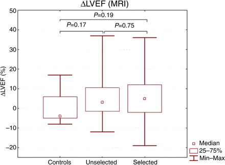
REGENT Study – Michał Tendera, et al. Intracoronary infusion of bone marrow-derived selected CD34+CXCR4+ cells and non-selected mononuclear cells in patients with acute STEMI and reduced left ventricular ejection fraction: results of randomized, multicentre Myocardial Regeneration by Intracoronary Infusion of Selected Population of Stem Cells in Acute Myocardial Infarction (REGENT) Trial. European Heart Journal 2009;30(11):1313-21. This study examined 200 patients who had experienced a heart attack, and seven days after the heart attack, they treated these patients with either unselected bone marrow cells (n = 80), selected bone marrow cells (n = 80), or a placebo (n = 40). This large study did not find statistically significant differences between the three groups, but the control group did not show an increase in the ejection fraction, but the unselected and selected bone marrow-treated patients did.
The figure shown below is from the Tendera et al., paper that shows the compiled changes in ejection fraction between the three groups:
As you can see, the control group patients experienced a decrease in their ejection fractions, but the two bone marrow-treated groups experienced an increase, even if it was slight. The figure below shows the data for the sickest patients.
As can be seen, for those patients with the sickest hearts there was a significant difference in the increase in the injection fraction and other heart-associated factors. For this reason, this study does not seem definitive. There were three deaths (one in each group), no strokes, four heart attacks (two in the controls and one in each experimental group), and a low rate of re-narrowing of the heart blood vessels. Since this is from 200 total patients, this is a very low rate of adverse events.
15. Jay H. Tendera, et al. Results of a phase 1, randomized, double-blind, placebo-controlled trial of bone marrow mononuclear stem cell administration in patients following ST-elevation myocardial infarction. American Heart Journal 2010;160:428-34. In this study forty patients were treated with either intracoronary bone marrow cells or a placebo. The two groups showed no significant differences in ejection fraction after six months, but the bone marrow-treated group showed no enlargement of the heart in response to the heart attack, whereas the control group did. No adverse heart events occurred.
This summarizes the clinical trials that used bone marrow to treat patients who had experienced recent heart attacks (acute myocardial infarctions). The preponderance of the data clearly shows that this procedure is safe, and effective to treat heart attacks. Secondly, several analyses that take the data from these trials and group them together into one gigantic study (meta-analysis) have been published, and these studies also show that bone marrow treatments for recent heart attacks are safe and effective (for example, see Meng Jiang, et al. Randomized controlled trials on the therapeutic effects of adult progenitor cells for myocardial infarction: meta-analysis. Expert Opinion on Biological Therapy 2010;10(5):667-80).
A new take on efficient delivery in regenerative medicine
Related articles
StemCells, Inc. Launches Alzheimer’s Disease Program Supported by California Institute for Regenerative Medicine (pharmaceuticalintelligence.com)
Introduction to Regenerative Therapy: How It Works (pharmaceuticalintelligence.com)
How nanotechnology can advance regenerative medicine (transbiotex.wordpress.com)
The Global Revolution in Biotechnology and the Impact of Regenerative Medicine (transbiotex.wordpress.com)
New Company Applies Regenerative Medicine to Corneal Transplants (transbiotex.wordpress.com)
Institute for Regenerative Medicine to Lead National Effort to Aid Wounded Warriors (transbiotex.wordpress.com)
The Alliance for Regenerative Medicine Gains Bipartisan Support for Gao Assessment of Federal Regenerative Medicine Activities (transbiotex.wordpress.com)
Joint development of the world’s first regenerative medicine/cell therapy business based on iPS cell technology
Healios & Dainippon Sumitomo Pharma in $50.6M Deal to form an alliance in regenerative medicine and cell therapy
Startup to Strengthen Synthetic Biology and Regenerative Medicine Industries with Cutting Edge Cell Products
Dr. Jon Rowley and Dr. Uplaksh Kumar, Co-Founders of RoosterBio, Inc., a newly formed biotech startup located in Frederick, are paving the way for even more innovation in the rapidly growing fields of Synthetic Biology and Regenerative Medicine
Dr. Jon Rowley and Dr. Uplaksh Kumar, Co-Founders of RoosterBio, Inc., a newly formed biotech startup located in Frederick, are paving the way for even more innovation in the rapidly growing fields of Synthetic Biology and Regenerative Medicine. Synthetic Biology combines engineering principles with basic science to build biological products, including regenerative medicines and cellular therapies. Regenerative medicine is a broad definition for innovative medical therapies that will enable the body to repair, replace, restore and regenerate damaged or diseased cells, tissues and organs. Regenerative therapies that are in clinical trials today may enable repair of damaged heart muscle following heart attack, replacement of skin for burn victims, restoration of movement after spinal cord injury, regeneration of pancreatic tissue for insulin production in diabetics and provide new treatments for Parkinson’s and Alzheimer’s diseases, to name just a few applications.
While the potential of the field is promising, the pace of development has been slow. One main reason for this is that the living cells required for these therapies are cost-prohibitive and not supplied at volumes that support many research and product development efforts. RoosterBio will manufacture large quantities of standardized primary cells at high quality and low cost, which will quicken the pace of scientific discovery and translation to the clinic. “Our goal is to accelerate the development of products that incorporate living cells by providing abundant, affordable and high quality materials to researchers that are developing and commercializing these regenerative technologies” says Dr. Rowley.
RoosterBio’s current focus is to supply high volume research-grade cells manufactured with processes consistent with current Good Manufacturing Practices (cGMP). These cells will be used for tissue engineering research and cell-based product development. This will position RoosterBio to quickly move on to producing clinical-grade cells to be used in translational R&D and clinical studies.
“We have spent almost 20 years as cell and tissue technologists and have lived with the pain of needing to generate large amounts of cells for experiments this whole time. RoosterBio was founded to address this problem for cell and tissue engineers, saving them time and money, and accelerating their path to the clinic,” says Dr. Rowley. RoosterBio will supply cells, starting with adult human bone marrow-derived stem cells, at volumes that will allow for a more rapid pace of experimentation in the lab.
“We will also offer paired media that has been engineered to quickly and efficiently expand the supplied cells to hundreds of millions or billions of cells within 1-2 weeks, something that would take 4-8 weeks using cell and media systems currently on the market,” adds Dr. Kumar. “We aim to usher in a new era of productivity to the field, and we believe that our products will at least triple the efficiency of the average laboratory”.
RoosterBio, Inc. is located in the Frederick Innovative Technology Center on Metropolitan Court in Frederick. Dr. Rowley entered into the incubation program in October of this year, and already gained four full time employees, and has several academic and industrial collaborators lined up. This team has made remarkable progress and are already poised for their official product launch for their human bone marrow-derived Mesenchymal Stem Cells (hBM-MSC), anticipated in March 2014.
RoosterBio’s product formats have been extraordinarily well received by the market, and RoosterBio has already secured customers who are anxiously awaiting their product launch. “I am excited to see that someone is taking on the challenge of providing a sufficient number of MSCs to immediately start experiments upon their receipt. This saves us several weeks of time upfront waiting for cells to expand to volumes that allow us to begin experiments,” says Todd McDevitt, Director of the Stem Cell Engineering Center at the Georgia Institute of Technology. “For tissue engineering folks like myself, this means we can focus our time on high priority research questions and not spend the majority of our time performing routine cell culture.”
The Tissue Engineering and Regenerative Medicine industry is one of the fastest growing in the life science sector with the total expenditure in 2011 at $17.1 billion. This number is expected to increase in 2020 to $40.5 billion. The sales of stem cell products accounted for $1.38 billion in 2010 and is expected to reach $3.9 billion by the year 2014 and $8 billion in annual revenues by 2020.
About RoosterBio
RoosterBio is focused on building a robust and sustainable Regenerative Medicine industry. Our products are affordable and standardized primary cells and media, manufactured and delivered with highest quality and in formats that simplify product development efforts. RoosterBio products will accelerate the translation of cell therapy and tissue engineering technologies into the clinic.
Related articles in Pharmaceutical Intelligence:
Like this:
Like Loading...


