Feeds:
Posts
Comments

Archive for the ‘Neuroscience’ Category

Visual math!

Larry H. Bernstein, MD, FCAP, Curator

LPBI


Eye cells may use math to detect motion

http://www.nih.gov/news-events/news-releases/eye-cells-may-use-math-detect-motion


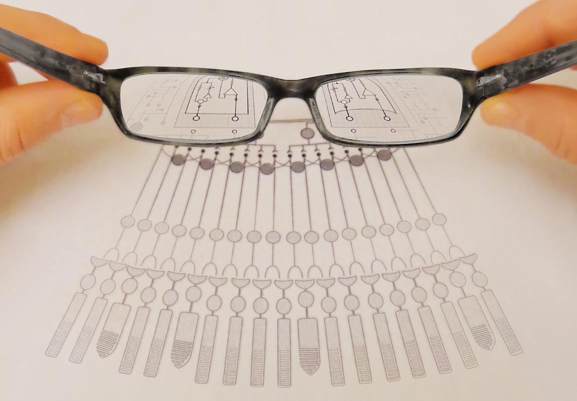
Image of glasses magnifying a diagram.

http://www.nih.gov/sites/default/files/styles/featured_media_breakpoint-large-extra/public/news-events/news-releases/2016/20160307-glasses.jpg


Our eyes constantly send bits of information about the world around us to our brains where the information is assembled into objects we recognize. Along the way, a series of neurons in the eye uses electrical and chemical signals to relay the information. In a study of mice, National Institutes of Health scientists showed how one type of neuron may do this to distinguish moving objects. The study suggests that the NMDA receptor, a protein normally associated with learning and memory, may help neurons in the eye and the brain relay that information.

“The eye is a window onto the outside world and the inner workings of the brain,” said Jeffrey S. Diamond, Ph.D., senior scientist at the NIH’s National Institute of Neurological Disorders and Stroke (NINDS), and the senior author of the study published in Neuron. “Our results show how neurons in the eye and the brain may use NMDA receptors to help them detect motion in a complex visual world.”

Our eyes constantly send bits of information about the world around us to our brains where the information is assembled into objects we recognize. Along the way, a series of neurons in the eye uses electrical and chemical signals to relay the information. In a study of mice, National Institutes of Health scientists showed how one type of neuron may do this to distinguish moving objects. The study suggests that the NMDA receptor, a protein normally associated with learning and memory, may help neurons in the eye and the brain relay that information.

“The eye is a window onto the outside world and the inner workings of the brain,” said Jeffrey S. Diamond, Ph.D., senior scientist at the NIH’s National Institute of Neurological Disorders and Stroke (NINDS), and the senior author of the study published in Neuron. “Our results show how neurons in the eye and the brain may use NMDA receptors to help them detect motion in a complex visual world.”

Vision begins when light enters the eye and hits the retina, which lines the back of the eyeball. Neurons in the retina convert light into nerve signals which are then sent to the brain. Using retinas isolated from mice, Dr. Alon Poleg-Polsky, Ph.D. a postdoctoral fellow in Dr. Diamond’s lab, studied neurons called directionally selective retinal ganglion cells (DSGCs), which are known to fire and send signals to the brain in response to objects moving in specific directions across the eye.

Electrical recordings showed that some of these cells fired when a bar of light passed across the retina from left to right, whereas others responded to light crossing in the opposite direction. Previous studies suggested these unique responses are controlled by incoming signals sent from neighboring cells at chemical communication points called synapses. In this study, Dr. Poleg-Polsky discovered that the activity of NMDA receptors at one set of synapses may regulate whether DSGCs sent direction-sensitive information to the brain.

NMDA receptors are proteins that generate electrical signals in response to the neurochemicals glutamate and glycine. When activated, they allow electrically charged ions to flow in and out of cells like water through an unlocked canal. In the early 1980s, studies in France and at the NIH showed that magnesium blocks the flow until the neuron is strongly activated and its electrical state rises above a certain voltage.  This regulation is thought to be critical for certain types of learning and memory, and in amplifying signals in neurons.

Further experiments by Dr. Poleg-Polsky examined how magnesium’s control of NMDA receptors may regulate the firing of DSGCs. To mimic realistic conditions, Dr. Poleg-Polsky passed bars of light across retinas while exposing them to various background lights. The results suggested that the variable magnesium block that ensured the cells consistently sent information to the brain in response to the passing bars of light despite the distracting incoming stream of signals generated by the background lights. The NMDA receptors did this by amplifying the cells’ responses to the bars in a process called multiplicative scaling.

“Cells in the eye can multiply,” said Dr. Poleg-Polsky. “The process may help these cells determine whether a tiger is sauntering by, or fast approaching as it’s looking for dinner.”

Neurons in the eye and brain receive a constant stream of information. The results of this study support a growing body of evidence suggesting that NMDA receptors play in critical role in how neurons relay information.

“Our results suggest that NMDA receptors help neurons distinguish relevant information from irrelevant background noise,” said Dr. Diamond. “In the future we plan to examine whether this process contributes to other aspects of vision.”

This work was supported by the NINDS Division of Intramural Research.

For more information, visit:
www.ninds.nih.gov
neuroscience.nih.gov/ninds/Home.aspx

The NINDS is the nation’s leading funder of research on the brain and nervous system. The mission of NINDS is to seek fundamental knowledge about the brain and nervous system and to use that knowledge to reduce the burden of neurological disease.

About the National Institutes of Health (NIH): NIH, the nation’s medical research agency, includes 27 Institutes and Centers and is a component of the U.S. Department of Health and Human Services. NIH is the primary federal agency conducting and supporting basic, clinical, and translational medical research, and is investigating the causes, treatments, and cures for both common and rare diseases. For more information about NIH and its programs, visit www.nih.gov.

NIH…Turning Discovery Into Health®

Reference

Poleg-Polsky et al. “NMDA receptors multiplicatively scale visual signals and enhance direction motion discrimination in retinal ganglion cells,” Neuron, March 3, 2016. http://dx.doi.org:/10.1016/j.neuron.2016.02.013

NMDA Receptors Multiplicatively Scale Visual Signals and Enhance Directional Motion Discrimination in Retinal Ganglion Cells
Alon Poleg-Polskycorrespondence, Jeffrey S. Diamond
http://dx.doi.org/10.1016/j.neuron.2016.02.013
Poleg-Polsky et al. report a novel form of multiplicative synaptic integration in retinal directionally selective ganglion cells that is mediated by NMDARs and directionally tuned synaptic inhibition. NMDAR-mediated multiplication enhances the accuracy of motion discrimination in noisy visual conditions.
Highlights
  • Novel form of multiplicative synaptic scaling in DSGCs
  • Multiplication depends on NMDARs and directionally tuned inhibitory signals
  • Multiplicative synaptic integration enhances accuracy of directional signaling


Summary

Postsynaptic responses in many CNS neurons are typically small and variable, often making it difficult to distinguish physiologically relevant signals from background noise. To extract salient information, neurons are thought to integrate multiple synaptic inputs and/or selectively amplify specific synaptic activation patterns. Here, we present evidence for a third strategy: directionally selective ganglion cells (DSGCs) in the mouse retina multiplicatively scale visual signals via a mechanism that requires both nonlinear NMDA receptor (NMDAR) conductances in DSGC dendrites and directionally tuned inhibition provided by the upstream retinal circuitry. Postsynaptic multiplication enables DSGCs to discriminate visual motion more accurately in noisy visual conditions without compromising directional tuning. These findings demonstrate a novel role for NMDARs in synaptic processing and provide new insights into how synaptic and network features interact to accomplish physiologically relevant neural computations.


Read Full Post »

Brain Cancer Vaccine in Development and other considerations

Larry H. Bernstein, MD, FCAP, Curator

LPBI


GEN News Highlights   Mar 3, 2016

Advanced Immunotherapeutic Method Shows Promise against Brain Cancer

http://www.genengnews.com/gen-news-highlights/advanced-immunotherapeutic-method-shows-promise-against-brain-cancer/81252433/


http://www.genengnews.com/Media/images/GENHighlight/Mar3_2016_LeuvenLab_CellDeathMouseBrain6232214015.jpg

The researchers induced a specific type of cell death in brain cancer cells from mice. The dying cancer cells were then incubated together with dendritic cells, which play a vital role in the immune system. The researchers discovered that this type of cancer cell killing releases “danger signals” that fully activate the dendritic cells. “We re-injected the activated dendritic cells into the mice as a therapeutic vaccine,” Professor Patrizia Agostinis explains. “That vaccine alerted the immune system to the presence of dangerous cancer cells in the body. As a result, the immune system could recognize them and start attacking the brain tumor.” [©KU Leuven Laboratory of Cell Death Research & Therapy, Dr. Abhishek D. Garg]


Scientists from KU Leuven in Belgium say they have shown that next-generation cell-based immunotherapy may offer new hope in the fight against brain cancer.

Cell-based immunotherapy involves the injection of a therapeutic anticancer vaccine that stimulates the patient’s immune system to attack the tumor. Thus far, the results of this type of immunotherapy have been mildly promising. However, Abhishek D. Garg and Professor Patrizia Agostinis from the KU Leuven department of cellular and molecular medicine believe they have found a novel way to produce more effective cell-based anticancer vaccines.

The researchers induced a specific type of cell death in brain cancer cells from mice. The dying cancer cells were then incubated together with dendritic cells, which play a vital role in the immune system. The investigators discovered that this type of cancer cell killing releases “danger signals” that fully activate the dendritic cells.

“We re-injected the activated dendritic cells into the mice as a therapeutic vaccine,” explains Prof. Agostinis. “That vaccine alerted the immune system to the presence of dangerous cancer cells in the body. As a result, the immune system could recognize them and start attacking the brain tumor.”

Combined with chemotherapy, this novel cell-based immunotherapy drastically increased the survival rates of mice afflicted with brain tumors. Almost 50% of the mice were completely cured. None of the mice treated with chemotherapy alone became long-term survivors.

“The major goal of any anticancer treatment is to kill all cancer cells and prevent any remaining malignant cells from growing or spreading again,” says Professor Agostinis. “This goal, however, is rarely achieved with current chemotherapies, and many patients relapse. That’s why the co-stimulation of the immune system is so important for cancer treatments. Scientists have to look for ways to kill cancer cells in a manner that stimulates the immune system. With an eye on clinical studies, our findings offer a feasible way to improve the production of vaccines against brain tumors.”

The team published its study (“Dendritic Cell Vaccines Based on Immunogenic Cell Death Elicit Danger Signals and T Cell–Driven Rejection of High-Grade Glioma”) in Science Translational Medicine.


Dendritic cell vaccines based on immunogenic cell death elicit danger signals and T cell–driven rejection of high-grade glioma


SLC7A11 expression is associated with seizures and predicts poor survival in patients with malignant glioma


Cortical GABAergic excitation contributes to epileptic activities around human glioma


Spherical Nucleic Acid Nanoparticle Conjugates as an RNAi-Based Therapy for Glioblastoma

Glioblastoma multiforme (GBM) is a neurologically debilitating disease that culminates in death 14 to 16 months after diagnosis. An incomplete understanding of how cataloged genetic aberrations promote therapy resistance, combined with ineffective drug delivery to the central nervous system, has rendered GBM incurable. Functional genomics efforts have implicated several oncogenes in GBM pathogenesis but have rarely led to the implementation of targeted therapies. This is partly because many “undruggable” oncogenes cannot be targeted by small molecules or antibodies. We preclinically evaluate an RNA interference (RNAi)–based nanomedicine platform, based on spherical nucleic acid (SNA) nanoparticle conjugates, to neutralize oncogene expression in GBM. SNAs consist of gold nanoparticles covalently functionalized with densely packed, highly oriented small interfering RNA duplexes. In the absence of auxiliary transfection strategies or chemical modifications, SNAs efficiently entered primary and transformed glial cells in vitro. In vivo, the SNAs penetrated the blood-brain barrier and blood-tumor barrier to disseminate throughout xenogeneic glioma explants. SNAs targeting the oncoprotein Bcl2Like12 (Bcl2L12)—an effector caspase and p53 inhibitor overexpressed in GBM relative to normal brain and low-grade astrocytomas—were effective in knocking down endogenous Bcl2L12 mRNA and protein levels, and sensitized glioma cells toward therapy-induced apoptosis by enhancing effector caspase and p53 activity. Further, systemically delivered SNAs reduced Bcl2L12 expression in intracerebral GBM, increased intratumoral apoptosis, and reduced tumor burden and progression in xenografted mice, without adverse side effects. Thus, silencing antiapoptotic signaling using SNAs represents a new approach for systemic RNAi therapy for GBM and possibly other lethal malignancies.


Rapid, Label-Free Detection of Brain Tumors with Stimulated Raman Scattering Microscopy

Surgery is an essential component in the treatment of brain tumors. However, delineating tumor from normal brain remains a major challenge. We describe the use of stimulated Raman scattering (SRS) microscopy for differentiating healthy human and mouse brain tissue from tumor-infiltrated brain based on histoarchitectural and biochemical differences. Unlike traditional histopathology, SRS is a label-free technique that can be rapidly performed in situ. SRS microscopy was able to differentiate tumor from nonneoplastic tissue in an infiltrative human glioblastoma xenograft mouse model based on their different Raman spectra. We further demonstrated a correlation between SRS and hematoxylin and eosin microscopy for detection of glioma infiltration (κ = 0.98). Finally, we applied SRS microscopy in vivo in mice during surgery to reveal tumor margins that were undetectable under standard operative conditions. By providing rapid intraoperative assessment of brain tissue, SRS microscopy may ultimately improve the safety and accuracy of surgeries where tumor boundaries are visually indistinct.


Neural Stem Cell–Mediated Enzyme/Prodrug Therapy for Glioma: Preclinical Studies


Magnetic Resonance Metabolic Imaging of Glioma


Exploiting the Immunogenic Potential of Cancer Cells for Improved Dendritic Cell Vaccines

Cancer immunotherapy is currently the hottest topic in the oncology field, owing predominantly to the discovery of immune checkpoint blockers. These promising antibodies and their attractive combinatorial features have initiated the revival of other effective immunotherapies, such as dendritic cell (DC) vaccinations. Although DC-based immunotherapy can induce objective clinical and immunological responses in several tumor types, the immunogenic potential of this monotherapy is still considered suboptimal. Hence, focus should be directed on potentiating its immunogenicity by making step-by-step protocol innovations to obtain next-generation Th1-driving DC vaccines. We review some of the latest developments in the DC vaccination field, with a special emphasis on strategies that are applied to obtain a highly immunogenic tumor cell cargo to load and to activate the DCs. To this end, we discuss the effects of three immunogenic treatment modalities (ultraviolet light, oxidizing treatments, and heat shock) and five potent inducers of immunogenic cell death [radiotherapy, shikonin, high-hydrostatic pressure, oncolytic viruses, and (hypericin-based) photodynamic therapy] on DC biology and their application in DC-based immunotherapy in preclinical as well as clinical settings.

Cancer immunotherapy has gained considerable momentum over the past 5 years, owing predominantly to the discovery of immune checkpoint inhibitors. These inhibitors are designed to release the brakes of the immune system that under physiological conditions prevent auto-immunity by negatively regulating cytotoxic T lymphocyte (CTL) function. Following the FDA approval of the anti-cytotoxic T lymphocyte-associated antigen-4 (CTLA-4) monoclonal antibody (mAb) ipilimumab (Yervoy) in 2011 for the treatment of metastatic melanoma patients (1), two mAbs targeting programed death (PD)-1 receptor signaling (nivolumab and pembrolizumab) have very recently joined the list of FDA-approved checkpoint blockers (respectively, for the treatment of metastatic squamous non-small cell lung cancer and relapsed/refractory melanoma patients) (2, 3).

However, the primary goal of cancer immunotherapy is to activate the immune system in cancer patients. This requires the induction of tumor-specific T-cell-mediated antitumor immunity. Checkpoint blockers are only able to abrogate the brakes of a functioning antitumoral immune response, implying that only patients who have pre-existing tumor-specific T cells will benefit most from checkpoint blockade. This is evidenced by the observation that ipilimumab may be more effective in patients who have pre-existing, albeit ineffective, antitumor immune responses (4). Hence, combining immune checkpoint blockade with immunotherapeutic strategies that prime tumor-specific T cell responses might be an attractive and even synergistic approach. This relatively new paradigm has lead to the revival of existing, and to date disappointing (as monotherapies), active immunotherapeutic treatment modalities. One promising strategy to induce priming of tumor-specific T cells is dendritic cell (DC)-based immunotherapy.

Dendritic cells are positioned at the crucial interface between the innate and adaptive immune system as powerful antigen-presenting cells capable of inducing antigen-specific T cell responses (5). Therefore, they are the most frequently used cellular adjuvant in clinical trials. Since the publication of the first DC vaccination trial in melanoma patients in 1995, the promise of DC immunotherapy is underlined by numerous clinical trials, frequently showing survival benefit in comparison to non-DC control groups (68). Despite the fact that most DC vaccination trials differ in several vaccine parameters (i.e., site and frequency of injection, nature of the DCs, choice of antigen), DC vaccination as a monotherapy is considered safe and rarely associates with immune-related toxicity. This is in sharp contrast with the use of mAbs or cytokine therapies. Ipilumumab has, for instance, been shown to induce immune-related serious adverse events in up to one-third of treated melanoma patients (1). The FDA approval of Sipuleucel-T (Provenge), an autologous DC-enriched vaccine for hormone-resistant metastatic prostate cancer, in 2010 is really considered as a milestone in the vaccination community (9). After 15 years of extensive clinical research, Sipileucel-T became the first cellular immunotherapy ever that received FDA approval, providing compelling evidence for the substantial socio-economic impact of DC-based immunotherapy. DC vaccinations have most often been applied in patients with melanoma, prostate cancer, high-grade glioma, and renal cell cancer. Although promising objective responses and tumor-specific T cell responses have been observed in all these cancer-types (providing proof-of-principle for DC-based immunotherapy), the clinical success of this treatment is still considered suboptimal (6). This poor clinical efficacy can in part be attributed to the severe tumor-induced immune suppression and the selection of patients with advanced disease status and poor survival prognostics (6, 1012).

There is a consensus in the field that step-by-step optimization and standardization of the production process of DC vaccines, to obtain a Th1-driven immune response, might enhance their clinical efficacy (13). In this review, we address some recent DC vaccine adaptations that impact DC biology. Combining these novel insights might bring us closer to an ideal DC vaccine product that can trigger potent CTL- and Th1-driven antitumor immunity.

One factor requiring more attention in this production process is the immunogenicity of the dying or dead cancer cells used to load the DCs. It has been shown in multiple preclinical cancer models that the methodology used to prepare the tumor cell cargo can influence the in vivo immunogenic potential of loaded DC vaccines (1419). Different treatment modalities have been described to enhance the immunogenicity of cancer cells in the context of DC vaccines. These treatments can potentiate antitumor immunity by inducing immune responses against tumor neo-antigens and/or by selectively increasing the exposure/release of particular damage-associated molecular patterns (DAMPs) that can trigger the innate immune system (14, 1719). The emergence of the concept of immunogenic cell death (ICD) might even further improve the immunogenic potential of DC vaccines. Cancer cells undergoing ICD have been shown to exhibit excellent immunostimulatory capacity owing to the spatiotemporally defined emission of a series of critical DAMPs acting as potent danger signals (20, 21). Thus far, three DAMPs have been attributed a crucial role in the immunogenic potential of nearly all ICD inducers: the surface-exposed “eat me” signal calreticulin (ecto-CRT), the “find me” signal ATP and passively released high-mobility group box 1 (HMGB1) (21). Moreover, ICD-experiencing cancer cells have been shown in various mouse models to act as very potent Th1-driving anticancer vaccines, already in the absence of any adjuvants (21, 22). The ability to reject tumors in syngeneic mice after vaccination with cancer cells (of the same type) undergoing ICD is a crucial hallmark of ICD, in addition to the molecular DAMP signature (21).

Here, we review the effects of three frequently used immunogenic modalities and four potent ICD inducers on DC biology and their application in DC vaccines in preclinical as well as clinical settings (Tables (Tables11 and and2).2). Moreover, we discuss the rationale for combining different cell death-inducing regimens to enhance the immunogenic potential of DC vaccines and to ensure the clinical relevance of the vaccine product.

A list of prominent enhancers of immunogenicity and ICD inducers applied in DC vaccine setups and their associations with DAMPs and DC biology.
A list of preclinical tumor models and clinical studies for evaluation of the in vivo potency of DC vaccines loaded with immunogenically killed tumor cells.
The Impact of DC Biology on the Efficacy of DC Vaccines

Over the past years, different DC vaccine parameters have been shown to impact the clinical effectiveness of DC vaccinations. In the next section, we will elaborate on some promising adaptations of the DC preparation protocol.

Given the labor-intensive ex vivo culturing protocol of monocyte-derived DCs and inspired by the results of the Provenge study, several groups are currently exploiting the use of blood-isolated naturally circulating DCs (7678). In this context, De Vries et al. evaluated the use of antigen-loaded purified plasmacytoid DCs for intranodal injection in melanoma patients (79). This strategy was feasible and induced only very mild side effects. In addition, the overall survival of vaccinated patients was greatly enhanced as compared to historical control patients. However, it still remains to be determined whether this strategy is more efficacious than monocyte-derived DC vaccine approaches (78). By contrast, experiments in the preclinical GL261 high-grade glioma model recently showed that vaccination with tumor antigen-loaded myeloid DCs resulted in more robust Th1 responses and a stronger survival benefit as compared to mice vaccinated with their plasmacytoid counterparts (80).

In view of their strong potential to stimulate cytotoxic T cell responses, several groups are currently exploring the use of Langerhans cell-like DCs as sources for DC vaccines (8183). These so-called IL-15 DCs can be derived from CD14+monocytes by culturing them with IL-15 (instead of the standard IL-4). Recently, it has been shown that in comparison to IL-4 DCs, these cells have an increased capacity to stimulate antitumor natural killer (NK) cell cytotoxicity in a contact- and IL-15-dependent manner (84). NK cells are increasingly being recognized as crucial contributors to antitumor immunity, especially in DC vaccination setups (85, 86). Three clinical trials are currently evaluating these Langerhans cell-type DCs in melanoma patients (NCT00700167, NCT 01456104, and NCT01189383).

Targeting cancer stem cells is another promising development, particularly in the setting of glioma (87). Glioma stem cells can foster tumor growth, radio- and chemotherapy-resistance, and local immunosuppression in the tumor microenvironment (87, 88). Furthermore, glioma stem cells may express higher levels of tumor-associated antigens and MHC complex molecules as compared to non-stem cells (89, 90). A preclinical study in a rodent orthotopic glioblastoma model has shown that DC vaccines loaded with neuropsheres enriched in cancer stem cells could induce more immunoreactivity and survival benefit as compared to DCs loaded with GL261 cells grown under standard conditions (91). Currently there are four clinical trials ongoing in high-grade glioma patients evaluating this approach (NCT00890032, NCT00846456, NCT01171469, and NCT01567202).

With regard to the DC maturation status of the vaccine product, a phase I/II clinical trial in metastatic melanoma patients has confirmed the superiority of mature antigen-loaded DCs to elicit immunological responses as compared to their immature counterparts (92). This finding was further substantiated in patients diagnosed with prostate cancer and recurrent high-grade glioma (93, 94). Hence, DCs need to express potent costimulatory molecules and lymph node homing receptors in order to generate a strong T cell response. In view of this finding, the route of administration is another vaccine parameter that can influence the homing of the injected DCs to the lymph nodes. In the context of prostate cancer and renal cell carcinoma it has been shown that vaccination routes with access to the draining lymph nodes (intradermal/intranodal/intralymphatic/subcutaneous) resulted in better clinical response rates as compared to intravenous injection (93). In melanoma patients, a direct comparison between intradermal vaccination and intranodal vaccination concluded that, although more DCs reached the lymph nodes after intranodal vaccination, the melanoma-specific T cells induced by intradermal vaccination were more functional (95). Furthermore, the frequency of vaccination can also influence the vaccine’s immunogenicity. Our group has shown in a cohort-comparison trial involving relapsed high-grade glioma patients that shortening the interval between the four inducer DC vaccines improved the progression-free survival curves (58, 96).

Another variable that has been systematically studied is the cytokine cocktail that is applied to mature the DCs. The current gold standard cocktail for DC maturation contains TNF-α, IL-1β, IL-6, and PGE2 (97, 98). Although this cocktail upregulates DC maturation markers and the lymph node homing receptor CCR7, IL-12 production by DCs could not be evoked (97, 98). Nevertheless, IL-12 is a critical Th1-driving cytokine and DC-derived IL-12 has been shown to associate with improved survival in DC vaccinated high-grade glioma and melanoma patients (99, 100). Recently, a novel cytokine cocktail, including TNF-α, IL-1β, poly-I:C, IFN-α, and IFN-γ, was introduced (101, 102). The type 1-polarized DCs obtained with this cocktail produced high levels of IL-12 and could induce strong tumor-antigen-specific CTL responses through enhanced induction of CXCL10 (99). In addition, CD40-ligand (CD40L) stimulation of DCs has been used to mature DCs in clinical trials (100, 103). Binding of CD40 on DCs to CD40L on CD4+ helper T cells licenses DCs and enables them to prime CD8+ effector T cells.

A final major determinant of the vaccine immunogenicity is the choice of antigen to load the DCs. Two main approaches can be applied: loading with selected tumor antigens (tumor-associated antigens or tumor-specific antigens) and loading with whole tumor cell preparations (13). The former strategy enables easier immune monitoring, has a lower risk of inducing auto-immunity, and can provide “off-the-shelf” availability of the antigenic cargo. Whole tumor cell-based DC vaccines, on the other hand, are not HLA-type dependent, have a reduced risk of inducing immune-escape variants, and can elicit immunity against multiple tumor antigens. Meta-analytical data provided by Neller et al. have demonstrated enhanced clinical efficacy in several tumor types of DCs loaded with whole tumor lysate as compared to DCs pulsed with defined tumor antigens (104). This finding was recently also substantiated in high-grade glioma patients, although this study was not set-up to compare survival parameters (105).

Toward a More Immunogenic Tumor Cell Cargo

The majority of clinical trials that apply autologous whole tumor lysate to load DC vaccines report the straightforward use of multiple freeze–thaw cycles to induce primary necrosis of cancer cells (8, 93). Freeze–thaw induced necrosis is, however, considered non-immunogenic and has even been shown to inhibit toll-like receptor (TLR)-induced maturation and function of DCs (16). To this end, many research groups have focused on tackling this roadblock by applying immunogenic modalities to induce cell death.

Immunogenic Treatment Modalities

Tables Tables11 and and22 list some frequently applied treatment methods to enhance the immunogenic potential of the tumor cell cargo that is used to load DC vaccines in an ICD-independent manner (i.e., these treatments do not meet the molecular and/or cellular determinants of ICD). Immunogenic treatment modalities can positively impact DC biology by inducing particular DAMPs in the dying cancer cells (Table (Table1).1). Table Table22 lists the preclinical and clinical studies that investigated their in vivo potential. Figure Figure11 schematically represents the application and the putative modes of action of these immunogenic enhancers in the setting of DC vaccines.

An external file that holds a picture, illustration, etc.
Object name is fimmu-06-00663-g001.jpg

http://www.ncbi.nlm.nih.gov/pmc/articles/PMC4712296/bin/fimmu-06-00663-g001.jpg

A schematic representation of immunogenic DC vaccines. Cancer cells show enhanced immunogenicity upon treatment with UV irradiation, oxidizing treaments, and heat shock, characterized by the release of particular danger signals and the (increased) production of tumor (neo-)antigens. Upon loading onto DCs, DCs undergo enhanced phagocytosis and antigen uptake and show phenotypic and partial functional maturation. Upon in vivo immunization, these DC vaccines elicit Th1- and cytotoxic T lymphocyte (CTL)-driven tumor rejection.

Ultraviolet Irradiation ….

Oxidation-Inducing Modalities

In recent years, an increasing number of data were published concerning the ability of oxidative stress to induce oxidation-associate molecular patterns (OAMPs), such as reactive protein carbonyls and peroxidized phospholipids, which can act as DAMPs (28, 29) (Table (Table1).1). Protein carbonylation, a surrogate indicator of irreversible protein oxidation, has for instance been shown to improve cancer cell immunogenicity and to facilitate the formation of immunogenic neo-antigens (30, 31).

One prototypical enhancer of oxidation-based immunogenicity is radiotherapy (21,23). In certain tumor types, such as high-grade glioma and melanoma, clinical trials that apply autologous whole tumor lysate to load DC vaccines report the random use of freeze–thaw cycles (to induce necrosis of cancer cells) or a combination of freeze–thaw cycles and subsequent high-dose γ-irradiation (8, 18) (Table (Table2).2). However, from the available clinical evidence, it is unclear which of both methodologies has superior immunogenic potential. In light of the oxidation-based immunogenicity that is associated with radiotherapy, we recently demonstrated the superiority of DC vaccines loaded with irradiated freeze–thaw lysate (in comparison to freeze–thaw lysate) in terms of survival advantage in a preclinical high-grade glioma model (18) (Table (Table2).2). ….

Heat Shock Treatment

Heat shock is a term that is applied when a cell is subjected to a temperature that is higher than that of the ideal body temperature of the organisms of which the cell is derived. Heat shock can induce apoptosis (41–43°C) or necrosis (>43°C) depending on the temperature that is applied (110). The immunogenicity of heat shock treated cancer cells largely resides within their ability to produce HSPs, such as HSP60, HSP70, and HSP90 (17, 32) (Table (Table1).1). …

An external file that holds a picture, illustration, etc.
Object name is fimmu-06-00663-g002.jpg

http://www.ncbi.nlm.nih.gov/core/lw/2.0/html/tileshop_pmc/tileshop_pmc_inline.html?title=Click%20on%20image%20to%20zoom&p=PMC3&id=4712296_fimmu-06-00663-g002.jpg

Figure 2

A schematic representation of immunogenic cell death (ICD)-based DC vaccines. ICD causes cancer cells to emit a spatiotemporally defined pattern of danger signals. Upon loading of these ICD-undergoing cancer cells onto DCs, they induce extensive phagocytosis and antigen uptake. Loaded DCs show enhanced phenotypic and functional maturation and immunization with these ICD-based DC vaccines instigates Th1-, Th17-, and cytotoxic T lymphocyte (CTL)-driven antitumor immunity in vivo.
Inducers of Immunogenic Cell Death

Immunogenic cell death is a cell death regimen that is associated with the spatiotemporally defined emission of immunogenic DAMPs that can trigger the immune system (20, 21, 113). ICD has been found to depend on the concomitant induction of reactive oxygen species (ROS) and activation of endoplasmatic reticulum (ER) stress (111). Besides the three DAMPs that are most crucial for ICD (ecto-CRT, ATP, and HMGB1), other DAMPs such as surface-exposed or released HSPs (notably HSP70 and HSP90) have also been shown to contribute to the immunogenic capacity of ICD inducers (20, 21). The binding of these DAMPs to their respective immune receptors (CD91 for HSPs/CRT, P2RX7/P2RY2 for ATP, and TLR2/4 for HMGB1/HSP70) leads to the recruitment and/or activation of innate immune cells and facilitates the uptake of tumor antigens by antigen-presenting cells and their cross-presentation to T cells eventually leading to IL-1β-, IL-17-, and IFN-γ-dependent tumor eradiation (22). This in vivo tumor rejecting capacity induced by dying cancer cells in the absence of any adjuvant, is considered as a prerequisite for an agent to be termed an ICD inducer. …

Although the list of ICD inducers is constantly growing (113), only few of these immunogenic modalities have been tested in order to generate an immunogenic tumor cell cargo to load DC vaccines (Tables (Tables11 and and2).2). Figure Figure22 schematically represents the preparation of ICD-based DC vaccines and their putative modes of action.

Radiotherapy

Ionizing X-ray or γ-ray irradiation exerts its anticancer effect predominantly via its capacity to induce DNA double-strand breaks leading to intrinsic cancer cell apoptosis (114). The idea that radiotherapy could also impact the immune system was derived from the observation that radiotherapy could induce T-cell-mediated delay of tumor growth in a non-irradiated lesion (115). This abscopal (ab-scopus, away from the target) effect of radiotherapy was later explained by the ICD-inducing capacity (116). Together with anthracyclines, γ-irradiation was one of the first treatment modalities identified to induce ICD. …

Shikonin

The phytochemical shikonin, a major component of Chinese herbal medicine, is known to inhibit proteasome activity. It serves multiple biological roles and can be applied as an antibacterial, antiviral, anti-inflammatory, and anticancer treatment. …

High-hydrostatic pressure

High-hydrostatic pressure (HHP) is an established method to sterilize pharmaceuticals, human transplants, and food. HHP between 100 and 250 megapascal (MPa) has been shown to induce apoptosis of murine and human (cancer) cells (121123). While DNA damage does not seem to be induced by HHP <1000 MPa, HHP can inhibit enzymatic functions and the synthesis of cellular proteins (122). Increased ROS production was detected in HHP-treated cancer cell lines and ER stress was evidenced by the rapid phosphorylation of eIF2α (42).  …

Oncolytic Viruses

Oncolytic viruses are self-replicating, tumor selective virus strains that can directly lyse tumor cells. Over the past few years, a new oncolytic paradigm has risen; entailing that, rather than utilizing oncolytic viruses solely for direct tumor eradication, the cell death they induce should be accompanied by the elicitation of antitumor immune responses to maximize their therapeutic efficacy (128). One way in which these oncolytic viruses can fulfill this oncolytic paradigm is by inducing ICD (128).

Thus far, three oncolytic virus strains can meet the molecular requirements of ICD; coxsackievirus B3 (CVB3), oncolytic adenovirus and Newcastle disease virus (NDV) (Table (Table1)1) (113). Infection of tumor cells with these viruses causes the production of viral envelop proteins that induce ER stress by overloading the ER. Hence, all three virus strains can be considered type II ICD inducers (113). …

Photodynamic therapy

Photodynamic therapy (PDT) is an established, minimally invasive anticancer treatment modality. It has a two-step mode of action involving the selective uptake of a photosensitizer by the tumor tissue, followed by its activation by light of a specific wavelength. This activation results in the photochemical production of ROS in the presence of oxygen (129131). One attractive feature of PDT is that the ROS-based oxidative stress originates in the particular subcellular location where the photosensitizer tends to accumulate, ultimately leading to the destruction of the tumor cell (132). …

Combinatorial Regimens

In DC vaccine settings, cancer cells are often not killed by a single treatment strategy but rather by a combination of treatments. In some cases, the underlying rationale lies within the additive or even synergistic value of combining several moderately immunogenic modalities. The combination of radiotherapy and heat shock has, for instance, been shown to induce higher levels of HSP70 in B16 melanoma cells than either therapy alone (16). In addition, a combination therapy consisting of heat shock, γ-irradiation, and UV irradiation has been shown to induce higher levels of ecto-CRT, ecto-HSP90, HMGB1, and ATP in comparison to either therapy alone or doxorubicin, a well-recognized inducer of ICD (57). ….

Triggering antitumor immune responses is an absolute requirement to tackle metastatic and diffusely infiltrating cancer cells that are resistant to standard-of-care therapeutic regimens. ICD-inducing modalities, such as PDT and radiotherapy, have been shown to be able to act as in situ vaccines capable of inducing immune responses that caused regression of distal untreated tumors. Exploiting these ICD inducers and other immunogenic modalities to obtain a highly immunogenic antigenic tumor cell cargo for loading DC vaccines is a highly promising application. In case of the two prominent ICD inducers, Hyp-PDT and HHP, preclinical studies evaluating this relatively new approach are underway and HHP-based DC vaccines are already undergoing clinical testing. In the preclinical testing phase, more attention should be paid to some clinically driven considerations. First, one should consider the requirement of 100% mortality of the tumor cells before in vivo application. A second consideration from clinical practice (especially in multi-center clinical trials) is the fact that most tumor specimens arrive in the lab in a frozen state. This implies that a significant number of cells have already undergone non-immunogenic necrosis before the experimental cell killing strategies are applied. ….


Read Full Post »

Synapse activity in neurotransmission

Larry H. Bernstein, MD, FCAP, Curator

LPBI


The case of the silent synapses: Why are only 20% of synapses active during neurotransmission?

Unknown information coding in the brain?
February 26, 2016   http://www.kurzweilai.net/study-finds-only-a-small-portion-of-synapses-may-be-active-during-neurotransmission

Using a fluorescent molecule to track neurotransmission of dopamine in mouse synapses, scientists made a puzzling discovery. … (credit: Sulzer Lab/Columbia University Medical Center)

Columbia University scientists recently tested a new optical technique to study how information is transmitted in the brains of mice and made a surprising discovery: When stimulated electrically to release dopamine (a neurotransmitteror chemical released by neurons, or nerve cells, to send signals to other nerve cells), only about 20 percent of synapses — the connections between cells that control brain activity — were active at any given time.

The effect had never been noticed. “Older techniques only revealed what was going on in large groups of synapses,” explained David Sulzer, PhD, professor of neurobiology in Psychiatry, Neurology, and Pharmacology at Columbia University Medical Center (CUMC). “We needed a way to observe the neurotransmitter activity of individual synapses, to help us better understand their intricate behavior.”

So Sulzer’s team turned to Dalibor Sames, PhD, associate professor of chemistry at Columbia, to develop a novel compound called “fluorescent false neurotransmitter 200″ (FFN200). When added to brain tissue or nerve cells from mice, FFN200 mimicked the brain’s natural neurotransmitters, allowing the researchers to spy on chemical messaging in action, focusing on complex tasks such as learning and memory.

[+]

Only 20% of synapses (red) were observed to transmit dopamine. The rest (green) were found to be silent. (credit: Sulzer Lab/Columbia University Medical Center)

Silent synapses: unknown information coding?

Using a fluorescence microscope, the researchers were able for the first time to view the release and re-uptake of dopamine — a neurotransmitter involved in motor learning, habit formation, and reward-seeking behavior — in individual synapses.

When all the neurons were electrically stimulated in a sample of brain tissue, the researchers expected all the synapses to release dopamine. Instead, they found that less than 20 percent of dopaminergic synapses were active following a pulse of electricity.

One possibility: these silent synapses hint at a mechanism of information coding in the brain that’s yet to be revealed, the researchers hypothesize.

The study’s authors plan to pursue that hypothesis in future experiments and examine how other neurotransmitters behave. “If we can work this out, we may learn a lot more about how alterations in dopamine levels are involved in brain disorders such as Parkinson’s disease, addiction, and schizophrenia,” Sulzer said.

The study was published in the latest issue of Nature Neuroscience.

The authors note in the paper that “the state of silent vesicle clusters may be important in disorders such as schizophrenia, which show striatal hyperdopaminergia [excessive release of dopamine in the brain’s reward center] and cortical hypodopaminergia [low amounts of dopamine in the cortex] and processes of  ‘unsilencing’ may have clinical applications for diseases such as Parkinson’s disease.”

https://youtu.be/apYAyypLlYg
Columbia Medical | Study Finds Only a Small Portion of Synapses May Be Active During Neurotransmission


Abstract of Fluorescent false neurotransmitter reveals functionally silent dopamine vesicle clusters in the striatum

Neurotransmission at dopaminergic synapses has been studied with techniques that provide high temporal resolution, but cannot resolve individual synapses. To elucidate the spatial dynamics and heterogeneity of individual dopamine boutons, we developed fluorescent false neurotransmitter 200 (FFN200), a vesicular monoamine transporter 2 (VMAT2) substrate that selectively traces monoamine exocytosis in both neuronal cell culture and brain tissue. By monitoring electrically evoked Ca2+ transients with GCaMP3 and FFN200 release simultaneously, we found that only a small fraction of dopamine boutons that exhibited Ca2+ influx engaged in exocytosis, a result confirmed with activity-dependent loading of the endocytic probe FM1-43. Thus, only a low fraction of striatal dopamine axonal sites with uptake-competent VMAT2 vesicles are capable of transmitter release. This is consistent with the presence of functionally ‘silent’ dopamine vesicle clusters and represents, to the best of our knowledge, the first report suggestive of presynaptically silent neuromodulatory synapses.

Read Full Post »

Genetic link to sleep and mood disorders

Larry H. Bernstein, MD, FCAP, Curator

LPBI


Scientists identify molecular link between sleep and mood

A poor night’s sleep is enough to put anyone in a bad mood, and although scientists have long suspected a link between mood and sleep, the molecular basis of this connection remained a mystery. Now, new research has found several rare genetic mutations on the same gene that definitively connect the two.

Sleep goes hand-in-hand with mood. People suffering from depression and mania, for example, frequently have altered sleeping patterns, as do those with seasonal affective disorder (SAD). And although no one knows exactly how these changes come about, in SAD sufferers they are influenced by changes in light exposure, the brain’s time-keeping cue. But is mood affecting sleep, is sleep affecting mood, or is there a third factor influencing both? Although a number of tantalizing leads have linked the circadian clock to mood, there is “no definitive factor that proves causality or indicates the direction of the relationship,” says Michael McCarthy, a neurobiologist at the San Diego Veterans’ Affairs Medical Center and the University of California (UC), San Diego.

To see whether they could establish a link between the circadian clock, sleep, and mood, scientists in the new study looked at the genetics of a family that suffers from abnormal sleep patterns and mood disorders, including SAD and something called advanced sleep phase, a condition in which people wake earlier and sleep earlier than normal. The scientists screened the family for mutations in key genes involved in the circadian clock, and identified two rare variants of the PERIOD3 (PER3) gene in members suffering from SAD and advanced sleep phase. “We found a genetic change in people who have both seasonal affective disorder and the morning lark trait” says lead researcher Ying-Hui Fu, a neuroscientist at UC San Francisco. When the team tested for these mutations in DNA samples from the general population, they found that they were extremely rare, appearing in less than 1% of samples.

Fu and her team then created mice that carried the novel genetic variants. These transgenic mice showed an unusual sleep-wake cycle and struggled less when handled by the researchers, a typical sign of depression. They also had lower levels of PER2, a protein involved in circadian rhythms, than unmutated mice, providing a possible molecular explanation for the unusual sleep patterns in the family. Fu says this supports the link between the PER3 mutations and both sleep and mood. “PER3’s role in mood regulation has never been demonstrated directly before,” she says. “Our results indicate that PER3 might function in helping us adjust to seasonal changes,” by modifying the body’s internal clock.

To investigate further, the team studied mice lacking a functional PER3 gene. They found that these mice showed symptoms of SAD, exhibiting more severe depression when the duration of simulated daylight in the laboratory was reduced. Because SAD affects between 2% and 9% of people worldwide, the novel variants can’t explain it fully. But understanding the function of PER3 could yield insights into the molecular basis of a wide range of sleep and mood disorders, Fu says.

Together, these experiments show that the PERIOD3 gene likely plays a key role in regulating the sleep-wake cycle, influencing mood and regulating the relationship between depression and seasonal changes in light availability, the team reports today in the Proceedings of the National Academy of Sciences. “The identification of a mutation in PER3 with such a strong effect on mood is remarkable,” McCarthy says. “It suggests an important role for the circadian clock in determining mood.”

The next step will be to investigate how well these results generalize to other people suffering from mood and sleep disorders. “It will be interesting to see if other rare variants in PER3 are found, or if SAD is consistently observed in other carriers,” McCarthy says. That could eventually lead to new drugs that selectively target the gene, which McCarthy says, “could be a strategy for treating mood or sleep disorders.”


http://dx.doi.org:/10.1126/science.aaf4095


Read Full Post »

High blood pressure can damage the retina’s blood vessels and limit the retina’s function. It can also put pressure on the optic nerve.

Sourced through Scoop.it from: www.healthline.com

See on Scoop.itCardiovascular Disease: PHARMACO-THERAPY

Read Full Post »

Cell-cell communication in brain

Larry H. Bernstein, MD, FCAP, Curator

LPBI


Adjustable Brain Cells

Neighboring neurons can manipulate astrocytes.

By Ruth Williams | February 18, 2016   http://www.the-scientist.com/?articles.view/articleNo/45376/title/Adjustable-Brain-Cells

http://www.the-scientist.com/images/News/February2016/astrocyte.jpg

Neurons in the adult mouse brain can shape the features and physiologies of nearby astroglial cells, according to a study published today (February 18) in Science. Researchers at McGill University in Montreal and their colleagues have identified a molecular signal called sonic hedgehog (Shh), secreted by neurons, that acts as the agent of change.

“What’s very exciting about the paper is this notion that a cell’s fate might be determined—after it has already established its morphology and location in the brain—based on interactions with its neighbors,” said neurologist Ed Ruthazer of the Montreal Neurological Institute at McGill who was not involved in the research. “And the conversion is not superficial,” he added, “it really does seem to fundamentally reorganize the transcriptome of the cell.”

Astroglia are non-neuronal cells in the central nervous system that generally support and modulate neuronal function. The mammalian brain has an assortment of astrocytes, which perform a variety of specialized functions. This diversity was largely thought to be established during embryogenesis and early postnatal development, said Keith Murai of McGill who led the new research. “But after that,” he said, “the properties of these cells were thought to be solidified . . . for the rest of their lives.”

Murai and his colleagues had a different view, however. “Some of these [astrocytes] are so specialized around certain neural circuits that it was hard to imagine that all of the properties of these cells could be determined by that point [in development],” he said. After all, the neural circuitry itself isn’t fully formed until much later.

To investigate whether astrocyte identity might continue to be shaped beyond the perinatal period, Murai’s team searched for gene products in adult neurons and astrocytes that might govern continuing development. To simplify matters, the researchers focused on the cerebellar cortex, where just two types of astrocyte exist—Bergmann glial cells (BGs), which encapsulate the impulse-receiving regions of Purkinji cell neurons (PCs), and velate astrocytes (VAs), which surround granule cell neurons (GCs). Their searches revealed many candidate factors, said Murai, but one pathway kept coming up: Shh signaling.

Shh is a developmental morphogen known to have many important roles in the developing embryo, including the specification of cells in the brain, explained Murai. “People thought that the pathway was shut down and eliminated from the brain after it developed,” he said, “but as it turns out, this pathway is very potent even in the adult brain.”

The team found that the Shh protein itself was produced by PC neurons in the cerebellum, and that Shh receptors were abundantly expressed in BG, but not VA cells.

Furthermore, BGs required Shh signals from PCs to maintain their identities. When transgenic techniques were used to switch off either Shh production in PCs or Shh signaling in BGs in adult mouse brains, the BG cells adopted a transcription profile similar to that of VAs. If Shh signaling in VAs was given a boost on the other hand, these cells became more like BGs.

We could “almost interconvert one type of astrocyte into another based upon the level of Shh signaling,” said Murai. And it wasn’t just a handful of factors, he added: “We’re talking about hundreds of genes that are either being turned on or turned off in response to this pathway.”

The team also found evidence that astrocytes in other brain regions were influenced by Shh manipulations, and that these cells’ electrophysiologies were altered as a result.

“The key message is that astrocytes’ molecular fate is not hardwired,” said cell biologist Cagla Eroglu of Duke University in Durham, North Carolina, who did not participate in the study. The shapes of these cells appear to be less malleable, however. While Shh signaling influenced astrocyte expression profiles and electrical behaviors, the cells’ morphologies remained largely unchanged.

The finding that astrocyte identity is considerably more plastic than previously thought is “exciting and interesting,” added Cagla, “but it remains to be seen what the exact function of this will be in terms of an animal’s behavior or its ability to learn.”

W.T. Farmer et al., “Neurons diversify astrocytes in the adult brain through sonic hedgehog signaling,” Science, 351:849-54, 2016.


Neurons diversify astrocytes in the adult brain through sonic hedgehog signaling

Glial cell properties dictated by neurons

Neurons in the brain coexist with astrocytes, a type of glial cell, which help support many functions of their neighboring nerve cells. Farmer et al. now show that the support goes both ways (see the Perspective by Stevens and Muthukumar). They explored the influence of neurons on two specialized types of astrocytes in the mouse cerebellar cortex. The neurons produced the morphogen known as Sonic Hedgehog. Hedgehog signaling adjusted distinctive gene expression within the two astrocyte cell types. Thus, mature neurons appear to promote and maintain specific properties of associated astrocytes.

Science, this issue p. 849; see also p. 813


Astrocytes are specialized and heterogeneous cells that contribute to central nervous system function and homeostasis. However, the mechanisms that create and maintain differences among astrocytes and allow them to fulfill particular physiological roles remain poorly defined. We reveal that neurons actively determine the features of astrocytes in the healthy adult brain and define a role for neuron-derived sonic hedgehog (Shh) in regulating the molecular and functional profile of astrocytes. Thus, the molecular and physiological program of astrocytes is not hardwired during development but, rather, depends on cues from neurons that drive and sustain their specialized properties.


Sonic hedgehog regulates discrete populations of astrocytes in the adult mouse forebrain.

Astrocytes are an essential component of the CNS, and recent evidence points to an increasing diversity of their functions. Identifying molecular pathways that mediate distinct astrocyte functions, is key to understanding how the nervous system operates in the intact and pathological states. In this study, we demonstrate that the Hedgehog (Hh) pathway, well known for its roles in the developing CNS, is active in astrocytes of the mature mouse forebrain in vivo. Using multiple genetic approaches, we show that regionally distinct subsets of astrocytes receive Hh signaling, indicating a molecular diversity between specific astrocyte populations. Furthermore, we identified neurons as a source of Sonic hedgehog (Shh) in the adult forebrain, suggesting that Shh signaling is involved in neuron-astrocyte communication. Attenuation of Shh signaling in postnatal astrocytes by targeted removal of Smoothened, an obligate Shh coreceptor, resulted in upregulation of GFAP and cellular hypertrophy specifically in astrocyte populations regulated by Shh signaling. Collectively, our findings demonstrate a role for neuron-derived Shh in regulating specific populations of differentiated astrocytes.
Emerging evidence indicates that astrocytes play active and diverse roles in the central nervous system (CNS). Astrocytes actively regulate cerebral blood flow (Takano et al., 2006), and respond to sensory stimuli in both the visual and somatosensory cortices (Wang et al., 2006; Schummers et al., 2008). During development, astrocytes play key roles in regulating synapse formation and function (Ullian et al., 2001), and promote maturation of dendritic spines (Nishida and Okabe, 2007). In addition, bidirectional communication between astrocytes and neurons is an important element of synaptic transmission (Zhang et al., 2003; Araque, 2008). Although it is becoming increasingly clear that astrocytes actively contribute to normal CNS function, the underlying molecular mechanisms that mediate the functional properties of astrocytes remain poorly understood.
Several lines of evidence suggest that Shh signaling could play a role in astrocyte development and/or function. In the developing optic nerve, Shh mediates proliferation of astrocyte precursors (Wallace and Raff, 1999), and application of Shh agonists to early postnatal cortical astrocyte cultures upregulates Shh target genes (Atkinson et al., 2009). In addition, the transcription factors Gli1, 2, and 3, essential components of the Shh signaling pathway, are enriched in postnatal and adult astrocyte cultures (Cahoy et al., 2008). Moreover, a recent study suggests that reactive astrocytes produce and respond to Shh following cortical freeze injury (Amankulor et al., 2009). However, it remains unknown whether astrocytes in the mature CNS participate in Shh signaling in vivo, under normal physiological conditions. Transduction of Hh signaling is mediated by the Gli family of transcription factors, Gli1, 2, and 3. Binding of Shh to the transmembrane receptor Patched (Ptc) relieves constitutive inhibition of a second transmembrane receptor, Smoothened (Smo), thereby initiating a cascade of events leading to induction of Gli1 transcription (Fuccillo et al., 2006). Gli1 transcription in the embryo is dependent on Hh signaling through Gli2 and Gli3 (Bai et al., 2004). Thus, the presence of Gli1 can be used as an indicator of cells actively responding to high levels of Hh signaling (Bai et al., 2002). In this study, we show that regionally distinct populations of mature, differentiated astrocytes are the primary cell type responding to Shh in the adult forebrain. We further identified neurons as a source of Shh, suggesting a novel role for Shh signaling in neuronastrocyte communication. Finally, we demonstrate that conditional deletion of Smo in postnatal astrocytes results in a mild astrogliosis in the cortex, suggesting that Shh is an important regulator of specific astrocyte populations.
Gli1 is expressed in many non-proliferating cells in the adult forebrain In order to examine the distribution and identity of Hh-responding cells in the adult forebrain, we used Gli1nlacZ/+ mice in which nuclear lacZ is expressed from the Gli1 locus (Bai et al., 2002). In the developing neural tube, Gli1 is expressed predominantly in ventral populations of proliferating neuronal and oligodendrocyte precursors (Platt et al., 1997; Jessell, 2000). In contrast, we found that Gli1-expressing cells were distributed in the adult forebrain as far dorsally as the cortex, where they were localized primarily to layers 3, 4, and 5 (Fig. 1A). Dense populations of Gli1-expressing cells were also found in multiple basal forebrain nuclei, including the septum (data not shown) and globus pallidus, as well as in the thalamus, and hypothalamus (Fig. 1B–D). These results are consistent with a previous report that Ptc and Smo mRNA can be detected in the globus pallidus, thalamus and hypothalamus of the adult rat (Traiffort et al., 1999). However, whereas low levels of Ptc transcript were detected only in the piriform cortex (Traiffort et al., 1999), our findings indicate that cells in the entorhinal, motor and somatosensory cortex express Gli1, suggesting a more widespread cortical distribution of Hh-responding cells. Shh signaling plays a critical role in regulating adult neural stem and progenitor cells in the adult forebrain (Han et al., 2008). Although constitutive neurogenesis does not occur outside the subependymal zone (SEZ) and subgranular zone (SGZ), glial progenitors proliferate throughout the parenchyma of the adult CNS (Nishiyama et al., 2002; Dawson et al., 2003). To examine whether Gli1-expressing cells outside the neurogenic regions correspond to glial progenitors, we used a second line of mice in which an inducible Cre recombinase (CreERT2) is expressed from the Gli1 locus (Gli1CreER/+; Ahn and Joyner, 2005). When combined with the Rosa26loxP-STOP-loxP-lacZ reporter allele (R26lacZ; Soriano, 1999), cells expressing CreERT2 from the Gli1 locus express lacZ following tamoxifen administration. LacZ expression is permanent and heritable, and because CreER is active for only ~36 hours (Nakamura et al., 2006), expression of the βGal reporter protein becomes a permanent marker of cells that were expressing Gli1 at the time of tamoxifen administration. Moreover, because βGal expression is cytoplasmic, it is possible to examine the morphology of marked cells.
The absence of an apparent expansion of marked cells between 1 and 6 months posttamoxifen suggested that Gli1-expressing cells are not proliferating. Consistent with this, analysis of double staining for BrdU and βGal in the cortex showed that βGal-expressing cells were not double labeled with BrdU at all time points examined (Fig. 2G). As expected, double labeled cells were readily observed in the SEZ (Fig. 2H), corresponding to adult neural stem and progenitor cells (Ahn and Joyner, 2005). However, since BrdU labeled cells were only rarely observed in the cortex, we could not rule out the possibility that some Gli1- expressing cells divided, but escaped detection due to insufficient BrdU. We therefore used a more extensive BrdU labeling protocol that has been shown to label glial progenitor cells throughout the adult forebrain and spinal cord (Horner et al., 2000). Adult Gli1nlacZ/+ animals were given 50mg/kg BrdU for 12 days, and examined 3 days after the last BrdU injection. Despite a greater number of BrdU-labeled cells than in the previous experiments, none of the cortical BrdU-positive cells co-expressed βGal (n=148 cells, Supp. Fig. 1). Glial progenitors in the adult forebrain and spinal cord express the proteoglycan, NG2 (Horner et al., 2000). Analysis of βGal and NG2 double labeling showed that none of the βGalexpressing cells corresponded to NG2-positive glial progenitor cells (Supp. Fig. 1). The apparent increase in βGal-expressing cells between day 8 and 1 month post-tamoxifen in Gli1CreER;R26lacZ forebrains therefore cannot be due to proliferation of glial progenitors, but instead must be due to accumulation of βGal protein within cells. Taken together, these data suggest that, with the exception of adult neural stem and progenitor cells, the vast majority of Gli1-expressing cells in the adult forebrain are terminally differentiated. Moreover, the regional distribution of cells expressing Gli1 indicates that Hh signaling occurs in discrete cell populations throughout the dorsal/ventral and anterior/posterior axes.
Regionally distinct populations of astrocytes express Gli1
We next examined the cell types that express Gli1 in the adult forebrain. We primarily analyzed Gli1CreER;R26lacZ animals because the cytoplasmic localization of βGal permitted morphological analysis. In addition, since our previous experiments indicated that reporter expression stabilizes by 3 months post-tamoxifen, we restricted our analysis to adult Gli1CreER;R26lacZ animals given tamoxifen 3 months earlier. Throughout the cortex, Gli1- expressing cells exhibited small cell bodies with an elaborate branching morphology (Fig. 1I and 3B). The processes were highly ramified and very fine, creating a bushy appearance, consistent with the morphology of protoplasmic astrocytes. In addition, some labeled cells extended processes to nearby blood vessels (Fig. 3B), further suggesting that these cells correspond to astrocytes.
Interestingly, although the majority of Gli1-positive cells are astrocytes, not all astrocytes express Gli1. In addition, the proportion of astrocytes that express Gli1 differs between specific forebrain regions. In the cortex of tamoxifen-treated Gli1CreER;R26lacZ mice, only 24% of the S100β-positive cells were marked with βGal (n=1932 cells), whereas 56% (n=1233 cells) and 80% (n=1489 cells) of S100β-expressing cells in the globus pallidus and hypothalamus respectively, were βGal-positive (Fig. 4). In contrast, the caudate putamen and area CA1 of the hippocampus exhibit few βGal-positive cells (Fig. 1), indicating that the vast majority of astrocytes in these regions do not express Gli1. Similarly, white matter astrocytes do not express Gli1, as indicated by the lack of βGal staining in the corpus callosum, anterior commissure, and fimbria (Fig. 1F–J).
Gli1 is a sensitive readout of Shh in the adult forebrain
Although transcriptional activation of Gli1 has been shown to be a reliable readout of Hh signaling in the developing CNS (Lee et al., 1997), it is possible that signaling pathways other than Hh might activate Gli1 transcription in the adult. In order to address whether Hh signaling is responsible for Gli1 expression in adult forebrain astrocytes, we took several approaches.


Astrocytes in Gli1-expressing regions of the forebrain show signs of mild reactive gliosis following postnatal interruption of Shh signaling
During development, Shh plays a critical role in specification of neuronal and oligodendrocyte precursors (Jessell, 2000). Shh has also been implicated in regulating proliferation of astrocytes in the developing optic nerve (Wallace and Raff, 1999; Dakubo et al., 2008).

Upregulation of GFAP and hypertrophy are hallmarks of reactive astrogliosis, which occurs in response to injury or disease (Eng and Ghirnikar, 1994). Reactive astrocytes have also been shown to undergo increased proliferation and synthesis of nestin and vimentin (Sofroniew, 2009). However BrdU labeling showed no change in proliferation between mutants and controls (data not shown), and staining for nestin and vimentin showed no change in expression of these intermediate filaments (data not shown). Taken together, these data show that the cellular response to interrupting Shh signaling includes key characteristics of reactive astrogliosis and indicates that Shh signaling plays an important role in mediating intracellular properties of specific astrocyte populations.


In this study, we show that high level Shh signaling in the adult CNS occurs in regionally distinct populations of mature, differentiated astrocytes. Our data demonstrate that neurons are a source of Shh, suggesting a novel signaling pathway involved in direct neuronastrocyte communication. Furthermore, we provide evidence that Shh signaling is required to maintain normal cellular functions in specific astrocyte populations. Taken together, our data are the first to demonstrate a critical role for Shh signaling in neuron-astrocyte communication in vivo, in the adult CNS.


An external file that holds a picture, illustration, etc.
Object name is nihms-244753-f0004.jpg

http://www.ncbi.nlm.nih.gov/pmc/articles/PMC2966838/bin/nihms-244753-f0004.jpg

Astrocytes that express Gli1 are differentially distributed between specific forebrain regions

(A–C) Double labeling immunohistochemistry for βGal (red) and S100β (green) in the cortex (A), globus pallidus (B), and hypothalamus (C) of adult Gli1CreER/+;R26lacZ animals given tamoxifen 3 months earlier. Counterstained with DAPI (blue). Scale bar, 25μm. (D) Quantitative analysis of the proportion of S100β-positive astrocytes within the cortex, globus pallidus, and hypothalamus that express Gli1. (mean ± SEM, n300 cells/region/animal, from 3 animals, *p < 0.05, **p < 0.01, one-way, repeated measures ANOVA and Tukey’s post-test.)


  1. Farmer WT, Abrahamsson T, Chierzi S, Lui C, Zaelzer C, Jones EV, Ponroy Bally B, Chen GG, Théroux JF, Peng J, Bourque CW, Charron F, Ernst C, Sjöström PJ, and Murai KK: Neurons Diversify Astrocytes in the Adult Brain Through Sonic Hedgehog Signaling. Science (2016) 351(6275):849-854 DOI: 10.1126/science.aab3103 [abstract] [perspective by Stevens and Muthukumar]
  2. Lalanne T, Oyrer J, Mancino A, Gregor E, Chung A, Huynh L, Burwell S, Maheux J, Farrant M, and Sjöström PJ: Synapse-specific expression of calcium-permeable AMPA receptors in neocortical layer 5. The Journal of Physiology (2016) 594(4):837-861. (Editor’s Choice) [Abstract]
  3. Larsen RS & Sjöström: Synapse-type-specific plasticity in local circuits. Current Opinion in Neurobiology (2015) 35:127-135. [Abstract]
  4. Costa RP, Froemke RC, Sjöström PJ, & van Rossum MCW: Unified pre- and postsynaptic long-term plasticity enables reliable and flexible learning. eLife(2015) DOI:10.7554/eLife.09457. [Abstract]
  5. Ferreira TA, Blackman AV, Oyrer J, Jayabal S, Chung AJ, Watt AJ, Sjöström PJ, & van Meyel DJ: Neuronal morphometry directly from bitmap images.Nature Methods (2014) 11(10):982-984, DOI:10.1038/nmeth.3125. [Abstract]
  6. Blackman AV, Grabuschnig S, Legenstein R, & Sjöström PJ: A comparison of manual neuronal reconstruction from biocytin histology or 2-photon imaging: morphometry and computer modeling. Frontiers in Neuroanatomy (2014) 8:65, DOI: 10.3389/fnana.2014.00065. [Abstract]
  7. Blackman AV, Abrahamsson T, Costa RP, Lalanne T, & Sjöström PJ: Target-cell-specific short-term plasticity in local circuits. Frontiers in Synaptic Neuroscience (2013) 5:11, DOI: 10.3389/fnsyn.2013.00011. [Abstract]
  8. Costa RP, Sjöström PJ, & van Rossum MCW: Probabilistic inference of short-term synaptic plasticity in neocortical microcircuits. Frontiers in Computational Neuroscience (2013) 7:75, DOI:10.3389/fncom.2013.00075. [Abstract] [ModelDB code]
  9. Costa RP, Watt AJ, & Sjöström PJ: How to train a neuron. eLife (2013) 2:e00491, DOI: 10.7554/eLife.00491. Commentary on Pawlak et al, eLife 2013.


Neurogenesis in the embryonic and adult brain: same regulators, different roles

Front. Cell. Neurosci., 27 November 2014 | http://dx.doi.org/10.3389/fncel.2014.00396

Neurogenesis persists in adult mammals in specific brain areas, known as neurogenic niches. Adult neurogenesis is highly dynamic and is modulated by multiple physiological stimuli and pathological states. There is a strong interest in understanding how this process is regulated, particularly since active neuronal production has been demonstrated in both the hippocampus and the subventricular zone (SVZ) of adult humans. The molecular mechanisms that control neurogenesis have been extensively studied during embryonic development. Therefore, we have a broad knowledge of the intrinsic factors and extracellular signaling pathways driving proliferation and differentiation of embryonic neural precursors. Many of these factors also play important roles during adult neurogenesis, but essential differences exist in the biological responses of neural precursors in the embryonic and adult contexts. Because adult neural stem cells (NSCs) are normally found in a quiescent state, regulatory pathways can affect adult neurogenesis in ways that have no clear counterpart during embryogenesis. BMP signaling, for instance, regulates NSC behavior both during embryonic and adult neurogenesis. However, this pathway maintains stem cell proliferation in the embryo, while it promotes quiescence to prevent stem cell exhaustion in the adult brain. In this review, we will compare and contrast the functions of transcription factors (TFs) and other regulatory molecules in the embryonic brain and in adult neurogenic regions of the adult brain in the mouse, with a special focus on the hippocampal niche and on the regulation of the balance between quiescence and activation of adult NSCs in this region.

Neural stem cells (NSCs) in the embryonic and early postnatal murine brain generate neurons and glia, including astrocytes and oligodendrocytes. The transition of proliferative and multipotent NSCs to fully differentiated neurons and glia is called neurogenesis and gliogenesis, respectively. Neurons are generated from early embryonic development until early postnatal stages, with only a few neurogenic zones remaining active in the adult (Götz and Huttner, 2005; Ming and Song, 2011;Paridaen and Huttner, 2014). In contrast, gliogenesis starts during late embryogenesis and continues in postnatal stages, with low but widespread production of both astrocytes and oligodendrocytes also occurring throughout the adult brain (Rowitch and Kriegstein, 2010; Gallo and Deneen, 2014; Guérout et al., 2014). The main neurogenic regions in the adult murine brain are the subependymal zone of the lateral ventricles, also called ventricular-subventricular Zone (V-SVZ) and the subgranular zone (SGZ) of the dentate gyrus (DG) in the hippocampus (Altman and Das, 1965; Doetsch et al., 1999; Ming and Song, 2011;Fuentealba et al., 2012). Both of these neurogenic regions have been shown to also be active in the adult human brain, with the V-SVZ thought to contribute new neurons to the striatum (whereas it produces neurons migrating to the olfactory bulb in mice) and the SGZ contributing neurons to the DG (Eriksson et al., 1998; Spalding et al., 2013; Ernst et al., 2014). The addition of new neurons to the complex circuitry of the adult brain is the focus of intensive research, which is uncovering crucial functions for the newly generated neurons in memory and behavior (Deng et al., 2010). In particular, the integration of adult-born granule cells to the hippocampus circuitry confers an extra degree of plasticity that is crucial for the acquisition of certain types of contextual memory (Jessberger et al., 2009; Sahay et al., 2011). Although adult neurogenesis is an ancient trait, with widespread neurogenesis occurring, for instance, in 16 different adult brain areas of zebrafish, the appearance of the DG as a structural and functional unit seems exclusive to mammals (Treves et al., 2008; Grandel and Brand, 2013). This fact, amongst others, has prompted the idea that hippocampal neurogenesis might be a newly evolved trait in some species, including humans, aimed to enhance adaptation to a continuously changing environment (Kempermann, 2012).

Significant advances have been made in our understanding of the regulation of mouse adult hippocampal neurogenesis in the last few years. Thus, our focus for the rest of the review will be on the mouse model of neurogenesis. The coordinated action of multiple signals acting on embryonic NSCs gives rise to the vast diversity of neuronal and glial populations that populate the mature brain. Embryonic neurogenesis is, thus, tightly linked to cell fate specification. In adult neurogenic regions, however, stem cells are tightly restricted to the generation of one (granule neurons of the DG) or a few types of neurons (granule neurons and periglomerular neurons in the V-SVZ) (Zhao et al., 2008; Ming and Song, 2011). Therefore signals and factors that specify subtype identities during development can control more subtle aspects of adult stem cell behavior.

In recent years, it has become evident that, at the single cell level, stem cells in the embryonic and the adult brain are not as versatile as previously thought. Instead of their classically attributed multipotency, they appear to be already committed to the generation of specific types of neural cells (Taverna et al., 2014). The causes and functions of the emerging heterogeneity of adult NSCs are among the most exciting questions remaining to be addressed in the field (DeCarolis et al., 2013; Encinas et al., 2013; Giachino et al., 2014b). In the case of the murine V-SVZ, different populations of adult NSCs, also called type-B cells, co-exist and give rise to distinct types of periglomerular cells and granule cells in the olfactory bulb. Different adult NSCs are characterized by the differential expression of specific transcription factors (TFs), including Nkx2.1, Pax6, Gsx2 and Nkx6.2, which also pattern the different domains of the embryonic telencephalon (Merkle et al., 2007; Brill et al., 2008;López-Juárez et al., 2013; Merkle et al., 2014). The distinct adult NSC populations are located in different regions along the V-SVZ and their distinct properties are acquired during development (Obernier et al., 2014). Despite the spatial separation of these stem cell populations, all their progeny follow the same long migratory path, the rostral migratory stream (RMS), towards their final destination in the olfactory bulb. In the hippocampus, adult NSCs, also called type-I cells or radial glial-like cells, generate exclusively granule neurons in the DG. The migration of granule neurons is very limited, as they settle, differentiate and integrate into the hippocampal circuitry in the granule cell layer (GCL) located just above the NSC from which they originated in the SGZ. While they appear uniform, adult NSCs in the DG respond to diverse and complex signals, raising the possibility that they are functionally heterogeneous.

Despite their many differences, adult NSCs in the two adult neurogenic niches share several key characteristics. Neural stem cells in both V-SVZ and SGZ, like radial glial stem cells in the embryo, express the molecules GFAP, Nestin and Sox2 and they directly contact blood vessels. Both NSC populations share a restricted potential, as just discussed, with each generating a unique neuronal subtype and one type of glia: in the V-SVZ they generate neurons and oligodendrocytes, while in the SGZ they generate neurons and astrocytes. Perhaps the two characteristics that distinguish adult NSCs most clearly from their embryonic counterparts are the acquisition of quiescence and their situation in a complex and stable cellular niche. While one of the main features of embryonic NSCs is their high proliferative rate, the opposite is true for adult NSCs, which remain for long periods out of the cell cycle, in G0. This is a characteristic that adult NSCs share with many stem cells in other mature tissues and one that is crucial to maintain tissue homeostasis and avoid stem cell exhaustion (Orford and Scadden, 2008;Simons and Clevers, 2011). The existence of adult neurogenic niches (complex cellular microenvironments surrounding adult NSCs) is also a characteristic shared with other tissues (Fuchs et al., 2004; Kuang et al., 2008; Mirzadeh et al., 2008; Ming and Song, 2011; Fuentealba et al., 2012; Goldstein and Horsley, 2012). The niche is comprised of diverse cell types and structures, such as astrocytes, neurons, axon projections and blood vessels, and one of its main functions is to create an appropriate environment that keeps the majority of stem cells quiescent and undifferentiated (Morrison and Spradling, 2008). The niche also provides a great variety of signals that modulate the behavior of adult stem cells and adjust the production of new cells to the needs of the tissue (Fuchs et al., 2004; Blank et al., 2008; Faigle and Song, 2013).

Embryonic and Adult Origin of Granule Cells

From a developmental point of view, the generation of the DG is unique. While the V-SVZ is seen as a continuation of the embryonic ventricular zone (VZ) of the telencephalon, the formation of the DG involves the generation of a dedicated progenitor cell source away from the VZ and in close proximity to the pial surface. This additional proliferative zone remains active during postnatal stages and eventually becomes the SGZ, which is the site of adult hippocampal neurogenesis (Figure 1;Bayer, 1980a,b; Altman and Bayer, 1990; Pleasure et al., 2000; Khalaf-Nazzal and Francis, 2013; Sugiyama et al., 2013).

http://www.frontiersin.org/files/Articles/118806/fncel-08-00396-r4/image_m/fncel-08-00396-g001.jpg

Development of the mouse hippocampus. Schematic representation of the dorsal telencephalon at different embryonic (E) stages and at birth (P0). The indicated area in each picture corresponds to the hippocampal region and is magnified on its right handside (blue squares). (A) At E12.5 the presumptive DNE is located between the HNE and the CH, which produces Cajal-Retzius cells (orange), shown lining the pial side of the cortex. (B) At E14.5 dentate precursors of the primary matrix (dark blue circles) are located in the VZ, and precursor cells start to migrate towards the pial side of the cortex forming the secondary matrix. In the VZ of the HNE, radial glial precursors (depicted in dark blue and triangular body shape) will give rise to hippocampal neurons. (C) At E17.5 the hippocampal fissure is formed and dentate precursor cells migrate to and accumulate there, forming the tertiary matrix (light blue). Cajal-Retzius cells are also present and follow the hippocampal fissure. At this stage the glial scaffold (not shown) extends from the CH to the hippocampal fissure and pial surface, directing the migration of dentate precursor cells. From the HNE, hippocampal neurons (red triangles) are born and migrate along radial glial cells towards their location in the hippocampal fields (CA1 and CA3 are shown). (D) At birth the blades of the DG start to form. Granule neurons in the DG (red triangles) appear first in the upper blade, below the hippocampal fissure. The continuous migration of Cajal-Retzius cells reaches the pial side and promotes the formation of the lower blade of the DG. Precursor cells in the primary and secondary matrix will soon disappear, but cells in the tertiary matrix continue actively dividing and producing granule neurons through postnatal DG development. HNE, hippocampal neuroepithelium; DNE, dentate neuroepithelium; CH, cortical hem; VZ, ventricular zone; 1ry, primary matrix; 2ry, secondary matrix; 3ry, tertiary matrix; DG, dentate gyrus; D, dorsal; M, medial; V, ventral; L, lateral.

Regulation of Adult Neurogenesis

The late maturation of the hippocampus, which spans late embryonic and early postnatal stages, means that the process of DG formation and the appearance of NSCs with adult characteristics are overlapping processes. It can therefore be difficult to distinguish between developmental and adult cues regulating hippocampal neurogenesis. However, several physiological and pathological situations, such as physical exercise, task learning, an enriched environment and seizures, have been shown to stimulate neurogenesis specifically in the adult DG (Rolando and Taylor, 2014). Although no direct link has been clearly established between those external stimuli and signaling pathways, numerous extracellular signaling molecules, including Bone Morphogenetic Proteins (BMPs), Notch, GABA, WNT, insulin growth factors (IGFs) and SHH, have been shown to regulate the rate of neurogenesis in the adult DG (Ming and Song, 2011; Faigle and Song, 2013). However, due to limitations of in vivo studies, little is known about the mechanisms by which these signals exert their effects. In the adult DG, NSCs generate granule cells via a well characterized cell lineage that includes a succession of transit amplifying or intermediate progenitor cells (IPCs), characterized by rapid divisions and the expression of a series of neurogenic TFs (Figure 2; Hsieh, 2012). Extrinsic stimuli can affect the proliferation and survival of NSCs but also of IPCs (typeIIa and typeIIb) or differentiating neuroblasts (typeIII) further along the lineage (Figure 2). The selective death of IPCs, for instance, is a major mechanism of regulation of neurogenesis in the DG, with as many as two thirds of these cells being actively eliminated by microglia (Sierra et al., 2010, 2014). Therefore, in order to understand the effects of signaling pathways and intrinsic factors on neurogenesis, it is crucial to determine the stages in the adult neurogenic lineage at which they act, and the cellular processes they regulate. In fact, one of the main difficulties faced by the adult neurogenesis field concerns the scarcity of markers for adult NSCs, which are often shared by other cell types (for instance, GFAP marks subpopulations of astrocytes and Nestin is expressed by early intermediate progenitors). This problem is only more evident in the case of distinguishing quiescent from activated adult NSCs, in which case there is an absolute lack of specific markers apart from the use of cell cycle genes. This issue has been partly addressed in a recent report in which an unbiased approach was used to identify genes differentially expressed by activated and quiescent adult NSCs isolated from the V-SVZ (Codega et al., 2014). This work demonstrates that the quiescent state is a much more complex state than simply the lack of proliferation markers, as the list of differentially expressed genes is enriched in genes related to very diverse cellular processes, such as lipids metabolism, signaling or adhesion. This quiescence signature is shared by adult quiescent stem cells from other organs, such as the blood, muscle or intestine (Cheung and Rando, 2013; Codega et al., 2014). It is thus likely that many of the general characteristics of quiescent stem cells will be shared between DG and SVZ, although no studies on the expression profile of adult DG NSCs have been performed to date.

Ageing of the brain is marked by a major decrease in the number of new neurons generated in the DG. This decrease has been attributed both to a reduction of the NSC pool and to an increased state of quiescence of the remaining stem cells (Lugert et al., 2010; Encinas et al., 2011; Jaskelioff et al., 2011; Seib et al., 2013). The possibility to increase neurogenesis in ageing mice by activating the quiescent stem cell pool is currently the focus of intensive research. In this regard, it was recently shown that systemic factors from young animals can re-activate neurogenesis in aged mice (Katsimpardi et al., 2014). However, disruption of quiescence signals can lead to a short-lived increase in neurogenesis, followed by a sharp decrease caused by a loss of quiescent NSCs (Ehm et al., 2010; Mira et al., 2010; Song et al., 2012). Assessing precisely how factors and signals affect stem cell behavior will be vital to understand their long-term effects on adult neurogenesis. Lineage tracing and particularly clonal analysis of NSCs in the DG have begun to provide evidence of the great diversity of responses of adult NSCs to stimuli, which can affect both their proliferation and differentiation potentials (Bonaguidi et al., 2011; Dranovsky et al., 2011; Song et al., 2012).

Notch Signaling

The functions of Notch signaling during embryonic brain development have been extensively reviewed elsewhere (Kageyama et al., 2008; Imayoshi and Kageyama, 2011). During development of the hippocampus, Notch does not seem to be involved in neural precursor specification or differentiation, but rather in broader decisions, including the regulation of neural lineage commitment, the tempo of neuronal and glial generation and the maintenance of stem cells. Notch receptors and ligands are broadly expressed during all stages of development of the hippocampus (Pleasure et al., 2000). Loss of the essential Notch signaling component RBPJk in the developing brain results in proliferation defects and premature differentiation of embryonic NSCs (Imayoshi et al., 2010). Similarly, loss of RBPJk or of the Notch ligand Jagged1 during hippocampal development leads to defects in proliferation and stem cell maintenance, although the formation of the DG is not prevented (Breunig et al., 2007; Lavado and Oliver, 2014). Therefore, the main function of the Notch pathway in embryonic NSCs is to maintain their proliferative and undifferentiated state.

Other Signaling Pathways: SHH, IGF and Neurotransmitters

We will briefly discuss here the roles of other key signaling pathways for which specific roles in hippocampal development or in adult neurogenesis have not been reported.

Sonic Hedgehog signaling has crucial roles in early patterning and cell fate specification in the embryonic brain. Recently, NSCs in the adult DG have been shown to originate from SHH-responsive progenitors in the ventral hippocampus (Li et al., 2013). Sonic Hedgehog signaling has been implicated in the proliferation and maintenance of both DG and V-SVZ adult NSCs (Machold et al., 2003; Álvarez-Buylla and Ihrie, 2014). Although the sources of SHH that regulate V-SVZ and SGZ neurogenesis have not been clearly identified yet, tracing the activity of SHH by the expression of the SHH-inducible geneGli1 in Gli1nLacZ mice has shown that NSCs in both adult neurogenic regions as well as a fraction of mature astrocytes express the beta galactosidase reporter protein and therefore receive SHH signals (Ahn and Joyner, 2005; Garcia et al., 2010;Ihrie et al., 2011; Petrova et al., 2013). Removal of SHH signaling from V-SVZ stem cells by deletion of the receptor Smoothened has revealed that SHH is necessary for the proliferation and long term maintenance of the stem cells, as well as the subtype specification of the neurons they generate (Palma et al., 2005; Balordi and Fishell, 2007; Kim et al., 2007; Ihrie et al., 2011; Petrova et al., 2013; Merkle et al., 2014). In adult DG stem cells, conditional disruption of primary cilia, which are required for SHH signaling, decreases the production of IPCs, supporting a role for SHH in NSC divisions in the DG as well (Breunig et al., 2008; Amador-Arjona et al., 2011). However, a more direct investigation of the role of SHH in adult DG neurogenesis has not yet been performed.

The Transition from Postnatal to Adult Neurogenesis: NFIX, Tlx, CcnD2 and Ascl1

Granule neurons in the adult DG are exclusively generated by NSCs located in the SGZ. During embryonic and postnatal development, in contrast, neurons are generated by a heterogeneous population of precursor cells in the dentate matrices (Figures 1, 2). The exact time at which the switch from embryonic to adult modes of neurogenesis occurs in the DG is still not well defined. Several independent pieces of evidence suggest that this happens around the second week of life in mice. At postnatal day 14 (P14), the blades of the DG are already formed and the source of new neurons in the DG becomes restricted to the tertiary matrix, which gradually becomes the SGZ (Pleasure et al., 2000; Sugiyama et al., 2013). At the same time, the first presumptive GFAP- and Nestin-positive NSCs adopt their characteristic location, with the nucleus residing in the SGZ and the basal process extending through the GCL (Li and Pleasure, 2005; Martynoga et al., 2013).

Conclusions

Adult neurogenic niches can be conceptualized as remnants of embryonic signaling centers (i.e., the septum/antihem giving rise to the V-SVZ and the CH generating the SGZ): they are the source of instructive signals that determine the fate of neighboring stem cells. However, in contrast with stem cells in the developing brain that must cope with a continuously changing environment, adult stem cells are surrounded by a relatively stable niche. The V-SVZ and the SGZ niches share many common features. However, while the cellular and molecular composition of the V-SVZ niche has been relatively well investigated, we lack a similar level of understanding of the SGZ niche. Further studies of the signals and cellular interactions that control NSC behavior in the DG will be required before we can appreciate the similarities and divergences in the regulation and function of stem cells in the two adult neurogenic niches (Figure 3).

http://www.frontiersin.org/files/Articles/118806/fncel-08-00396-r4/image_m/fncel-08-00396-g003.jpg

Niche regulation of mouse adult stem cells in the dentate gyrus. (A) Representation of a neural stem cell (blue) in the adult subgranular zone of the dentate gyrus and some of its interactions with the niche. Granule neurons (yellow), interneurons (red), intermediate precursors (green) and astrocytes (purple) are shown providing quiescence cues, while blood vessels and astrocytes are shown providing activation cues. (B) How quiescence and activation signals are interpreted by adult stem cells is still not known. Here we show several intracellular factors that have been linked to the quiescent (left, Hes5, p. 57, FoxO3 and REST) or active (right, Tlx, Ascl1 and CcnD2) state of stem cells in the adult DG. We also show other factors expressed in NSCs with no clear function in the switch from quiescence to activation (Sox2, Pax6, GFAP and GLAST) in the central part of the schematized cell.

Read Full Post »

Alzheimer’s Disease – tau art thou, or amyloid

Larry H. Bernstein, MD, FCAP, Curator

LPBI


Alzheimer’s Disease and Tau  

http://www.nyas.org/Publications/Ebriefings/Detail.aspx

Pathogenic Mechanisms and Therapeutic Approaches

Organizers: Robert Martone (St. Jude Children’s Research Hospital) and Sonya Dougal (The New York Academy of Sciences)Presented by the Brain Dysfunction Discussion Group

Reported by Caitlin McOmish | Posted February 2, 2016


http://www.nyas.org/image.axd?id=7391dacc-ddda-4b9a-ad45-06a05953b756&t=635690168670270000


Microtubule-associated protein tau helps maintain the stability and flexibility of microtubules in neuronal axons. Alternative splicing of the tau gene, MAPT, produces 6 isoforms of tau in the brain and many more in the peripheral nervous system. Tau can be phosphorylated at over 30 sites, and it undergoes many posttranslational modifications to operate as a substrate for multiple enzymes. However, tau also mediates pathological functions including neuroinflammatory response, seizure, and amyloid-β (Aβ) toxicity, and tau pathology is a hallmark of conditions including frontotemporal dementia, traumatic brain injury (TBI), Down syndrome, focal cortical dysplasia, and Alzheimer’s disease (AD), as well as some tumors and infections. On September 18, 2015, speakers at the Brain Dysfunction Discussion Group’s Alzheimer’s Disease and Tau: Pathogenic Mechanisms and Therapeutic Approaches symposium discussed the mechanisms by which tau becomes pathological and how the pathology spreads. They also described emerging therapeutic strategies for AD focused on tau.


http://www.nyas.org/image.axd?id=4a8bd710-d75a-4655-8bee-ef48fac6a783&t=635888888349400000

Microtubule-associated protein tau has a complex biology, including multiple splice variants and phosphorylation sites. Tau is a key component of microtubules, which contribute to neuronal stability. In AD, tau changes, causing microtubules to collapse, and tau proteins clump together to form neurofibrillary tangles. (Image presented by Robert Martone courtesy of the National Institute on Aging)


Tau is ubiquitous in the brain, with widespread effects, but has historically been overlooked as a driving force in AD. In his introduction to the symposium, Robert Martone from St. Jude Children’s Research Hospital highlighted tau’s activity and emergence as a treatment target for this devastating disorder. Hyperphosphorylated tau (p-tau) has long been recognized as a principle component of neurofibrillary tangles in AD; tau monomers are misfolded into oligomers that form tau filaments. As Hartmuth Kolb from Johnson & Johnson explained, the development in 2012 of a tau-specific positron emission tomography (PET) tracer led to important insights into the presence and spread of tau pathology over the course of tauopathies, including AD, in humans. Notably, researchers demonstrated that tau pathology propagates through the brain in a predictable pattern, corresponding to the Braak stages of AD.

http://www.nyas.org/image.axd?id=16b77dfd-391b-4495-913f-95a2fba38458&t=635888888240470000

Tau pathology spreads through the brain in a predictable pattern. Abnormal tau protein is first observed in the transentorhinal region (stages I and II) and spreads to the limbic regions in stages III and IV, when early signs of AD begin to be observed. Pathology subsequently extends throughout the neocortex, driving fully developed AD. This staging was first described by Braak and Braak in 1991. (Image courtesy of Hartmuth Kolb)


It is likely that the symptoms of AD are produced by the combined effects of tau and Aβ pathologies. George Bloom from the University of Virginia described how Aβ and tau interact to cause mature neurons to reenter the cell cycle, leading to cell death. In a healthy brain, insulin acts as a gatekeeper that maintains adult neurons in the G0 phase after the cells permanently exit the cell cycle. In AD, amyloid oligomers sequester neuronal insulin receptors, causing insulin resistance. In parallel, tau phosphorylation at key sites—pY18 (fyn site), pS409 (PKA site), pS416 (CAM Kinase site), and pS262—drives mTOR signaling at the plasma membrane but not at the lysosome, resulting in cell cycle reentry. In a normal cell, activation of mTOR at the lysosome overrides the cell cycle reentry signal—creating an important regulatory mechanism for maintaining healthy neurons. However, lysosomal activation of mTOR is insulin dependent and thus affected by Aβ-induced insulin insensitivity. Amyloid oligomers, via insulin regulation, release the brakes on a cascade of events driven by p-tau that leads to cell cycle reentry and cell death.

http://www.nyas.org/image.axd?id=35ed7190-cb27-4bed-bc0d-1fa27091e733&t=635888888067800000

Hallmark dysfunction produced by Aβ is dependent on tau. Pathological Aβ drives the formation of p-tau in the brain, resulting in synaptic dysfunction, cell death, and broad neurocognitive symptoms. This process can be influenced by a range of factors including genetic predisposition, environmental risk factors, and biochemical signaling pathways. (Image courtesy of George Bloom)


Khalid Iqbal from the New York State Institute for Basic Research in Developmental Disabilities described research showing that p-tau spreads through the brain in a rodent model, well beyond the injection site, in a prion-like manner, and that the spread of pathology can be mitigated by the addition of PP2A—a phosphatase known to be decreased in gray and white matter in AD. PP2A regulation is affected in AD, stroke, and brain acidosis, providing a link between these disorders and tau pathology.

Discussion of the pathophysiology of AD commonly focuses on Aβ plaques and neurofibrillary tangles (NFTs) composed of misassembled hyperphosphorylated tau; it has generally been thought that these plaques and tangles are the primary causes of symptoms. However, recent evidence indicates that oligomeric variants of tau are actually far more toxic than the form of tau present in NFTs. Michael Hutton from Eli Lilly and Company studies the properties needed for tau to become pathological. He used animal models to show that the abnormal p-tau “seed,” from which a prion-like spread develops, must be of a high molecular weight (with at minimum three tau units) and highly phosphorylated to induce healthy tau to become pathological. These characteristics are necessary but not sufficient for effective seeding. There is also evidence that tau pathology propagates via an autocatalytic cycle of seeded aggregation and fragmentation.

Propagation, in addition to requiring a large number of p-tau units in aggregates, may be affected by the isomerization of those monomers. Kun Ping Lu from Harvard Medical School provided data suggesting that cis but not trans pT231-tau is a precursor of tauopathy, linking TBI to the later development of neurodegenerative diseases such as chronic traumatic encephalopathy and AD. He demonstrated a role for Pin1, a phosphorylation-specific prolyl isomerase, in this process using animal models of TBI and AD. Pin1, which is regulated in response to stress, prevents the accumulation of toxic cis p-tau by converting it to the trans isoform, but this process is inhibited in AD and TBI. Lu showed that cis p-tau’s ability to cause and spread neurodegeneration can be blocked by a cis p-tau monoclonal antibody in vitro and in animal models, pointing to the therapeutic potential of targetingcis p-tau for treatment of TBI and AD.

Culturing p-tau seeds in vitro produces a broad array of tau aggregate structures. Marc Diamond from the University of Texas Southwestern Medical Center discussed the diverse structures produced by different tau seeds, which his team has studied in a series of experiments using in vitro models, animal models, and human postmortem analyses. His lab showed that distinct conformations of aggregate seeds propagate stably, infecting normal cells and leading them to acquire abnormal tau aggregates with distinct, reproducible structures and different biochemical properties. In another study, the team showed that the morphology of the p-tau aggregates was related to diagnosis. Seeds sourced from postmortem human tissue produced reliable phenotypes in culture, which tracked with different diagnoses, retroactively predicting biological outcome. Thus, the characteristics of the p-tau seed have a large influence on the biological outcome, providing a new prospect for presymptomatic diagnosis.


http://www.nyas.org/image.axd?id=53f0c661-b6ca-4366-b289-a610fa12572f&t=635888888166270000

Tau seeds obtained from postmortem brain tissue from AD, argyrophilic grain disease (AGD), corticobasal degeneration (CBD), Pick’s disease (PiD), and progressive supranuclear palsy (PSP) produce unique aggregate pathologies in cell culture, including toxic, mosaic, ordered, disordered, and speckled. AD-derived seeds largely produce the speckled phenotype. (Images courtesy of Marc Diamond)


With the mechanisms by which p-tau forms, converts healthy tau, and seeds dysfunction established, the question of how p-tau exits the cell and moves through the brain arises. The pattern of spread and the speed with which the pathology progresses suggests that p-tau propagates trans-synaptically. Nicole Leclerc from the University of Montreal provided evidence to support this view. It is likely, her lab has shown, that tau is secreted and taken up by neurons in an active process, in response to neuronal activity. Tau secretion in vitro increases under conditions such as starvation and lysosomal dysfunction, phenomena found in the early stages of AD. Moreover, hyperphosphorylation appears to increase the targeting of tau to the secretory pathway, potentially accelerating the spread of p-tau. Intriguingly, however, the extracellular tau is hypophosphorylated, suggesting large-scale dephosphorylation during the secretory process. This hypo-tau may activate muscarinic acetylcholine receptors, increasing intracellular Ca2+ and promoting cell death.

These findings suggest that the synapse plays a critical role in the development of AD; the extrasynaptic environment is known to be exquisitely regulated by microglia. The focus of studies into neurodegenerative disorders is often neurons, but genetic studies have repeatedly identified changes in expression of microglial genes in AD, including in one of the leading AD candidate genes, TREM2, demonstrating a fundamental contribution of these cells to AD. Richard Ransohoff of Biogen discussed the importance of this cell type. Microglia enter the brain at around embryonic day (E) 9.5 in rodents and are crucially involved in maintaining brain health. During development the cells play a major role in large-scale synaptic pruning required for effective neural maturation. They are also highly responsive to the environment, and stress in adulthood can reengage microglial synaptic pruning—a process that is adaptive during development but maladaptive in adulthood. The process is regulated by complement system cascades. TGF-β expressed by astrocytes drives neurons to express C1q presynaptically, initiating complement elements to accumulate at the site, ultimately activating microglia to prune the synaptic connection. In AD, inappropriate activation of this cascade may lead to the removal of otherwise healthy connections. Ransohoff described a role for CXCR3, the fractalkine receptor, in regulating reactivity of microglia, and thus mitigating pruning of adult synapses. Regulation of microglia reactivity is driven by epigenetically induced changes in inflammatory response genes. Correspondingly, in the absence of CXCR3, tau pathology is aggravated in htau mice (which express human tau isoforms), suggesting a protective effect of the CXCR3 pathway. Ransohoff closed with the caveat that microglia are not intrinsically helpful or harmful; their properties are context dependent and must be unraveled by empirical observations in appropriate models.

Peter Davies from the Feinstein Institute for Medical Research discussed the need to better incorporate current knowledge into research model design, particularly to develop monoclonal antibodies for the treatment of AD. Monoclonal antibodies are a promising strategy, but translating preclinical findings into successful clinical outcomes will require careful consideration of the context of the early research. Most transgenic animal models for AD express p-tau in all neurons, but such extensive p-tau spread is not found in human AD brains. There are several hurdles to determine the drugs’ efficacy and safety in humans; it is difficult to assess specificity and find appropriate dosages. In a series of studies with a focus on external reproducibility, Davies presented evidence from animal models showing that immunotherapy can block the spread of p-tau but cannot undo pathology already present in the brain. In the htau mouse model several putative antibodies lacked efficacy and in some cases appeared to worsen pathology. These findings underscore the need for both better models and improved understanding of mechanisms of action before moving drugs to the clinic.


The New York Academy of Sciences. Alzheimer’s Disease and Tau: Pathogenic Mechanisms and Therapeutic Approaches. Academy eBriefings. 2015. Available at: www.nyas.org/Tau2015-eB


Read Full Post »

brain implants without wires

Larry H. Bernstein, MD, FCAP, Curator

LPBI


Powering brain implants without wires with thin-film wireless power transmission system

Avoids risk of infections through skull opening and leakage of cerebrospinal fluid, and allows for free-moving subjects and more flexible uses of brain-computer interfaces
February 8, 2016

http://www.kurzweilai.net/powering-brain-implants-without-wires-with-thin-film-wireless-power-transmission-system


Schematic of proposed architecture of an implantable wireless-powered neural interface system that can provide power to implanted devices. Adding a transmitter chip could allow for neural signals to be transmitted via the antenna for external processing. (credit: Toyohashi University Of Technology)


A research team at Toyohashi University of Technology in Japan has fabricated an implanted wireless power transmission (WPT) device to deliver power to an implanted neural interface system, such as a brain-computer interface (BCI) device.

Described in an open-access paper in Sensors journal, the system avoids having to connect an implanted device to an external power source via wires through a hole in the skull, which can cause infections through the opening and risk of infection and leakage of the cerebrospinal fluid during long-term measurement. The system also allows for free-moving subjects, allowing for more natural behavior in experiments.


Photographs of fabricated flexible antenna and bonded CMOS rectifier chip with RF transformer (credit: Kenji Okabe et al./Sensors)


The researchers used a wafer-level packaging technique to integrate a silicon large-scale integration (LSI) chip in a thin (5 micrometers), flexible parylene film, using flip-chip (face-down) bonding to the film. The system includes a thin-film antenna and a rectifier to convert a radio-frequency signal to DC voltage (similar to how an RFID chip works). The entire system measures 27 mm × 5 mm, and the flexible film can conform to the surface of the brain.


http://www.kurzweilai.net/images/Warwick-turns-on-light.jpg

Coventry University prof. Kevin Warwick turns on a light with a double-click of his finger, which triggers an implant in his arm (wired to a computer connected to the light). Adding an RF transmitter chip (and associated processing) to the Toyohashi system could similarly allow for controlling devices, but without wires. (credit: Kevin Warwick/element14)


The researchers plan to integrate additional functions, including amplifiers, analog-to-digital converters, signal processors, and  a radio frequency circuit for transmitting (and receiving) data.

Tethered Braingate brain-computer interface for paralyzed patients (credit: Brown University)


Such a system could perform some of the functions of the Braingate system, which allows paralyzed patients to communicate (see “People with paralysis control robotic arms using brain-computer interface“).

This work is partially supported by Grants-in-Aid for Scientific Research, Young Scientists, and the Japan Society for the Promotion of Science.

https://youtu.be/LW6tcuBJ6-w

element14 | Kevin Warwick’s BrainGate Implant


Abstract of Co-Design Method and Wafer-Level Packaging Technique of Thin-Film Flexible Antenna and Silicon CMOS Rectifier Chips for Wireless-Powered Neural Interface Systems

In this paper, a co-design method and a wafer-level packaging technique of a flexible antenna and a CMOS rectifier chip for use in a small-sized implantable system on the brain surface are proposed. The proposed co-design method optimizes the system architecture, and can help avoid the use of external matching components, resulting in the realization of a small-size system. In addition, the technique employed to assemble a silicon large-scale integration (LSI) chip on the very thin parylene film (5 μm) enables the integration of the rectifier circuits and the flexible antenna (rectenna). In the demonstration of wireless power transmission (WPT), the fabricated flexible rectenna achieved a maximum efficiency of 0.497% with a distance of 3 cm between antennas. In addition, WPT with radio waves allows a misalignment of 185% against antenna size, implying that the misalignment has a less effect on the WPT characteristics compared with electromagnetic induction.


Read Full Post »

Neuron welding

Neuron welding

Larry H. Bernstein, MD, FCAP, Curator

LPBI


How to ‘weld’ neurons with a laser

February 9, 2016

http://www.kurzweilai.net/how-to-weld-neurons-with-a-laser


An illustration of how a femtosecond laser pulse is delivered to the target point between an axon and a neuronal soma (cell body) (credit: the authors)

University of Alberta researchers have developed a method of connecting neurons using ultrashort laser pulses. The technique gives researchers complete control over the cell connection process and could lead to new research and treatment methods, including physical reattachment of severed neurons right after injury, the researchers say.

The team’s findings are published in the open-access Nature journal Scientific Reports.

Procedure

After putting two neurons in a special solution that prevents them from sticking together, the researchers brought them into contact with each other and delivered femtosecond (10-15 seconds) laser pulses to the meeting point of the two cells, causing them to establish solid bonds and form a common membrane at the targeted area.


(Left): An illustration of the phospholipid bilayers of the neuron soma and axon (the attachment region is designated with a circular spot — not the laser focal point). (Center): The laser pulse’s high intensity causes a reversible destabilization of both phospholipid layers. The generated free ions (red) and free electrons (orange) cross the center nonpolar region and break bonds between the fatty acid hydrophobic tails. (Right): The relaxation process results in the formation of new stable bonds and formation of singular, hemifused, cell membrane only at the targeted connection point. (credit: Nir Katchinskiy et al./ Scientific Reports)


The cells remained viable and the connection strong. It took the neurons just 15 milliseconds to stick to each other; the process would have taken hours to occur naturally.

“The preservation of the viability of the neural network will allow researchers to study new complex pathophysiological processes, such as neurogenesis, Wallerian degeneration, segmental demyelination, and axonal degeneration,” the authors note.


Abstract of Novel Method  for Neuronal Nanosurgical Connection

Neuronal injury may cause an irreversible damage to cellular, organ and organism function. While preventing neural injury is ideal, it is not always possible. There are multiple etiologies for neuronal injury including trauma, infection, inflammation, immune mediated disorders, toxins and hereditary conditions. We describe a novel laser application, utilizing femtosecond laser pulses, in order to connect neuronal axon to neuronal soma. We were able to maintain cellular viability, and demonstrate that this technique is universal as it is applicable to multiple cell types and media.

Read Full Post »

Brain-motion mobility enhancement

Larry H. Bernstein, MD, FCAP, Curator

LPBI


Implantable ‘stentrode’ to allow paralyzed patients to control an exoskeleton with their mind

UC Berkeley spinoff also announces lighter, lower-cost Phoenix exoskeleton
February 10, 2016

The “stentrode,” created by the University of Melbourne’s Vascular Bionics Laboratory, adapts an off-the-shelf self-expanding stent to include a recording electrode array. The device is delivered to the brain through blood vessels in the neck, thus avoiding many of the risks associated with traditional placement of neural implants through open-brain surgery. (credit: University of Melbourne)

A DARPA-funded research team has created a novel minimally invasive brain-machine interface and recording device that can be implanted into the brain through blood vessels, reducing the need for invasive surgery and the risks associated with breaching the blood-brain barrier when treating patients for physical disabilities and neurological disorders.


(credit: University of Melbourne)


A DARPA-funded research team has created a novel minimally invasive brain-machine interface and recording device that can be implanted into the brain through blood vessels, reducing the need for invasive surgery and the risks associated with breaching the blood-brain barrier when treating patients for physical disabilities and neurological disorders.


The new technology, developed by University of Melbourne medical researchers under DARPA’s Reliable Neural-Interface Technology (RE-NET) program, promises to give people with spinal cord injuries new hope to walk again.

The brain-machine interface consists of a stent-based electrode (stentrode), which is implanted within a blood vessel next to the brain, and records the type of neural activity that has been shown in pre-clinical trials to move limbs through an exoskeleton or to control bionic limbs.

The new device is the size of a small paperclip and will be implanted in the first in-human trial at The Royal Melbourne Hospital in 2017.

The research results, published Monday Feb. 8 in Nature Biotechnology, show the device is capable of recording high-quality signals emitted from the brain’s motor cortex without the need for open brain surgery.

“We have been able to create the world’s only minimally invasive device that is implanted into a blood vessel in the brain via a simple day procedure, avoiding the need for high risk open brain surgery,” said Thomas Oxley, principal author and neurologist at The Royal Melbourne Hospital and Research Fellow at The Florey Institute of Neurosciences and the University of Melbourne.

Stroke and spinal cord injuries are leading causes of disability, affecting 1 in 50 people. There are 20,000 Australians with spinal cord injuries, with the typical patient a 19-year old male, and about 150,000 Australians left severely disabled after stroke.


https://youtu.be/hB3H3wHwO24

The University of Melbourne | Stentrode in action


Stentrode with 8 × 750 micrometer electrode discs (yellow arrow) self-expanding during deployment from catheter (green arrow). Scale bar, 3 mm. (credit: Thomas J. Oxley et al./Nature Biotechnology)


“The electrode array self-expands to stick to the inside wall of a vein, enabling the researchers to record local brain activity. By extracting the recorded neural signals, we can use these as commands to control wheelchairs, exoskeletons, prosthetic limbs or computers. In our first-in-human trial, that we anticipate will begin within two years, we are hoping to achieve direct brain control of an exoskeleton for three people with paralysis,” he said.

Thought control

“Currently, exoskeletons are controlled by manual manipulation of a joystick to switch between the various elements of walking — stand, start, stop, turn. The stentrode will be the first device that enables direct thought control of these devices.”

Professor Clive May, neurophysiologist at The Florey, said the data from the pre-clinical study highlighted that the implantation of the device was safe for long-term use. “Our study also showed that it was safe and effective to implant the device via angiography, which is minimally invasive compared with the high risks associated with open-brain surgery.

The authors note that “avoiding direct contact with cortical neurons may mitigate brain trauma and chronic local inflammation,” subject to additional evaluation.


https://youtu.be/kYbPb4XtAVI

The University of Melbourne | Stentrode: Moving with the power of thought


In addition to DARPA, the research was supported by Australia’s National Health and Medical Research Council, the U.S. Office of Naval Research Global, The Australian Defence Health Foundation, The Brain Foundation, and The Royal Melbourne Hospital Neuroscience Foundation.

Lighter, more agile exoskeleton helps the paralyzed to walk

http://www.kurzweilai.net/images/SuitXPhoenix-exoskeleton.jpg

Steven Sanchez, who was paralyzed from the waist down after a BMX accident, wears SuitX’s light, more agile Phoenix exoskeleton. (credit: SuitX)


Meanwhile, in related research (also based on initial funding from DARPA), SuitX, a spinoff of UC Berkeley’s Robotics and Human Engineering Laboratory robotics lab, introduced last week the Phoenix — a new lighter, more agile and lower-cost manually controlled exoskeleton.

The Phoenix is lightweight and has two motors at the hips and electrically controlled tension settings that tighten when the wearer is standing and swing freely when they’re walking. Users can control the movement of each leg and walk up to 1.1 miles per hour by pushing buttons integrated into a pair of crutches. It’s powered for up to eight hours by a battery pack worn in a backpack.

Developed from the Berkeley Lower Extremity Exoskeleton (BLEEX), the Phoenix is one of the lightest and most accessible exoskeletons available, according to SuitX. It can be adjusted to fit varied weights, heights, and leg sizes and can be used for a range of mobility hindrances. At $40,000, it’s about the half the cost of other exoskeletons that help restore mobility.


Abstract of Minimally invasive endovascular stent-electrode array for high-fidelity, chronic recordings of cortical neural activity

High-fidelity intracranial electrode arrays for recording and stimulating brain activity have facilitated major advances in the treatment of neurological conditions over the past decade. Traditional arrays require direct implantation into the brain via open craniotomy, which can lead to inflammatory tissue responses, necessitating development of minimally invasive approaches that avoid brain trauma. Here we demonstrate the feasibility of chronically recording brain activity from within a vein using a passive stent-electrode recording array (stentrode). We achieved implantation into a superficial cortical vein overlying the motor cortex via catheter angiography and demonstrate neural recordings in freely moving sheep for up to 190 d. Spectral content and bandwidth of vascular electrocorticography were comparable to those of recordings from epidural surface arrays. Venous internal lumen patency was maintained for the duration of implantation. Stentrodes may have wide ranging applications as a neural interface for treatment of a range of neurological conditions.


Read Full Post »

« Newer Posts - Older Posts »