Feeds:
Posts
Comments

Archive for the ‘Cognition’ Category

Beyond tau and amyloid

Larry H. Bernstein, MD, FCAP, Curator

LPBI


BEYOND AΒ AND TAU: OTHER TOXIC INSULTS AND AD PATHOLOGY


Neurovascular pathways to neurodegeneration in Alzheimer’s disease and other disorders.

Berislav V. Zlokovic

Nature Reviews Neuroscience 12, 723-738 (December 2011) |   http:dx.doi.org:/10.1038/nrn3114

The neurovascular unit (NVU) comprises brain endothelial cells, pericytes or vascular smooth muscle cells, glia and neurons. The NVU controls blood–brain barrier (BBB) permeability and cerebral blood flow, and maintains the chemical composition of the neuronal ‘milieu’, which is required for proper functioning of neuronal circuits. Recent evidence indicates that BBB dysfunction is associated with the accumulation of several vasculotoxic and neurotoxic molecules within brain parenchyma, a reduction in cerebral blood flow, and hypoxia. Together, these vascular-derived insults might initiate and/or contribute to neuronal degeneration. This article examines mechanisms of BBB dysfunction in neurodegenerative disorders, notably Alzheimer’s disease, and highlights therapeutic opportunities relating to these neurovascular deficits.


Summary

The neurovascular unit comprises vascular cells (endothelial cells, pericytes and vascular smooth muscle cells (VSMCs)), glial cells (astrocytes, microglia and oliogodendroglia) and neurons.
Neurodegenerative disorders such as Alzheimer’s disease and amyotrophic lateral sclerosis (ALS) are associated with microvascular dysfunction and/or degeneration in the brain, neurovascular disintegration, defective blood–brain barrier (BBB) function and/or vascular factors.
The interactions between endothelial cells and pericytes are crucial for the formation and maintenance of the BBB. Indeed, pericyte deficiency leads to BBB breakdown and extravasation of multiple vasculotoxic and neurotoxic circulating macromolecules, which can contribute to neuronal dysfunction, cognitive decline and neurodegenerative changes.
Alterations in cerebrovascular metabolic functions can also lead to the secretion of multiple neurotoxic and inflammatory factors.
BBB dysfunction and/or breakdown and cerebral blood flow (CBF) reductions and/or dysregulation may occur in sporadic Alzheimer’s disease and experimental models of this disease before cognitive decline, amyloid-β deposition and brain atrophy. In patients with ALS and in some experimental models of ALS, CBF dysregulation, blood–spinal cord barrier breakdown and spinal cord hypoperfusion have been reported prior to motor neuron cell death.
Several studies in animal models of Alzheimer’s disease and, more recently, in patients with this disorder have shown diminished amyloid-β clearance from brain tissue. The recognition of amyloid-β clearance pathways opens exciting new therapeutic opportunities for this disease.
‘Multiple-target, multiple-action’ agents will stand a better chance of controlling the complex disease mechanisms that mediate neurodegeneration in disorders such as Alzheimer’s disease than will agents that have only one target. According to the vasculo-neuronal-inflammatory triad model of neurodegenerative disorders, in addition to neurons, brain endothelium, VSMCs, pericytes, astrocytes and activated microglia all represent important therapeutic targets.


Neurons depend on blood vessels for their oxygen and nutrient supplies, and for the removal of carbon dioxide and other potentially toxic metabolites from the brain’s interstitial fluid (ISF). The importance of the circulatory system to the human brain is highlighted by the fact that although the brain comprises ~2% of total body mass, it receives up to 20% of cardiac output and is responsible for ~20% and ~25% of the body’s oxygen consumption and glucose consumption, respectively1. To underline this point, when cerebral blood flow (CBF) stops, brain functions end within seconds and damage to neurons occurs within minutes2.

Neurodegenerative disorders such as Alzheimer’s disease and amyotrophic lateral sclerosis (ALS) are associated with microvascular dysfunction and/or degeneration in the brain, neurovascular disintegration, defective blood–brain barrier (BBB) function and/or vascular factors1, 3, 4, 5, 6, 7, 8, 9, 10, 11, 12. Microvascular deficits diminish CBF and, consequently, the brain’s supply of oxygen, energy substrates and nutrients. Moreover, such deficits impair the clearance of neurotoxic molecules that accumulate and/or are deposited in the ISF, non-neuronal cells and neurons. Recent evidence suggests that vascular dysfunction leads to neuronal dysfunction and neurodegeneration, and that it might contribute to the development of proteinaceous brain and cerebrovascular ‘storage’ disorders. Such disorders include cerebral β-amyloidosis and cerebral amyloid angiopathy (CAA), which are caused by accumulation of the peptide amyloid-β in the brain and the vessel wall, respectively, and are features of Alzheimer’s disease1.

In this Review, I will discuss neurovascular pathways to neurodegeneration, placing a focus on Alzheimer’s disease because more is known about neurovascular dysfunction in this disease than in other neurodegenerative disorders. The article first examines transport mechanisms for molecules to cross the BBB, before exploring the processes that are involved in BBB breakdown at the molecular and cellular levels, and the consequences of BBB breakdown, hypoperfusion, and hypoxia and endothelial metabolic dysfunction for neuronal function. Next, the article reviews evidence for neurovascular changes during normal ageing and neurovascular BBB dysfunction in various neurodegenerative diseases, including evidence suggesting that vascular defects precede neuronal changes. Finally, the article considers specific mechanisms that are associated with BBB dysfunction in Alzheimer’s disease and ALS, and therapeutic opportunities relating to these neurovascular deficits.

The neurovascular unit

The neurovascular unit (NVU) comprises vascular cells (that is, endothelium, pericytes and vascular smooth muscle cells (VSMCs)), glial cells (that is, astrocytes, microglia and oliogodendroglia) and neurons1,2, 13 (Fig. 1). In the NVU, the endothelial cells together form a highly specialized membrane around blood vessels. This membrane underlies the BBB and limits the entry of plasma components, red blood cells (RBCs) and leukocytes into the brain. The BBB also regulates the delivery into the CNS of circulating energy metabolites and essential nutrients that are required for proper neuronal and synaptic function. Non-neuronal cells and neurons act in concert to control BBB permeability and CBF. Vascular cells and glia are primarily responsible for maintenance of the constant ‘chemical’ composition of the ISF, and the BBB and the blood–spinal cord barrier (BSCB) work together with pericytes to prevent various potentially neurotoxic and vasculotoxic macromolecules in the blood from entering the CNS, and to promote clearance of these substances from the CNS1.

In the brain, pial arteries run through the subarachnoid space (SAS), which contains the cerebrospinal fluid (CSF). These vessels give rise to intracerebral arteries, which penetrate into brain parenchyma. Intracerebral arteries are separated from brain parenchyma by a single, interrupted layer of elongated fibroblast-like cells of the pia and the astrocyte-derived glia limitans membrane that forms the outer wall of the perivascular Virchow–Robin space. These arteries branch into smaller arteries and subsequently arterioles, which lose support from the glia limitans and give rise to pre-capillary arterioles and brain capillaries. In an intracerebral artery, the vascular smooth muscle cell (VSMC) layer occupies most of the vessel wall. At the brain capillary level, vascular endothelial cells and pericytes are attached to the basement membrane. Pericyte processes encase most of the capillary wall, and they communicate with endothelial cells directly through synapse-like contacts containing connexins and N-cadherin. Astrocyte end-foot processes encase the capillary wall, which is composed of endothelium and pericytes. Resting microglia have a ‘ramified’ shape and can sense neuronal injury.

Figure 2 | Blood–brain barrier transport mechanisms.

Small lipophilic drugs, oxygen and carbon dioxide diffuse across the blood–brain barrier (BBB), whereas ions require ATP-dependent transporters such as the (Na++K+)ATPase. Transporters for nutrients include the glucose transporter 1 (GLUT1; also known as solute carrier family 2, facilitated glucose transporter member 1 (SLC2A1)), the lactate transporter monocarboxylate transporter 1 (MCT1) and the L1 and y+ transporters for large neutral and cationic essential amino acids, respectively. These four transporters are expressed at both the luminal and albuminal membranes. Non-essential amino acid transporters (the alanine, serine and cysteine preferring system (ASC), and the alanine preferring system (A)) and excitatory amino acid transporter 1 (EAAT1), EAAT2 and EAAT3 are located at the abluminal side. The ATP-binding cassette (ABC) efflux transporters that are found in the endothelial cells include multidrug resistance protein 1 (ABCB1; also known as ATP-binding cassette subfamily B member 1) and solute carrier organic anion transporter family member 1C1 (OATP1C1). Finally, transporters for peptides or proteins include the endothelial protein C receptor (EPCR) for activated protein C (APC); the insulin receptors (IRs) and the transferrin receptors (TFRs), which are associated with caveolin 1 (CAV1); low-density lipoprotein receptor-related protein 1 (LRP1) for amyloid-β, peptide transport system 1 (PTS1) for encephalins; and the PTS2 and PTS4–vasopressin V1a receptor (V1AR) for arginine vasopressin.


Transport across the blood–brain barrier. The endothelial cells that form the BBB are connected by tight and adherens junctions, and it is the tight junctions that confer the low paracellular permeability of the BBB1. Small lipophilic molecules, oxygen and carbon dioxide diffuse freely across the endothelial cells, and hence the BBB, but normal brain endothelium lacks fenestrae and has limited vesicular transport.

The high number of mitochondria in endothelial cells reflects a high energy demand for active ATP-dependent transport, conferred by transporters such as the sodium pump ((Na++K+)ATPase) and the ATP-binding cassette (ABC) efflux transporters. Sodium influx and potassium efflux across the abluminal side of the BBB is controlled by (Na++K+)ATPase (Fig. 2). Changes in sodium and potassium levels in the ISF influence the generation of action potentials in neurons and thus directly affect neuronal and synaptic functions1, 12.

Brain endothelial cells express transporters that facilitate the transport of nutrients down their concentration gradients, as described in detail elsewhere1, 14 (Fig. 2). Glucose transporter 1 (GLUT1; also known as solute carrier family 2, facilitated glucose transporter member 1 (SLC2A1)) — the BBB-specific glucose transporter — is of special importance because glucose is a key energy source for the brain.

Monocarboxylate transporter 1 (MCT1), which transports lactate, and the L1 and y+ amino acid transporters are expressed at the luminal and abluminal membranes12, 14. Sodium-dependent excitatory amino acid transporter 1 (EAAT1), EAAT2 and EAAT3 are expressed at the abluminal side of the BBB15 and enable removal of glutamate, an excitatory neurotransmitter, from the brain (Fig. 2). Glutamate clearance at the BBB is essential for protecting neurons from overstimulation of glutaminergic receptors, which is neurotoxic16.

ABC transporters limit the penetration of many drugs into the brain17. For example, multidrug resistance protein 1 (ABCB1; also known as ATP-binding cassette subfamily B member 1) controls the rapid removal of ingested toxic lipophilic metabolites17 (Fig. 2). Some ABC transporters also mediate the efflux of nutrients from the endothelium into the ISF. For example, solute carrier organic anion transporter family member 1C1 (OATP1C1) transports thyroid hormones into the brain. MCT8 mediates influx of thyroid hormones from blood into the endothelium18 (Fig. 2).

The transport of circulating peptides across the BBB into the brain is restricted or slow compared with the transport of nutrients19. Carrier-mediated transport of neuroactive peptides controls their low levels in the ISF20, 21, 22, 23, 24 (Fig. 2). Some proteins, including transferrin, insulin, insulin-like growth factor 1 (IGF1), leptin25, 26, 27 and activatedprotein C (APC)28, cross the BBB by receptor-mediated transcytosis (Fig. 2).

Circumventricular organs. Several small neuronal structures that surround brain ventricles lack the BBB and sense chemical changes in blood or the cerebrospinal fluid (CSF) directly. These brain areas are known as circumventricular organs (CVOs). CVOs have important roles in multiple endocrine and autonomic functions, including the control of feeding behaviour as well as regulation of water and salt metabolism29. For example, the subfornical organ is one of the CVOs that are capable of sensing extracellular sodium using astrocyte-derived lactate as a signal for local neurons to initiate neural, hormonal and behavioural responses underlying sodium homeostasis30. Excessive sodium accumulation is detrimental, and increases in plasma sodium above a narrow range are incompatible with life, leading to cerebral oedema (swelling), seizures and death29.

Vascular-mediated pathophysiology

The key pathways of vascular dysfunction that are linked to neurodegenerative diseases include BBB breakdown, hypoperfusion–hypoxia and endothelial metabolic dysfunction (Fig. 3). This section examines processes that are involved in BBB breakdown at the molecular and cellular levels, and explores the consequences of all three pathways for neuronal function and viability.

Figure 3 | Vascular-mediated neuronal damage and neurodegeneration.

a | Blood–brain barrier (BBB) breakdown that is caused by pericyte detachment leads to leakage of serum proteins and focal microhaemorrhages, with extravasation of red blood cells (RBCs). RBCs release haemoglobin, which is a source of iron. In turn, this metal catalyses the formation of toxic reactive oxygen species (ROS) that mediate neuronal injury. Albumin promotes the development of vasogenic oedema, contributing to hypoperfusion and hypoxia of the nervous tissue, which aggravates neuronal injury. A defective BBB allows several potentially vasculotoxic and neurotoxic proteins (for example, thrombin, fibrin and plasmin) to enter the brain. b | Progressive reductions in cerebral blood flow (CBF) lead to increasing neuronal dysfunction. Mild hypoperfusion, oligaemia, leads to a decrease in protein synthesis, whereas more-severe reductions in CBF, leading to hypoxia, cause an array of detrimental effects.


Blood–brain barrier breakdown. Disruption to tight and adherens junctions, an increase in bulk-flow fluid transcytosis, and/or enzymatic degradation of the capillary basement membrane cause physical breakdown of the BBB.

The levels of many tight junction proteins, their adaptor molecules and adherens junction proteins decrease in Alzheimer’s disease and other diseases that cause dementia1, 9, ALS31, multiple sclerosis32 and various animal models of neurological disease8, 33. These decreases might be partly explained by the fact that vascular-associated matrix metalloproteinase (MMP) activity rises in many neurodegenerative disorders and after ischaemic CNS injury34, 35; tight junction proteins and basement membrane extracellular matrix proteins are substrates for these enzymes34. Lowered expression of messenger RNAs that encode several key tight junction proteins, however, has also been reported in some neurodegenerative disorders, such as ALS31.

Endothelial cell–pericyte interactions are crucial for the formation36, 37and maintenance of the BBB33, 38. Pericyte deficiency can lead to a reduction in expression of certain tight junction proteins, including occludin, claudin 5 and ZO1 (Ref. 33), and to an increase in bulk-flow transcytosis across the BBB, causing BBB breakdown38. Both processes can lead to extravasation of multiple small and large circulating macromolecules (up to 500 kDa) into the brain parenchyma33, 38. Moreover, in mice, an age-dependent progressive loss of pericytes can lead to BBB disruption and microvasular degeneration and, subsequently, neuronal dysfunction, cognitive decline and neurodegenerative changes33. In their lysosomes, pericytes concentrate and degrade multiple circulating exogenous39 and endogenous proteins, including serum immunoglobulins and fibrin33, which amplify BBB breakdown in cases of pericyte deficiency.

BBB breakdown typically leads to an accumulation of various molecules in the brain. The build up of serum proteins such as immunoglobulins and albumin can cause brain oedema and suppression of capillary blood flow8, 33, whereas high concentrations of thrombin lead to neurotoxicity and memory impairment40, and accelerate vascular damage and BBB disruption41. The accumulation of plasmin (derived from circulating plasminogen) can catalyse the degradation of neuronal laminin and, hence, promote neuronal injury42, and high fibrin levels accelerate neurovascular damage6. Finally, an increase in the number of RBCs causes deposition of haemoglobin-derived neurotoxic products including iron, which generates neurotoxic reactive oxygen species (ROS)8, 43(Fig. 3a). In addition to protein-mediated vasogenic oedema, local tissue ischaemia–hypoxia depletes ATP stores, causing (Na++K+)ATPase pumps and Na+-dependent ion channels to stop working and, consequently, the endothelium and astrocytes to swell (known as cytotoxic oedema)44. Upregulation of aquaporin 4 water channels in response to ischaemia facilitates the development of cytotoxic oedema in astrocytes45.

Hypoperfusion and hypoxia. CBF is regulated by local neuronal activity and metabolism, known as neurovascular coupling46. The pial and intracerebral arteries control the local increase in CBF that occurs during brain activation, which is termed ‘functional hyperaemia’. Neurovascular coupling requires intact pial circulation, and for VSMCs and pericytes to respond normally to vasoactive stimuli33, 46, 47. In addition to VSMC-mediated constriction and vasodilation of cerebral arteries, recent studies have shown that pericytes modulate brain capillary diameter through constriction of the vessel wall47, which obstructs capillary flow during ischaemia48. Astrocytes regulate the contractility of intracerebral arteries49, 50.

Progressive CBF reductions have increasingly serious consequences for neurons (Fig. 3b). Briefly, mild hypoperfusion — termed oligaemia — affects protein synthesis, which is required for the synaptic plasticity mediating learning and memory46. Moderate to severe CBF reductions and hypoxia affect ATP synthesis, diminishing (Na++K+)ATPase activity and the ability of neurons to generate action potentials9. In addition, such reductions can lower or increase pH, and alter electrolyte balances and water gradients, leading to the development of oedema and white matter lesions, and the accumulation of glutamate and proteinaceous toxins (for example, amyloid-β and hyperphopshorylated tau) in the brain. A reduction of greater than 80% in CBF results in neuronal death2.

The effect of CBF reductions has been extensively studied at the molecular and cellular levels in relation to Alzheimer’s disease. Reduced CBF and/or CBF dysregulation occurs in elderly individuals at high risk of Alzheimer’s disease before cognitive decline, brain atrophy and amyloid-β accumulation10, 46, 51, 52, 53, 54. In animal models, hypoperfusion can induce or amplify Alzheimer’s disease-like neuronal dysfunction and/or neuropathological changes. For example, bilateral carotid occlusion in rats causes memory impairment, neuronal dysfunction, synaptic changes and amyloid-β oligomerization55, leading to accumulation of neurotoxic amyloid-β oligomers56. In a mouse model of Alzheimer’s disease, oligaemia increases neuronal amyloid-β levels and neuronal tau phosphophorylation at an epitope that is associated with Alzheimer’s disease-type paired helical filaments57. In rodents, ischaemia leads to the accumulation of hyperphosphorylated tau in neurons and the formation of filaments that resemble those present in human neurodegenerative tauopathies and Alzheimer’s disease58. Mice expressing amyloid-β precursor protein (APP) and transforming growth factor β1 (TGFβ1) develop deficient neurovascular coupling, cholinergic denervation, enhanced cerebral and cerebrovascular amyloid-β deposition, and age-dependent cognitive decline59.

Recent studies have shown that ischaemia–hypoxia influences amyloidogenic APP processing through mechanisms that increase the activity of two key enzymes that are necessary for amyloid-β production; that is, β-secretase and γ-secretase60, 61, 62, 63. Hypoxia-inducible factor 1α (HIF1α) mediates transcriptional increase in β-secretase expression61. Hypoxia also promotes phosphorylation of tau through the mitogen-activated protein kinase (MAPK; also known as extracellular signal-regulated kinase (ERK)) pathway64, downregulates neprilysin — an amyloid-β-degrading enzyme65 — and leads to alterations in the expression of vascular-specific genes, including a reduction in the expression of the homeobox protein MOX2 gene mesenchyme homeobox 2 (MEOX2) in brain endothelial cells5 and an increase in the expression of the myocardin gene (MYOCD) in VSMCs66. In patients with Alzheimer’s disease and in models of this disorder, these changes cause vessel regression, hypoperfusion and amyloid-β accumulation resulting from the loss of the key amyloid-β clearance lipoprotein receptor (see below). In addition, hypoxia facilitates alternative splicing of Eaat2 mRNA in Alzheimer’s disease transgenic mice before amyloid-β deposition67 and suppresses glutamate reuptake by astrocytes independently of amyloid formation68, resulting in glutamate-mediated neuronal injury that is independent of amyloid-β.

In response to hypoxia, mitochondria release ROS that mediate oxidative damage to the vascular endothelium and to the selective population of neurons that has high metabolic activity. Such damage has been suggested to occur before neuronal degeneration and amyloid-β deposition in Alzheimer’s disease69, 70. Although the exact triggers of hypoxia-mediated neurodegeneration and the role of HIF1α in neurodegeneration versus preconditioning-mediated neuroprotection remain topics of debate, mitochondria-generated ROS seem to have a primary role in the regulation of the HIF1α-mediated transcriptional switch that can activate an array of responses, ranging from mechanisms that increase cell survival and adaptation to mechanisms inducing cell cycle arrest and death71. Whether inhibition of hypoxia-mediated pathogenic pathways will delay onset and/or control progression in neurodegenerative conditions such as Alzheimer’s disease remains to be determined.

When comparing the contributions of BBB breakdown and hypoperfusion to neuronal injury, it is interesting to consider Meox2+/− mice. Such animals have normal pericyte coverage and an intact BBB but a substantial perfusion deficit5 that is comparable to that found in pericyte-deficient mice that develop BBB breakdown33 Notably, however, Meox2+/− mice show less pronounced neurodegenerative changes than pericyte-deficient mice, indicating that chronic hypoperfusion–hypoxia alone can cause neuronal injury, but not to the same extent as hypoperfusion–hypoxia combined with BBB breakdown.

Endothelial neurotoxic and inflammatory factors. Alterations in cerebrovascular metabolic functions can lead to the secretion of multiple neurotoxic and inflammatory factors72, 73. For example, brain microvessels that have been isolated from individuals with Alzheimer’s disease (but not from neurologically normal age-matched and young individuals) and brain microvessels that have been treated with inflammatory proteins release neurotoxic factors that kill neurons74, 75. These factors include thrombin, the levels of which increase with the onset of Alzheimer’s disease76. Thrombin can injure neurons directly40and indirectly by activating microglia and astrocytes73. Compared with those from age-matched controls, brain microvessels from individuals with Alzheimer’s disease secrete increased levels of multiple inflammatory mediators, such as nitric oxide, cytokines (for example, tumour necrosis factor (TNF), TGFβ1, interleukin-1β (IL-1β) and IL-6), chemokines (for example, CC-chemokine ligand 2 (CCL2; also known as monocyte chemoattractant protein 1 (MCP1)) and IL-8), prostaglandins, MMPs and leukocyte adhesion molecules73. Endothelium-derived neurotoxic and inflammatory factors together provide a molecular link between vascular metabolic dysfunction, neuronal injury and inflammation in Alzheimer’s disease and, possibly, in other neurodegenerative disorders.

Neurovascular changes

This section examines evidence for neurovascular changes during normal ageing and for neurovascular and/or BBB dysfunction in various neurodegenerative diseases, as well as the possibility that vascular defects can precede neuronal changes.

Age-associated neurovascular changes. Normal ageing diminishes brain circulatory functions, including a detectable decay of CBF in the limbic and association cortices that has been suggested to underlie age-related cognitive changes77. Alterations in the cerebral microvasculature, but not changes in neural activity, have been shown to lead to age-dependent reductions in functional hyperaemia in the visual system in cats78 and in the sensorimotor cortex in pericyte-deficient mice33. Importantly, a recent longitudinal CBF study in neurologically normal individuals revealed that people bearing the apolipoprotein E (APOE) ɛ4allele — the major genetic risk factor for late-onset Alzheimer’s disease79, 80, 81 — showed greater regional CBF decline in brain regions that are particularly vulnerable to pathological changes in Alzheimer’s disease than did people without this allele82.

A meta-analysis of BBB permeability in 1,953 individuals showed that neurologically healthy humans had an age-dependent increase in vascular permeability83. Moreover, patients with vascular or Alzheimer’s disease-type dementia and leucoaraiosis — a small-vessel disease of the cerebral white matter — had an even greater age-dependent increase in vascular permeability83. Interestingly, an increase in BBB permeability in brain areas with normal white matter in patients with leukoaraiosis has been suggested to play a causal part in disease and the development of lacunar strokes84. Age-related changes in the permeability of the blood–CSF barrier and the choroid plexus have been reported in sheep85.

Vascular pathology. Patients with Alzheimer’s disease or other dementia-causing diseases frequently show focal changes in brain microcirculation. These changes include the appearance of string vessels (collapsed and acellular membrane tubes), a reduction in capillary density, a rise in endothelial pinocytosis, a decrease in mitochondrial content, accumulation of collagen and perlecans in the basement membrane, loss of tight junctions and/or adherens junctions3, 4, 5, 6, 9,46, 86, and BBB breakdown with leakage of blood-borne molecules4, 6,7, 9. The time course of these vascular alterations and how they relate to dementia and Alzheimer’s disease pathology remain unclear, as no protocol that allows the development of the diverse brain vascular pathology to be scored, and hence to be tracked with ageing, has so far been developed and widely validated87. Interestingly, a recent study involving 500 individuals who died between the ages of 69 and 103 years showed that small-vessel disease, infarcts and the presence of more than one vascular pathological change were associated with Alzheimer’s disease-type pathological lesions and dementia in people aged 75 years of age87. These associations were, however, less pronounced in individuals aged 95 years of age, mainly because of a marked ageing-related reduction in Alzheimer’s disease neuropathology relative to a moderate but insignificant ageing-related reduction in vascular pathology87.

Accumulation of amyloid-β and amyloid deposition in pial and intracerebral arteries results in CAA, which is present in over 80% of Alzheimer’s disease cases88. In patients who have Alzheimer’s disease with established CAA in small arteries and arterioles, the VSMC layer frequently shows atrophy, which causes a rupture of the vessel wall and intracerebral bleeding in about 30% of these patients89, 90. These intracerebral bleedings contribute to, and aggravate, dementia. Patients with hereditary cerebral β-amyloidosis and CAA of the Dutch, Iowa, Arctic, Flemish, Italian or Piedmont L34V type have accelerated VSMC degeneration resulting in haemorrhagic strokes and dementia91. Duplication of the gene encoding APP causes early-onset Alzheimer’s disease dementia with CAA and intracerebral haemorrhage92.

Early studies of serum immunoglobulin leakage reported that patients with ALS had BSCB breakdown and BBB breakdown in the motor cortex93. Microhaemorrhages and BSCB breakdown have been shown in the spinal cord of transgenic mice expressing mutant variants of human superoxide dismutase 1 (SOD1), which in mice cause an ALS-like disease8, 94, 95. In mice with ALS-like disease and in patients with ALS, BSCB breakdown has been shown to occur before motor neuron degeneration or brain atrophy8, 11, 95.

BBB breakdown in the substantia nigra and the striatum has been detected in murine models of Parkinson’s disease that are induced by administration of 1-methyl-4-phenyl-1,2,3,6-tetrahydropyridine (MPTP)96, 97, 98. However, the temporal relationship between BBB breakdown and neurodegeneration in Parkinson’s disease is currently unknown. Notably, the prevalence of CAA and vascular lesions increases in Parkinson’s disease99, 100. Vascular lesions in the striatum and lacunar infarcts can cause vascular parkinsonism syndrome101. A recent study reported BBB breakdown in a rat model of Huntington’s disease that is induced with the toxin 3-nitropropionic acid102.

Several studies have established disruption of BBB with a loss of tight junction proteins during neuroinflammatory conditions such as multiple sclerosis and its murine model, experimental allergic encephalitis. Such disruption facilitates leukocyte infiltration, leading to oliogodendrocyte death, axonal damage, demyelination and lesion development32.

Functional changes in the vasculature. In individuals with Alzheimer’s disease, GLUT1 expression at the BBB decreases103, suggesting a shortage in necessary metabolic substrates. Studies using18F-fluorodeoxyglucose (FDG) positron emission tomography (PET) have identified reductions in glucose uptake in asymptomatic individuals with a high risk of dementia104, 105. Several studies have suggested that reduced glucose uptake across the BBB, as seen by FDG PET, precedes brain atrophy104, 105, 106, 107, 108.

Amyloid-β constricts cerebral arteries109. In a mouse model of Alzheimer’s disease, impairment of endothelium-dependent regulation of neocortical microcirculation110, 111 occurs before amyloid-β accumulation. Recent studies have shown that CD36, a scavenger receptor that binds amyloid-β, is essential for the vascular oxidative stress and diminished functional hyperaemia that occurs in response to amyloid-β exposure112. Neuroimaging studies in patients with Alzheimer’s disease have shown that neurovascular uncoupling occurs before neurodegenerative changes10, 51, 52, 53. Moreover, cognitively normal APOE ɛ4 carriers at risk of Alzheimer’s disease show impaired CBF responses to brain activation in the absence of neurodegenerative changes or amyloid-β accumulation54. Recently, patients with Alzheimer’s disease as well as mouse models of this disease with high cerebrovascular levels of serum response factor (SRF) and MYOCD, the two transcription factors that control VSMC differentiation, have been shown to develop a hypercontractile arterial phenotype resulting in brain hypoperfusion, diminished functional hyperaemia and CAA66, 113. More work is needed to establish the exact role of SRF and MYOCD in the vascular dysfunction that results in the Alzheimer’s disease phenotype and CAA.

PET studies with 11C-verapamil, an ABCB1 substrate, have indicated that the function of ABCB1, which removes multiple drugs and toxins from the brain, decreases with ageing114 and is particularly compromised in the midbrain of patients with Parkinson’s disease, progressive supranuclear palsy or multiple system atrophy115. More work is needed to establish the exact roles of ABC BBB transporters in neurodegeneration and whether their failure precedes the loss of dopaminergic neurons that occurs in Parkinson’s disease.

In mice with ALS-like disease and in patients with ALS, hypoperfusion and/or dysregulated CBF have been shown to occur before motor neuron degeneration or brain atrophy8, 116. Reduced regional CBF in basal ganglia and reduced blood volume have been reported in pre-symptomatic gene-tested individuals at risk for Huntington’s disease117. Patients with Huntington’s disease display a reduction in vasomotor activity in the cerebral anterior artery during motor activation118.

Vascular and neuronal common growth factors. Blood vessels and neurons share common growth factors and molecular pathways that regulate their development and maintenance119, 120. Angioneurins are growth factors that exert both vasculotrophic and neurotrophic activities121. The best studied angioneurin is vascular endothelial growth factor (VEGF). VEGF regulates vessel formation, axonal growth and neuronal survival120. Ephrins, semaphorins, slits and netrins are axon guidance factors that also regulate the development of the vascular system121. During embryonic development of the neural tube, blood vessels and choroid plexus secrete IGF2 into the CSF, which regulates the proliferation of neuronal progenitor cells122. Genetic and pharmacological manipulations of angioneurin activity yielded various vascular and cerebral phenotypes121. Given the dual nature of angioneurin action, these studies have not been able to address whether neuronal dysfunction results from a primary insult to neurons and/or whether it is secondary to vascular dysfunction.

Increased levels of VEGF, a hypoxia-inducible angiogenic factor, were found in the walls of intraparenchymal vessels, perivascular deposits, astrocytes and intrathecal space of patients with Alzheimer’s disease, and were consistent with the chronic cerebral hypoperfusion and hypoxia that were observed in these individuals73. In addition to VEGF, brain microvessels in Alzheimer’s disease release several molecules that can influence angiogenesis, including IL-1β, IL-6, IL-8, TNF, TGFβ, MCP1, thrombin, angiopoietin 2, αVβ3 and αVβ5 integrins, and HIF1α73. However, evidence for increased vascularity in Alzheimer’s disease is lacking. On the contrary, several studies have reported that focal vascular regression and diminished microvascular density occur in Alzheimer’s disease4, 5, 73 and in Alzheimer’s disease transgenic mice123. The reason for this discrepancy is not clear. The anti-angiogenic activity of amyloid-β, which accumulates in the brains of individuals with Alzheimer’s disease and Alzheimer’s disease models, may contribute to hypovascularity123. Conversely, genome-wide transcriptional profiling of brain endothelial cells from patients with Alzheimer’s disease revealed that extremely low expression of vascular-restricted MEOX2 mediates aberrant angiogenic responses to VEGF and hypoxia, leading to capillary death5. This finding raises the interesting question of whether capillary degeneration in Alzheimer’s disease results from unsuccessful vascular repair and/or remodelling. Moreover, mice that lack one Meox2 allele have been shown to develop a primary cerebral endothelial hypoplasia with chronic brain hypoperfusion5, resulting in secondary neurodegenerative changes33.

Does vascular dysfunction cause neuronal dysfunction? In summary, the evidence that is discussed above clearly indicates that vascular dysfunction is tightly linked to neuronal dysfunction. There are many examples to illustrate that primary vascular deficits lead to secondary neurodegeneration, including CADASIL (cerebral autosomal dominant arteriopathy with subcortical infarcts), an hereditary small-vessel brain disease resulting in multiple small ischaemic lesions, neurodegeneration and dementia124; mutations in SLC2A1 that cause dysfunction of the BBB-specific GLUT1 transporter in humans resulting in seizures; cognitive impairment and microcephaly125; microcephaly and epileptiform discharges in mice with genetic deletion of a single Slc2a1allele126; and neurodegeneration mediated by a single Meox2 homebox gene deletion restricted to the vascular system33. Patients with hereditary cerebral β-amyloidosis and CAA of the Dutch, Iowa, Arctic, Flemish, Italian or Piedmont L34V type provide another example showing that primary vascular dysfunction — which in this case is caused by deposition of vasculotropic amyloid-β mutants in the arterial vessel wall — leads to dementia and intracerebral bleeding. Moreover, as reviewed in the previous sections, recent evidence suggests that BBB dysfunction and/or breakdown, and CBF reductions and/or dysregulation may occur in sporadic Alzheimer’s disease and experimental models of this disease before cognitive decline, amyloid-β deposition and brain atrophy. In patients with ALS and in some experimental models of ALS, CBF dysregulation, BSCB breakdown and spinal cord hypoperfusion have been reported to occur before motor neuron cell death. Whether neurological changes follow or precede vascular dysfunction in Parkinson’s disease, Huntington’s disease and multiple sclerosis remains less clear. However, there is little doubt that vascular injury mediates, amplifies and/or lowers the threshold for neuronal dysfunction and loss in several neurological disorders.

Disease-specific considerations

This section examines how amyloid-β levels are kept low in the brain, a process in which the BBB has a central role, and how faulty BBB-mediated clearance mechanisms go awry in Alzheimer’s disease. On the basis of this evidence and the findings discussed elsewhere in the Review, a new hypothesis for the pathogenesis of Alzheimer’s disease that incorporates the vascular evidence is presented. ALS-specific disease mechanisms relating to the BBB are then examined.

Alzheimer’s disease. Amyloid-β clearance from the brain by the BBB is the best studied example of clearance of a proteinaceous toxin from the CNS. Multiple pathways regulate brain amyloid-β levels, including its production and clearance (Fig. 4). Recent studies127, 128, 129 have confirmed earlier findings in multiple rodent and non-human primate models demonstrating that peripheral amyloid-β is an important precursor of brain amyloid-β130, 131, 132, 133, 134, 135, 136. Moreover, peripheral amyloid-β sequestering agents such as soluble LRP1 (ref.137), anti-amyloid-β antibodies138, 139, 140, gelsolin and the ganglioside GM1 (Ref. 141), or systemic expression of neprilysin142, 143have been shown to reduce the amyloid burden in Alzheimer’s disease mice by eliminating contributions of the peripheral amyloid-β pool to the total brain pool of this peptide.

Figure 4 | The role of blood–brain barrier transport in brain homeostasis of amyloid-β.

Amyloid-β (Aβ) is produced from the amyloid-β precursor protein (APP), both in the brain and in peripheral tissues. Clearance of amyloid-β from the brain normally maintains its low levels in the brain. This peptide is cleared across the blood–brain barrier (BBB) by the low-density lipoprotein receptor-related protein 1 (LRP1). LRP1 mediates rapid efflux of a free, unbound form of amyloid-β and of amyloid-β bound to apolipoprotein E2 (APOE2), APOE3 or α2-macroglobulin (not shown) from the brain’s interstitial fluid into the blood, and APOE4 inhibits such transport. LRP2 eliminates amyloid-β that is bound to clusterin (CLU; also known as apolipoprotein J (APOJ)) by transport across the BBB, and shows a preference for the 42-amino-acid form of this peptide. ATP-binding cassette subfamily A member 1 (ABCA1; also known as cholesterol efflux regulatory protein) mediates amyloid-β efflux from the brain endothelium to blood across the luminal side of the BBB (not shown). Cerebral endothelial cells, pericytes, vascular smooth muscle cells, astrocytes, microglia and neurons express different amyloid-β-degrading enzymes, including neprilysin (NEP), insulin-degrading enzyme (IDE), tissue plasminogen activator (tPA) and matrix metalloproteinases (MMPs), which contribute to amyloid-β clearance. In the circulation, amyloid-β is bound mainly to soluble LRP1 (sLRP1), which normally prevents its entry into the brain. Systemic clearance of amyloid-β is mediated by its removal by the liver and kidneys. The receptor for advanced glycation end products (RAGE) provides the key mechanism for influx of peripheral amyloid-β into the brain across the BBB either as a free, unbound plasma-derived peptide and/or by amyloid-β-laden monocytes. Faulty vascular clearance of amyloid-β from the brain and/or an increased re-entry of peripheral amyloid-β across the blood vessels into the brain can elevate amyloid-β levels in the brain parenchyma and around cerebral blood vessels. At pathophysiological concentrations, amyloid-β forms neurotoxic oligomers and also self-aggregates, which leads to the development of cerebral β-amyloidosis and cerebral amyloid angiopathy.


The receptor for advanced glycation end products (RAGE) mediates amyloid-β transport in brain and the propagation of its toxicity. RAGE expression in brain endothelium provides a mechanism for influx of amyloid-β144, 145 and amyloid-β-laden monocytes146 across the BBB, as shown in Alzheimer’s disease models (Fig. 4). The amyloid-β-rich environment in Alzheimer’s disease and models of this disorder increases RAGE expression at the BBB and in neurons147, 148, amplifying amyloid-β-mediated pathogenic responses. Blockade of amyloid-β–RAGE signalling in Alzheimer’s disease is a promising strategy to control self-propagation of amyloid-β-mediated injury.

Several studies in animal models of Alzheimer’s disease and, more recently, in patients with this disorder149 have shown that diminished amyloid-β clearance occurs in brain tissue in this disease. LRP1 plays an important part in the three-step serial clearance of this peptide from brain and the rest of the body150 (Fig. 4). In step one, LRP1 in brain endothelium binds brain-derived amyloid-β at the abluminal side of the BBB, initiating its clearance to blood, as shown in many animal models151, 152, 153, 154, 155, 156 and BBB models in vitro151, 157,158. The vasculotropic mutants of amyloid-β that have low binding affinity for LRP1 are poorly cleared from the brain or CSF151, 159, 160. APOE4, but not APOE3 or APOE2, blocks LRP1-mediated amyloid-β clearance from the brain and, hence, promotes its retention161, whereas clusterin (also known as apolipoprotein J (APOJ)) mediates amyloid-β clearance across the BBB via LRP2 (Ref. 153). APOE and clusterin influence amyloid-β aggregation162, 163. Reduced LRP1 levels in brain microvessels, perhaps in addition to altered levels of ABCB1, are associated with amyloid-β cerebrovascular and brain accumulation during ageing in rodents, non-human primates, humans, Alzheimer’s disease mice and patients with Alzheimer’s disease66, 151, 152, 164, 165, 166. Moreover, recent work has shown that brain LRP1 is oxidized in Alzheimer’s disease167, and may contribute to amyloid-β retention in brain because the oxidized form cannot bind and/or transport amyloid-β137. LRP1 also mediates the removal of amyloid-β from the choroid plexus168.

In step two, circulating soluble LRP1 binds more than 70% of plasma amyloid-β in neurologically normal humans137. In patients with Alzheimer’s disease or mild cognitive impairment (MCI), and in Alzheimer’s disease mice, amyloid-β binding to soluble LRP1 is compromised due to oxidative changes137, 169, resulting in elevated plasma levels of free amyloid-β isoforms comprising 40 or 42 amino acids (amyloid-β1–40 and amyloid-β1–42). These peptides can then re-enter the brain, as has been shown in a mouse model of Alzheimer’s disease137. Rapid systemic removal of amyloid-β by the liver is also mediated by LRP1 and comprises step three of the clearance process170.

In brain, amyloid-β is enzymatically degraded by neprilysin171, insulin-degrading enzyme172, tissue plasminogen activator173 and MMPs173,174 in various cell types, including endothelial cells, pericytes, astrocytes, neurons and microglia. Cellular clearance of this peptide by astrocytes and VSMCs is mediated by LRP1 and/or another lipoprotein receptor66, 175. Clearance of amyloid-β aggregates by microglia has an important role in amyloid-β-directed immunotherapy176 and reduction of the amyloid load in brain177. Passive ISF–CSF bulk flow and subsequent clearance through the CSF might contribute to 10–15% of total amyloid-β removal152, 153, 178. In the injured human brain, increasing soluble amyloid-β concentrations in the ISF correlated with improvements in neurological status, suggesting that neuronal activity might regulate extracellular amyloid-β levels179.

The role of BBB dysfunction in amyloid-β accumulation, as discussed above, underlies the contribution of vascular dysfunction to Alzheimer’s disease (see Fig. 5 for a model of vascular damage in Alzheimer’s disease). The amyloid hypothesis for the pathogenesis of Alzheimer’s disease maintains that this peptide initiates a cascade of events leading to neuronal injury and loss and, eventually, dementia180, 181. Here, I present an alternative hypothesis — the two-hit vascular hypothesis of Alzheimer’s disease — that incorporates the vascular contribution to this disease, as discussed in this Review (Box 1). This hypothesis states that primary damage to brain microcirculation (hit one) initiates a non-amyloidogenic pathway of vascular-mediated neuronal dysfunction and injury, which is mediated by BBB dysfunction and is associated with leakage and secretion of multiple neurotoxic molecules and/or diminished brain capillary flow that causes multiple focal ischaemic or hypoxic microinjuries. BBB dysfunction also leads to impairment of amyloid-β clearance, and oligaemia leads to increased amyloid-β generation. Both processes contribute to accumulation of amyloid-β species in the brain (hit two), where these peptides exert vasculotoxic and neurotoxic effects. According to the two-hit vascular hypothesis of Alzheimer’s disease, tau pathology develops secondary to vascular and/or amyloid-β injury.

Figure 5 | A model of vascular damage in Alzheimer’s disease.

a | In the early stages of Alzheimer’s disease, small pial and intracerebral arteries develop a hypercontractile phenotype that underlies dysregulated cerebral blood flow (CBF). This phenotype is accompanied by diminished amyloid-β clearance by the vascular smooth muscle cells (VSMCs). In the later phases of Alzheimer’s disease, amyloid deposition in the walls of intracerebral arteries leads to cerebral amyloid angiopathy (CAA), pronounced reductions in CBF, atrophy of the VSMC layer and rupture of the vessels causing microbleeds. b | At the level of capillaries in the early stages of Alzheimer’s disease, blood–brain barrier (BBB) dysfunction leads to a faulty amyloid-β clearance and accumulation of neurotoxic amyloid-β oligomers in the interstitial fluid (ISF), microhaemorrhages and accumulation of toxic blood-derived molecules (that is, thrombin and fibrin), which affect synaptic and neuronal function. Hyperphosphorylated tau (p-tau) accumulates in neurons in response to hypoperfusion and/or rising amyloid-β levels. At this point, microglia begin to sense neuronal injury. In the later stages of the disease in brain capillaries, microvascular degeneration leads to increased deposition of basement membrane proteins and perivascular amyloid. The deposited proteins and amyloid obstruct capillary blood flow, resulting in failure of the efflux pumps, accumulation of metabolic waste products, changes in pH and electrolyte composition and, subsequently, synaptic and neuronal dysfunction. Neurofibrillary tangles (NFTs) accumulate in response to ischaemic injury and rising amyloid-β levels. Activation of microglia and astrocytes is associated with a pronounced inflammatory response. ROS, reactive oxygen species.


Amyotrophic lateral sclerosis. The cause of sporadic ALS, a fatal adult-onset motor neuron neurodegenerative disease, is not known182. In a relatively small number of patients with inherited SOD1 mutations, the disease is caused by toxic properties of mutant SOD1 (Ref. 183). Mutations in the genes encoding ataxin 2 and TAR DNA-binding protein 43 (TDP43) that cause these proteins to aggregate have been associated with ALS182, 184. Some studies have suggested that abnormal SOD1 species accumulate in sporadic ALS185. Interestingly, studies in ALS transgenic mice expressing a mutant version of human SOD1 in neurons, and in non-neuronal cells neighbouring these neurons, have shown that deletion of this gene from neurons does not influence disease progression186, whereas deletion of this gene from microglia186 or astrocytes187 substantially increases an animal’s lifespan. According to an emerging hypothesis of ALS that is based on studies in SOD1 mutant mice, the toxicity that is derived from non-neuronal neighbouring cells, particularly microglia and astrocytes, contributes to disease progression and motor neuron degeneration186, 187, 188, 189, 190, whereas BBB dysfunction might be critical for disease initiation8, 43, 94, 95. More work is needed to determine whether this concept of disease initiation and progression may also apply to cases of sporadic ALS.

Human data support a role for angiogenic factors and vessels in the pathogenesis of ALS. For example, the presence of VEGF variations (which were identified in large meta-analysis studies) has been linked to ALS191. Angiogenin is another pro-angiogenic gene that is implicated in ALS because heterozygous missense mutations in angiogenin cause familial and sporadic ALS192. Moreover, mice with a mutation that eliminates hypoxia-responsive induction of the Vegf gene (Vegfδ/δ mice) develop late-onset motor neuron degeneration193. Spinal cord ischaemia worsens motor neuron degeneration and functional outcome in Vegfδ/δmice, whereas the absence of hypoxic induction of VEGF in mice that develop motor neuron disease from expression of ALS-linked mutant SOD1G93A results in substantially reduced survival191.

Therapeutic opportunities

Many investigators believe that primary neuronal dysfunction resulting from an intrinsic neuronal disorder is the key underlying event in human neurodegenerative diseases. Thus, most therapeutic efforts for neurodegenerative diseases have so far been directed at the development of so-called ‘single-target, single-action’ agents to target neuronal cells directly and reverse neuronal dysfunction and/or protect neurons from injurious insults. However, most preclinical and clinical studies have shown that such drugs are unable to cure or control human neurological disorders2, 181, 183, 194, 195. For example, although pathological overstimulation of glutaminergic NMDA receptors (NMDARs) has been shown to lead to neuronal injury and death in several disorders, including stroke, Alzheimer’s disease, ALS and Huntington’s disease16, NMDAR antagonists have failed to show a therapeutic benefit in the above-mentioned human neurological disorders.

Recently, my colleagues and I coined the term vasculo-neuronal-inflammatory triad195 to indicate that vascular damage, neuronal injury and/or neurodegeneration, and neuroinflammation comprise a common pathological triad that occurs in multiple neurological disorders. In line with this idea, it is conceivable that ‘multiple-target, multiple-action’ agents (that is, drugs that have more than one target and thus have more than one action) will have a better chance of controlling the complex disease mechanisms that mediate neurodegeneration than agents that have only one target (for example, neurons). According to the vasculo-neuronal-inflammatory triad model, in addition to neurons, brain endothelium, VSMCs, pericytes, astrocytes and activated microglia are all important therapeutic targets.

Here, I will briefly discuss a few therapeutic strategies based on the vasculo-neuronal-inflammatory triad model. VEGF and other angioneurins may have multiple targets, and thus multiple actions, in the CNS120. For example, preclinical studies have shown that treatment of SOD1G93A rats with intracerebroventricular VEGF196 or intramuscular administration of a VEGF-expressing lentiviral vector that is transported retrogradely to motor neurons in SOD1G93A mice197 reduced pathology and extended survival, probably by promoting angiogenesis and increasing the blood flow through the spinal cord as well as through direct neuronal protective effects of VEGF on motor neurons. On the basis of these and other studies, a phase I–II clinical trial has been initiated to evaluate the safety of intracerebroventricular infusion of VEGF in patients with ALS198. Treatment with angiogenin also slowed down disease progression in a mouse model of ALS199.

IGF1 delivery has been shown to promote amyloid-β vascular clearance and to improve learning and memory in a mouse model of Alzheimer’s disease200. Local intracerebral implantation of VEGF-secreting cells in a mouse model of Alzheimer’s disease has been shown to enhance vascular repair, reduce amyloid burden and improve learning and memory201. In contrast to VEGF, which can increase BBB permeability, TGFβ, hepatocyte growth factor and fibroblast growth factor 2 promote BBB integrity by upregulating the expression of endothelial junction proteins121 in a similar way to APC43. However, VEGF and most growth factors do not cross the BBB, so the development of delivery strategies such as Trojan horses is required for their systemic use25.

A recent experimental approach with APC provides an example of a neurovascular medicine that has been shown to favourably regulate multiple pathways in non-neuronal cells and neurons, resulting in vasculoprotection, stabilization of the BBB, neuroprotection and anti-inflammation in several acute and chronic models of the CNS disorders195 (Box 2).

The recognition of amyloid-β clearance pathways (Fig. 4), as discussed above, opens exciting new therapeutic opportunities for Alzheimer’s disease. Amyloid-β clearance pathways are promising therapeutic targets for the future development of neurovascular medicines because it has been shown both in animal models of Alzheimer’s disease1 and in patients with sporadic Alzheimer’s disease149 that faulty clearance from brain and across the BBB primarily determines amyloid-β retention in brain, causing the formation of neurotoxic amyloid-β oligomers56 and the promotion of brain and cerebrovascular amyloidosis3. The targeting of clearance mechanisms might also be beneficial in other diseases; for example, the clearance of extracellular mutant SOD1 in familial ALS, the prion protein in prion disorders and α-synuclein in Parkinson’s disease might all prove beneficial. However, the clearance mechanisms for these proteins in these diseases are not yet understood.

Conclusions and perspectives

Currently, no effective disease-modifying drugs are available to treat the major neurodegenerative disorders202, 203, 204. This fact leads to a question: are we close to solving the mystery of neurodegeneration? The probable answer is yes, because the field has recently begun to recognize that, first, damage to neuronal cells is not the sole contributor to disease initiation and progression, and that, second, correcting disease pathways in vascular and glial cells may offer an array of new approaches to control neuronal degeneration that do not involve targeting neurons directly. These realizations constitute an important shift in paradigm that should bring us closer to a cure for neurodegenerative diseases. Here, I raise some issues concerning the existing models of neurodegeneration and the new neurovascular paradigm.

The discovery of genetic abnormalities and associations by linkage analysis or DNA sequencing has broadened our understanding of neurodegeneration204. However, insufficient effort has been made to link genetic findings with disease biology. Another concern for neurodegenerative research is how we should interpret findings from animal models202. Genetically engineered models of human neurodegenerative disorders in Drosophila melanogaster andCaenorhabditis elegans have been useful for dissecting basic disease mechanisms and screening compounds. However, in addition to having much simpler nervous systems, insects and avascular species do not have cerebrovascular and immune systems that are comparable to humans and, therefore, are unlikely to replicate the complex disease pathology that is found in people.

For most neurodegenerative disorders, early steps in the disease processes remain unclear, and biomarkers for these stages have yet to be identified. Thus, it is difficult to predict whether mammalian models expressing human genes and proteins that we know are implicated in the intermediate or later stages of disease pathophysiology, such as amyloid-β or tau in Alzheimer’s disease7, 181, will help us to discover therapies for the early stages of disease and for disease prevention, because the exact role of these pathological accumulations during disease onset remains uncertain. According to the two-hit vascular hypothesis of Alzheimer’s disease, incorporating vascular factors that are associated with Alzheimer’s disease into current models of this disease may more faithfully replicate dementia events in humans. Alternatively, by focusing on the comorbidities and the initial cellular and molecular mechanisms underlying early neurovascular dysfunction that are associated with Alzheimer’s disease, new models of dementia and neurodegeneration may be developed that do not require the genetic manipulation of amyloid-β or tau expression.

The proposed neurovascular triad model of neurodegenerative diseases challenges the traditional neurocentric view of such disorders. At the same time, this model raises a set of new important issues that require further study. For example, the molecular basis of the neurovascular link with neurodegenerative disorders is poorly understood, in terms of the adhesion molecules that keep the physical association of various cell types together, the molecular crosstalk between different cell types (including endothelial cells, pericytes and astrocytes) and how these cellular interactions influence neuronal activity. Addressing these issues promises to create new opportunities not only to better understand the molecular basis of the neurovascular link with neurodegeneration but also to develop novel neurovascular-based medicines.

The construction of a human BBB molecular atlas will be an important step towards understanding the role of the BBB and neurovascular interactions in health and disease. Achievement of this goal will require identifying new BBB transporters by using genomic and proteomic tools, and by cloning some of the transporters that are already known. Better knowledge of transporters at the human BBB will help us to better understand their potential as therapeutic targets for disease.

Development of higher-resolution imaging methods to evaluate BBB integrity, key transporters’ functions and CBF responses in the microregions of interest (for example, in the entorhinal region of the hippocampus) will help us to understand how BBB dysfunction correlates with cognitive outcomes and neurodegenerative processes in MCI, Alzheimer’s disease and related disorders.

The question persists: are we missing important therapeutic targets by studying the nervous system in isolation from the influence of the vascular system? The probable answer is yes. However, the current exciting and novel research that is based on the neurovascular model has already begun to change the way that we think about neurodegeneration, and will continue to provide further insights in the future, leading to the development of new neurovascular therapies.

References

  1. Zlokovic, B. V. The blood–brain barrier in health and chronic neurodegenerative disorders. Neuron 57, 178–201 (2008).

  2. Moskowitz, M. A., Lo, E. H. & Iadecola, C. The science of stroke: mechanisms in search of treatments. Neuron 67, 181–198 (2010).
    A comprehensive review describing mechanisms of ischaemic injury to the neurovascular unit.

  3. Zlokovic, B. V. Neurovascular mechanisms of Alzheimer’s neurodegeneration. Trends Neurosci. 28, 202–208 (2005).

  4. Brown, W. R. & Thore, C. R. Review: cerebral microvascular pathology in ageing and neurodegeneration. Neuropathol. Appl. Neurobiol. 37, 56–74 (2011).

  5. Wu, Z. et al. Role of the MEOX2 homeobox gene in neurovascular dysfunction in Alzheimer disease. Nature Med. 11, 959–965 (2005).
    A study demonstrating that low expression of MEOX2 in brain endothelium leads to aberrant angiogenesis and vascular regression in Alzheimer’s disease.

  6. Paul, J., Strickland, S. & Melchor, J. P. Fibrin deposition accelerates neurovascular damage and neuroinflammation in mouse models of Alzheimer’s disease. J. Exp. Med. 204, 1999–2008 (2007).
    A study showing BBB breakdown in models of Alzheimer’s disease.

  7. Zipser, B. D. et al. Microvascular injury and blood–brain barrier leakage in Alzheimer’s disease. Neurobiol. Aging 28, 977–986 (2007).

  8. Zhong, Z. et al. ALS-causing SOD1 mutants generate vascular changes prior to motor neuron degeneration. Nature Neurosci. 11, 420–422 (2008).
    A study demonstrating that BSCB defects precede motor neuron degeneration in mice that develop an ALS-like disease.

  9. Kalaria, R. N. Vascular basis for brain degeneration: faltering controls and risk factors for dementia. Nutr. Rev. 68, S74–S87 (2010).

  10. Knopman, D. S. & Roberts, R. Vascular risk factors: imaging and neuropathologic correlates. J. Alzheimers Dis. 20, 699–709 (2010).

  11. Miyazaki, K. et al. Disruption of neurovascular unit prior to motor neuron degeneration in amyotrophic lateral sclerosis. J. Neurosci. Res. 89, 718–728 (2011).

  12. Neuwelt, E. A. et al. Engaging neuroscience to advance translational research in brain barrier biology. Nature Rev. Neurosci. 12, 169–182 (2011).

  13. Guo, S. & Lo, E. H. Dysfunctional cell–cell signaling in the neurovascular unit as a paradigm for central nervous system disease.Stroke 40, S4–S7 (2009).

  14. Redzic, Z. Molecular biology of the blood–brain and the blood–cerebrospinal fluid barriers: similarities and differences. Fluids Barriers CNS 8, 3 (2011).

  15. O’Kane, R. L., Martinez-Lopez, I., DeJoseph, M. R., Vina, J. R. & Hawkins, R. A. Na+-dependent glutamate transporters (EAAT1, EAAT2, and EAAT3) of the blood–brain barrier. A mechanism for glutamate removal. J. Biol. Chem. 274, 31891–31895 (1999).

………   212

Author affiliations

  1. Department of Physiology and Biophysics, and Center for Neurodegeneration and Regeneration at the Zilkha Neurogenetic Institute, University of Southern California, Keck School of Medicine, 1501 San Pablo Street, Los Angeles, California 90089, USA.
    Email: bzlokovi@usc.edu


Retromer in Alzheimer disease, Parkinson disease and other neurological disorders.

Scott A. Small and Gregory A. Petsko

Nature Reviews Neuroscience  2015; 16:126-132.   http://dx.doi.org:/10.1038/nrn3896


Retromer is a protein assembly that has a central role in endosomal trafficking, and retromer dysfunction has been linked to a growing number of neurological disorders. First linked to Alzheimer disease, retromer dysfunction causes a range of pathophysiological consequences that have been shown to contribute to the core pathological features of the disease. Genetic studies have established that retromer dysfunction is also pathogenically linked to Parkinson disease, although the biological mechanisms that mediate this link are only now being elucidated. Most recently, studies have shown that retromer is a tractable target in drug discovery for these and other disorders of the nervous system.

Yeast has proved to be an informative model organism in cell biology and has provided early insight into much of the molecular machinery that mediates the intracellular transport of proteins1,2. Indeed, the term ‘retromer’ was first introduced in a yeast study in 1998 (Ref. 3). In this study, retromer was referred to as a complex of proteins that was dedicated to transporting cargo in a retrograde direction, from the yeast endosome back to the Golgi.

By 2004, a handful of studies had identified the molecular4 and the functional5, 6 homologies of the mammalian retromer, and in 2005 retromer was linked to its first human disorder, Alzheimer disease (AD)7. At the time, the available evidence suggested that the mammalian retromer might match the simplicity of its yeast homologue. Since then, a dramatic and exponential rise in research focusing on retromer has led to more than 300 publications. These studies have revealed the complexity of the mammalian retromer and its functional diversity in endosomal transport, and have implicated retromer in a growing number of neurological disorders.

New evidence indicates that retromer is a ‘master conductor’ of endosomal sorting and trafficking8. Synaptic function heavily depends on endosomal trafficking, as it contributes to the presynaptic release of neurotransmitters and regulates receptor density in the postsynaptic membrane, a process that is crucial for neuronal plasticity9. Therefore, it is not surprising that a growing number of studies are showing that retromer has an important role in synaptic biology10, 11, 12, 13. These observations may account for why the nervous system seems particularly sensitive to genetic and other defects in retromer. In this Progress article, we briefly review the molecular organization and the functional role of retromer, before discussing studies that have linked retromer dysfunction to several neurological diseases — notably, AD and Parkinson disease (PD).

Function and organization

The endosome is considered a hub for intracellular transport. From the endosome, transmembrane proteins can be actively sorted and trafficked to various intracellular sites via distinct transport routes (Fig. 1a). Studies have shown that the mammalian retromer mediates two of the three transport routes out of endosomes. First, retromer is involved in the retrieval of cargos from endosomes and in their delivery, in a retrograde direction, to the trans-Golgi network (TGN)5,6. Retrograde transport has many cellular functions but, as we describe, it is particularly important for the normal delivery of hydrolases and proteases to the endosomal–lysosomal system. The second transport route in which retromer functions is the recycling of cargos from endosomes back to the cell surface14, 15 (Fig. 1a). It is this transport route that is particularly important for neurons, as it mediates the normal delivery of glutamate and other receptors to the plasma membrane during synaptic remodelling and plasticity10, 11, 12, 13.

Figure 1: Retromer’s endosomal transport function and molecular organization.
Retromer's endosomal transport function and molecular organization.

a | Retromer mediates two transport routes out of endosomes via tubules that extend out of endosomal membranes. The first is the retrograde pathway in which cargo is retrieved from the endosome and trafficked to the trans-Golgi network (TGN). The second is the recycling pathway in which cargo is trafficked back from the endosome to the cell surface. The degradation pathway, which is not mediated by retromer, involves the trafficking of cargo from endosomes to lysosomes for degradation. b | The retromer assembly of proteins can be organized into distinct functional modules, all of which work together as part of retromer’s transport role. The ‘cargo-recognition core’ is the central module of the retromer assembly and comprises a trimer of proteins, in which vacuolar protein sorting-associated protein 26 (VPS26) and VPS29 bind VPS35. The ‘tubulation’ module includes protein complexes that bind the cargo-recognition core and aid in the formation and stabilization of tubules that extend out of endosomes, directing the transport of cargos towards their final destinations. The ‘membrane-recruiting’ proteins recruit the cargo-recognition core to the endosomal membrane. The WAS protein family homologue (WASH) complex of proteins also binds the cargo-recognition core and is involved in endosomal ‘actin remodelling’ to form actin patches, which are important for directing cargos towards retromer’s transport pathways. Retromer cargos includes a range of receptors — which bind the cargo-recognition core — and their ligands. PtdIns3P, phosphatidylinositol-3-phosphate.

As well as extending the endosomal transport routes, recent studies have considerably expanded the number of molecular constituents and what is known about the functional organization of the mammalian retromer. Following this expansion in knowledge of the molecular diversity and organizational complexity, retromer might be best described as a multimodular protein assembly. The protein or group of proteins that make up each module can vary, but each module is defined by its distinct function, and the modules work in unison in support of retromer’s transport role.

Two modules are considered central to the retromer assembly. First and foremost is a trimeric complex that functions as a ‘cargo-recognition core’, which selects and binds to the transmembrane proteins that need to be transported and that reside in endosomal membranes5, 6. This trimeric core comprises vacuolar protein sorting-associated protein 26 (VPS26), VPS29 and VPS35; VPS35 functions as the core’s backbone to which the other two proteins bind16. VPS26 is the only member of the core that has been found to have two paralogues, VPS26a and VPS26b17,18, and studies suggest that VPS26b might be differentially expressed in the brain19, 20. Some studies suggest that VPS26a and VPS26b are functionally redundant21, whereas others suggest that they might form distinct cargo-recognition cores20, 22.

The second central module of the retromer assembly is the ‘tubulation’ module, which is made up of proteins that work together in the formation and the stabilization of tubules that extend out of endosomes and that direct the transport of cargo towards its final destination (Fig. 1b). The proteins in this module, which directly binds the cargo-recognition core, are members of the subgroup of the sorting nexin (SNX) family that are characterized by the inclusion of a carboxy-terminal BIN–amphiphysin–RVS (BAR) domain23. These members include SNX1, SNX2, SNX5 and SNX6 (Refs 24,25). As part of the tubulation module, these SNX-BAR proteins exist in different dimeric combinations, but typically SNX1 interacts with SNX5 or SNX6, and SNX2 interacts with SNX5 or SNX6 (Refs 26,27). The EPS15-homology domain 1 (EHD1) protein can be included in this module, as it is involved in stabilizing the tubules formed by the SNX-BAR proteins28.

A third module of the retromer assembly functions to recruit the cargo-recognition core to endosomal membranes and to stabilize the core once it is there (Fig. 1b). Proteins that are part of this ‘membrane-recruiting’ module include SNX3 (Ref. 29), the RAS-related protein RAB7A30, 31,32 and TBC1 domain family member 5 (TBC1D5), which is a member of the TRE2–BUB2–CDC16 (TBC) family of RAB GTPase-activating proteins (GAPs)28. In addition, the lipid phosphatidylinositol-3-phosphate (PtdIns3P), which is found on endosomal membranes, contributes to recruiting most of the retromer-related SNXs through their phox homology domains33. Interestingly, another SNX with a phox homology domain, SNX27, was recently linked to retromer and its function15, 34. SNX27 functions as an adaptor for binding to PDZ ligand-containing cargos that are destined for transport to the cell surface via the recycling pathway. Thus, according to the functional organization of the retromer assembly, SNX27 belongs to the module that engages in cargo recognition and selection.

Recent studies have identified a fourth module of the retromer assembly. The five proteins in this module — WAS protein family homologue 1 (WASH1), FAM21, strumpellin, coiled-coil domain-containing protein 53 (CCDC53) and KIAA1033 (also known as WASH complex subunit 7) — form the WASH complex and function as an ‘actin-remodelling’ module28, 35, 36 (Fig. 1b). Specifically, the WASH complex functions in the rapid polymerization of actin to create patches of actin filaments on endosomal membranes. The complex is recruited to endosomal membranes by binding VPS35 (Ref. 28), and together they divert cargo towards retromer transport pathways and away from the degradation pathway.

The cargos that are transported by retromer include the receptors that directly bind the cargo-recognition core and the ligands of these receptors that are co-transported with the receptors. The receptors that are transported by retromer that have so far been identified to be the most relevant to neurological diseases are the family of VPS10 domain-containing receptors (including sortilin-related receptor 1 (SORL1; also known as SORLA), sortilin, and SORCS1, SORCS2 and SORCS3)7; the cation-independent mannose-6-phosphate receptor (CIM6PR)6, 5; glutamate receptors10; and phagocytic receptors that mediate the clearing function of microglia37. The most disease-relevant ligand to be identified that is trafficked as retromer cargo is the β-amyloid precursor protein (APP)7, 38, 39, 40, 41, which binds SORL1 and perhaps other VPS10 domain-containing receptors42 at the endosomal membrane.

Retromer dysfunction

Guided by retromer’s established function, and on the basis of empirical evidence, there are three well-defined pathophysiological consequences of retromer dysfunction that have proven to be relevant to AD and nervous system disorders. First, retromer dysfunction can cause cargos that typically transit rapidly through the endosome to reside in the endosome for longer than normal durations, such that they can be pathogenically processed into neurotoxic fragments (for example, APP, when stalled in the endosome, is more likely to be processed into amyloid-β, which is implicated in AD43 (Fig. 2a)). Second, by reducing endosomal outflow via impairment of the recycling pathway, retromer dysfunction can lead to a reduction in the number of cell surface receptors that are important for brain health (for example, microglia phagocytic receptors37 (Fig. 2b)).

Figure 2: The pathophysiology of retromer dysfunction.
The pathophysiology of retromer dysfunction.

Retromer dysfunction has three established pathophysiological consequences. In the examples shown, the left graphic represents a cell with normal retromer function and the right graphic represents a cell with a deficit in retromer function. a | Retromer dysfunction causes increased levels of cargo to reside in endosomes. For example, in primary neurons, retromer transports the β-amyloid precursor protein (APP) out of endosomes. Accordingly, retromer dysfunction increases APP levels in endosomes, leading to accelerated APP processing, resulting in an accumulation of neurotoxic fragments of APP (namely, β-carboxy-terminal fragment (βCTF) and amyloid-β) that are pathogenic in Alzheimer disease. b | Retromer dysfunction causes decreased cargo levels at the cell surface. For example, in microglia, retromer mediates the transport of phagocytic receptors to the cell surface and retromer dysfunction results in a decrease in the delivery of these receptors. Studies suggest that this cellular phenotype might have a pathogenic role in Alzheimer disease. c | Retromer dysfunction causes decreased delivery of proteases to the endosome. Retromer is required for the normal retrograde transport of the cation-independent mannose-6-phosphate receptor (CIM6PR) from the endosome back to the trans-Golgi network (TGN). It is in the TGN that this receptor binds cathepsin D and other proteases, and transports them to the endosome, to support the normal function of the endosomal–lysosomal system. By impairing the retrograde transport of the receptor, retromer dysfunction ultimately leads to reduced delivery of cathepsin D to this system. Cathepsin D deficiency has been shown to disrupt the endosomal–lysosomal system and to trigger tau pathology either within endosomes or secondarily in the cytosol.

The third consequence (Fig. 2c) is a result of the established role that retromer has in the retrograde transport of receptors, such as CIM6PR5, 6 or sortilin44, after these receptors transport proteases from the TGN to the endosome. Once at the endosome, the proteases disengage from the receptors, are released into endosomes and migrate to lysosomes. These proteases function in the endosomal–lysosomal system to degrade proteins, protein oligomers and aggregates45. Retromer functions to transfer the ‘naked’ receptor from the endosome back to the TGN via the retrograde pathway5, 6, allowing the receptors to continue in additional rounds of protease delivery. Accordingly, by reducing the normal retrograde transport of these receptors, retromer dysfunction has been shown to reduce the proper delivery of proteases to the endosomal–lysosomal system5,6, which, as discussed below, is a pathophysiological state linked to several brain disorders.

Although requiring further validation, recent studies suggest that retromer dysfunction might be involved in two other mechanisms that have a role in neurological disease. One study suggested that retromer might be involved in trafficking the transmembrane protein autophagy-related protein 9A (ATG9A) to recycling endosomes, from where it can then be trafficked to autophagosome precursors — a trafficking step that is crucial in the formation and the function of autophagosomes46. Autophagy is an important mechanism by which neurons clear neurotoxic aggregates that accumulate in numerous neurodegenerative diseases47. A second study has suggested that retromer dysfunction might enhance the seeding and the cell-to-cell spread of intracellular neurotoxic aggregates48, which have emerged as novel pathophysiological mechanisms that are relevant to AD49, PD50 and other neurodegenerative diseases.

Alzheimer disease

Retromer was first implicated in AD in a molecular profiling study that relied on functional imaging observations in patients and animal models to guide its molecular analysis7. Collectively, neuroimaging studies confirmed that the entorhinal cortex is the region of the hippocampal circuit that is affected first in AD, even in preclinical stages, and suggested that this effect was independent of ageing (as reviewed in Ref. 51). At the same time, neuroimaging studies identified a neighbouring hippocampal region, the dentate gyrus, that is relatively unaffected in AD52. Guided by this information, a study was carried out in which the two regions of the brain were harvested post mortem from patients with AD and from healthy individuals, intentionally covering a broad range of ages. A statistical analysis was applied to the determined molecular profiles of the regions that was designed to address the following question: among the thousands of profiled molecules, which are the ones that are differentially affected in the entorhinal cortex versus the dentate gyrus, in patients versus controls, but that are not affected by age? The final results led to the determination that the brains of patients with AD are deficient in two core retromer proteins — VPS26 and VPS35 (Ref. 7).

Little was known about the receptors of the neuronal retromer, so to understand how retromer deficiency might be mechanistically linked to AD, an analysis was carried out on the molecular data set that looked for transmembrane molecules for which expression levels correlated with VPS35 expression. The top ‘hit’ was the transcript encoding the transmembrane protein SORL1 (Ref. 43). As SORL1 belongs to the family of VPS10-containing receptors and as VPS10 is the main retromer receptor in yeast3, it was postulated that SORL1 and the family of other VPS10-containing proteins (sortillin, SORCS1, SORCS2 and SORCS3) might function as retromer receptors in neurons7. In addition, SORL1 had recently been reported to bind APP53, so if SORL1 was assumed to be a receptor that is trafficked by retromer, then APP might be the cargo that is co-trafficked by retromer. This led to a model in which retromer traffics APP out of endosomes7, which are the organelles in which APP is most likely to be cleaved by βAPP-cleaving enzyme 1 (BACE1; also known as β-secretase 1)43; this is the initial enzymatic step in the pathogenic processing of APP.

Subsequent studies were required to further establish the pathogenic link between retromer and AD, and to test the proposed model. The pathogenic link was further supported by human genetic studies. First, a genetic study investigating the association between AD, the genes encoding the components of the retromer cargo-recognition core and the family of VPS10-containing receptors found that variants of SORL1 increase the risk of developing AD38. This finding was confirmed by numerous studies, including a recent large-scale AD genome-wide association study54. Other genetic studies identified AD-associated variants in genes encoding proteins that are linked to nearly all modules of the retromer assembly55, including genes encoding proteins of the retromer tubulation module (SNX1), genes encoding proteins of the retromer membrane-recruiting module (SNX3 and RAB7A) and genes encoding proteins of the retromer actin-remodelling module (KIAA1033). In addition, nearly all of the genes encoding the family of VPS10-containing retromer receptors have been found to have variants that associate with AD56. Finally, a study found that brain regions that are differentially affected in AD are deficient in PtdIns3P, which is the phospholipid required for recruiting many sorting nexins to endosomal membranes57. Thus, together with the observation that the brains of patients with AD are deficient in VPS26a and VPS35 (Refs 7,37), all modules in the retromer assembly are implicated in AD.

Studies in mice39, 58, 59, flies39 and cells in culture34, 40, 41, 60, 61 have investigated how retromer dysfunction leads to the pathogenic processing of APP. Although rare discrepancies have been observed among these studies62, when viewed in total, the most consistent findings are that retromer dysfunction causes increased pathogenic processing of APP by increasing the time that APP resides in endosomes. Moreover, these studies have confirmed that SORL1 and other VPS10-containing proteins function as APP receptors that mediate APP trafficking out of endosomes.

Retromer has unexpectedly been linked to microglial abnormalities37 — another core feature of AD — which, on the basis of recent genetic findings, seem to have an upstream role in disease pathogenesis54, 63. A recent study found that microglia harvested from the brains of individuals with AD are deficient in VPS35 and provided evidence suggesting that retromer’s recycling pathway regulates the normal delivery of various phagocytic receptors to the cell surface of microglia37, including the phagocytic receptor triggering receptor expressed on myeloid cells 2 (TREM2) (Fig. 2b). Mutations in TREM2 have been linked to AD63, and a recent study indicates that these mutations cause a reduction in its cell surface delivery and accelerate TREM2 degradation, which suggests that the mutations are linked to a recycling defect64. While they are located at the microglial cell surface, these phagocytic receptors function in the clearance of extracellular proteins and other molecules from the extracellular space65. Taken together, these recent studies suggest that defects in the retromer’s recycling pathway can, at least in part, account for the microglial defects observed in the disease.

The microtubule-associated protein tau is the key element of neurofibrillary tangles, which are the other hallmark histological features of AD. Although a firm link between retromer dysfunction and tau toxicity remains to be established, recent insight into tau biology suggests several plausible mechanisms that are worth considering. Tau is a cytosolic protein, but nonetheless, through mechanisms that are still undetermined, it is released into the extracellular space from where it gains access to neuronal endosomes via endocytosis66, 67. In fact, recent studies suggest that the pathogenic processing of tau is triggered after it is endocytosed into neurons and while it resides in endosomes67. Of note, it still remains unknown which specific tau processing step — its phosphorylation, cleavage or aggregation — is an obligate step towards tau-related neurotoxicity. Accordingly, if defects in microglia or in other phagocytic cells reduce their capacity to clear extracellular tau, this would accelerate tau endocytosis in neurons and its pathogenic processing.

A second possibility comes from the established role retromer has in the proper delivery of cathepsin D and other proteases to the endosomal–lysosomal system via CIM6PR or sortilin (Fig. 2c). Studies in sheep, mice and flies68 have shown that cathepsin D deficiency can enhance tau toxicity and that this is mediated by a defective endosomal–lysosomal system68. Whether this mechanism leads to abnormal processing of tau within endosomes or in the cytosol via caspase activation68 remains unclear. As discussed above, retromer dysfunction will lead to a decrease in the normal delivery of cathepsin D to the endosome and will result in endosomal–lysosomal system defects. Retromer dysfunction can therefore be considered as a functional phenocopy of cathepsin D deficiency, which suggests a plausible link between retromer dysfunction and tau toxicity. Nevertheless, although these recent insights establish plausibility and support further investigation into the link between retromer and tau toxicity, whether this link exists and how it may be mediated remain open and outstanding questions.

Parkinson disease

The pathogenic link between retromer and PD is singular and straightforward: exome sequencing has identified autosomal-dominant mutations in VPS35 that cause late-onset PD69, 70, one of a handful of genetic causes of late-onset disease. However, the precise mechanism by which these mutations cause the disease is less clear.

Among a group of recent studies, all46, 48, 71, 72, 73, 74, 75, 76 but one77 strongly suggest that these mutations cause a loss of retromer function. At the molecular level, the mutations do not seem to disrupt mutant VPS35 from interacting normally with VPS26 and VPS29, and from forming the cargo-recognition core. Rather, two studies suggest that the mutations have a restricted effect on the retromer assembly but reduce the ability of VPS35 to associate with the WASH complex46, 75. Studies disagree about the pathophysiological consequences of the mutations. Four studies suggest that the mutations affect the normal retrograde transport of CIM6PR71, 73, 75, 76 from the endosome back to the TGN (Fig. 2c). In this scenario, the normal delivery of cathepsin D to the endosomal–lysosomal system should be reduced and this has been empirically shown73. Cathepsin D has been shown to be the dominant endosomal–lysosomal protease for the normal processing of α-synuclein76, and mutations could therefore lead to abnormal α-synuclein processing and to the formation of α-synuclein aggregates, which are thought to have a key pathogenic role in PD.

A separate study suggested that the mutation might cause a mistrafficking of ATG9, and thereby, as discussed above, reduce the formation and the function of autophagosomes46. Autophagosomes have also been implicated as an intracellular site in which α-synuclein aggregates are cleared. Thus, although future studies are needed to resolve these discrepant findings (which may in fact not be mutually exclusive), these studies are generally in agreement that retromer defects will probably increase the neurotoxic levels of α-synuclein aggregates48.

Several studies in flies71, 74 and in rat neuronal cultures71 provide strong evidence that increasing retromer function by overexpressing VPS35 rescues the neurotoxic effects of the most common PD-causing mutations in leucine-rich repeat kinase 2 (LRRK2). Moreover, a separate study has shown that increasing retromer levels rescues the neurotoxic effect of α-synuclein aggregates in a mouse model48. These findings have immediate therapeutic implications for drugs that increase VPS35 and retromer function, as discussed in the next section, but they also offer mechanistic insight. LRRK2 mutations were found to phenocopy the transport defects caused either by theVPS35 mutations or by knocking down VPS35 (Ref. 71). Together, this and other studies78suggest that LRRK2 might have a role in retromer-dependent transport, but future studies are required to clarify this role.

Other neurological disorders

Besides AD and PD, in which a convergence of findings has established a strong pathogenic link, retromer is being implicated in an increasing number of other neurological disorders. Below, we briefly review three disorders for which the evidence of the involvement of retromer in their pathophysiology is currently the most compelling.

The first of these disorders is Down syndrome (DS), which is caused by an additional copy of chromosome 21. Given the hundreds of genes that are duplicated in DS, it has been difficult to identify which ones drive the intellectual impairments that characterize this condition. A recent elegant study provides strong evidence that a deficiency in the retromer cargo-selection protein SNX27 might be a primary driver for some of these impairments79. This study found that the brains of individuals with DS were deficient in SNX27 and that this deficiency may be caused by an extra copy of a microRNA (miRNA) encoded by human chromosome 21 (the miRNA is produced at elevated levels and thereby decreases SNX27 expression). Consistent with the known role of SNX27 in retromer function, decreased expression of this protein in mice disrupted glutamate receptor recycling in the hippocampus and led to dendritic dysfunction. Importantly, overexpression of SNX27 rescued cognitive and other defects in animal models79, which not only strengthens the causal link between retromer dysfunction and cognitive impairment in DS but also has important therapeutic implications.

Hereditary spastic paraplegia (HSP) is another disorder linked to retromer. HSP is caused by genetic mutations that affect upper motor neurons and is characterized by progressive lower limb spasticity and weakness. Although there are numerous mutations that cause HSP, most are unified by their effects on intracellular transport80. One HSP-associated gene in particular encodes strumpellin81, which is a member of the WASH complex.

The third disorder linked to retromer is neuronal ceroid lipofuscinosis (NCL). NCL is a young-onset neurodegenerative disorder that is part of a larger family of lysosomal storage diseases and is caused by mutations in one of ten identified genes — nine neuronal ceroid lipofuscinosis (CLN) genes and the gene encoding cathepsin D82. Besides cathepsin D, for which the link to retromer has been discussed above, CLN3 seems to function in the normal trafficking of CIM6PR83. However, the most direct link to retromer has been recently described for CLN5, which seems to function, at least in part, as a retromer membrane-recruiting protein84.

Retromer as a therapeutic target

As suggested by the first study implicating retromer in AD7, and in several subsequent studies71,85, increasing the levels of retromer’s cargo-recognition core enhances retromer’s transport function. Motivated by this observation and after a decade-long search86, we identified a novel class of ‘retromer pharmacological chaperones’ that can bind and stabilize retromer’s cargo-recognition core and increase retromer levels in neurons61.

Validating the motivating hypothesis, the chaperones were found to enhance retromer function, as shown by the increased transport of APP out of endosomes and a reduction in the accumulation of APP-derived neurotoxic fragments61. Although there are numerous other pharmacological approaches for enhancing retromer function, this success provides the proof-of-principle that retromer is a tractable therapeutic target.

As retromer functions in all cells, a general concern is whether enhancing its function will have toxic adverse effects. However, studies have found that in stark contrast to even mild retromer deficiencies, increasing retromer levels has no obvious negative consequences in yeast, neuronal cultures, flies or mice40, 48, 61, 71. This might make sense because unlike drugs that, for example, function as inhibitors, simply increasing the normal flow of transport through the endosome might not be cytotoxic.

If retromer drugs are safe and can effectively enhance retromer function in the nervous system — which are still outstanding issues — there are two general indications for considering their clinical application. One rests on the idea that these agents will only be efficacious in patients who have predetermined evidence of retromer dysfunction. The most immediate example is that of individuals with PD that is caused by LRRK2 mutations. As discussed above, several ‘preclinical’ studies in flies and neuronal cultures have already established that increasing retromer levels71, 74can reverse the neurotoxic effects of such mutations and, thus, if this approach is proven to be safe, LRRK2-linked PD might be an appropriate indication for clinical trials.

Alternatively, the pathophysiology of a disease might be such that retromer-enhancing drugs would be efficacious regardless of whether there is documented evidence of retromer dysfunction. AD illustrates this point. As reviewed above, current evidence suggests that retromer-enhancing drugs will, at the very least, decrease pathogenic processing of APP in neurons and enhance microglial function, even if there are no pre-existing defects in retromer.

More generally, histological studies comparing the entorhinal cortex of patients with sporadic AD to age-matched controls have documented that enlarged endosomes are a defining cellular abnormality in AD87, 88. Importantly, enlarged endosomes are uniformly observed in a broad range of patients with sporadic AD, which suggests that enlarged endosomes reflect an intracellular site at which molecular aetiologies converge87. In addition, because they are observed in early stages of the disease in regions of the brain without evidence of amyloid pathology87, enlarged endosomes are thought to be an upstream event. Mechanistically, the most likely cause of enlarged endosomes is either too much cargo flowing into endosomes — as occurs, for example, with apolipoprotein E4 (APOE4), which has been shown to accelerate endocytosis89, 90 — or too little cargo flowing out, as observed in retromer dysfunction40, 61 and related transport defects57. By any mechanism, retromer-enhancing drugs might correct this unifying cellular defect and might be expected to be beneficial regardless of the specific aetiology.

Conclusions

The fact that retromer defects, including those derived from bona fide genetic mutations, seem to differentially target the nervous system suggests that the nervous system is differentially dependent on retromer for its normal function. We think that this reflects the unique cellular properties of neurons and how synaptic biology heavily depends on endosomal transport and trafficking. Although plausible, future studies are required to confirm and to test the details of this hypothesis.

However, currently, it is the clinical rather than the basic neuroscience of retromer that is much better understood, with the established pathophysiological consequences of retromer dysfunction providing a mechanistic link to the disorders in which retromer has been implicated. Nevertheless, many questions remain. The two most interesting questions, which are in fact inversions of each other, relate to regional vulnerability in the nervous system. First, why does retromer dysfunction target specific neuronal populations? Second, how can retromer dysfunction cause diseases that target different regions of the nervous system? Recent evidence hints at answers to both questions, which must somehow be rooted in the functional and molecular diversity of retromer.

The type and the extent of retromer defects linked to different disorders might provide pathophysiological clues as well as reasons for differential vulnerability. As discussed, in AD there seem to be across-the-board defects in retromer, such that each module of the retromer assembly as well as multiple retromer cargos have been pathogenically implicated. By contrast, the profile of retromer defects in PD seems to be more circumscribed, involving selective disruption of the interaction between VPS35 and the WASH complex. These insights might agree with histological87, 88 and large-scale genetic studies54 that suggest that endosomal dysfunction is a unifying focal point in the cellular pathogenesis of AD. In contrast, genetics and other studies91suggest that the cellular pathobiology of PD is more distributed, implicating the endosome but other organelles as well, in particular the mitochondria.

Interestingly, studies suggest that the entorhinal cortex — a region that is differentially vulnerable to AD — has unique dendritic structure and function92, which are highly dependent on endosomal transport. We speculate that it is the unique synaptic biology of the entorhinal cortex that can account for why it might be particularly sensitive to defects in endosomal transport in general and retromer dysfunction in particular, and for why this region is the early site of disease. Future studies are required to investigate this hypothesis, as well as to understand why the substantia nigra or other regions that are differentially vulnerable to PD would be particularly sensitive to the more circumscribed defect in retromer.

Perhaps the most important observation for clinical neuroscience is the now well-established fact that increasing levels of retromer proteins enhances retromer function and has already proved capable of reversing defects associated with AD, PD and DS in either cell culture or in animal models. The relationships between protein levels and function are not always simple, but emerging pharmaceutical technologies that selectively and safely increase protein levels are now a tractable goal in drug discovery93. With the evidence mounting that retromer has a pathogenic role in two of the most common neurodegenerative diseases, we think that targeting retromer to increase its functional activity is an important goal that has strong therapeutic promise.

References

  • Schekman, R. Charting the secretory pathway in a simple eukaryote. Mol. Biol. Cell 21,37813784 (2010).
  • Henne, W. M., Buchkovich, N. J. & Emr, S. D. The ESCRT pathway. Dev. Cell 21, 7791(2011).
  • Seaman, M. N., McCaffery, J. M. & Emr, S. D. A membrane coat complex essential for endosome-to-Golgi retrograde transport in yeast. J. Cell Biol. 142, 665681 (1998).
  • Haft, C. R.et al. Human orthologs of yeast vacuolar protein sorting proteins Vps26, 29, and 35: assembly into multimeric complexes. Mol. Biol. Cell 11, 41054116 (2000).
  • Seaman, M. N. Cargo-selective endosomal sorting for retrieval to the Golgi requires retromer. J. Cell Biol. 165, 111122 (2004).
  • Arighi, C. N., Hartnell, L. M., Aguilar, R. C., Haft, C. R. & Bonifacino, J. S. Role of the mammalian retromer in sorting of the cation-independent mannose 6-phosphate receptor. J. Cell Biol. 165, 123133 (2004).
  • Small, S. A.et al. Model-guided microarray implicates the retromer complex in Alzheimer’s disease. Ann. Neurol. 58, 909919 (2005).
  • Burd, C. & Cullen, P. J. Retromer: a master conductor of endosome sorting. Cold Spring Harb. Perspect. Biol. 6, a016774 (2014).
  • Carroll, R. C., Beattie, E. C., von Zastrow, M. & Malenka, R. C. Role of AMPA receptor endocytosis in synaptic plasticity. Nature Rev. Neurosci. 2, 315324 (2001).
  • Choy, R. W.et al. Retromer mediates a discrete route of local membrane delivery to dendrites. Neuron 82, 5562 (2014).
  • Zhang, D.et al. RAB-6.2 and the retromer regulate glutamate receptor recycling through a retrograde pathway. J. Cell Biol. 196, 85101 (2012).
  • Hussain, N. K., Diering, G. H., Sole, J., Anggono, V. & Huganir, R. L. Sorting nexin 27 regulates basal and activity-dependent trafficking of AMPARs. Proc. Natl Acad. Sci. USA111, 1184011845 (2014).
  • Loo, L. S., Tang, N., Al-Haddawi, M., Dawe, G. S. & Hong, W. A role for sorting nexin 27 in AMPA receptor trafficking. Nature Commun. 5, 3176 (2014).
  • Feinstein, T. N.et al. Retromer terminates the generation of cAMP by internalized PTH receptors. Nature Chem. Biol. 7, 278284 (2011).
  • Temkin, P.et al. SNX27 mediates retromer tubule entry and endosome-to-plasma membrane trafficking of signalling receptors. Nature Cell Biol. 13, 715721 (2011).
  • Seaman, M. N. Recycle your receptors with retromer. Trends Cell Biol. 15, 6875 (2005).
  • Kerr, M. C.et al. A novel mammalian retromer component, Vps26B. Traffic 6, 9911001(2005).
  • Collins, B. M.et al. Structure of Vps26B and mapping of its interaction with the retromer protein complex. Traffic 9, 366379 (2008).
  • Kim, E.et al. Identification of novel retromer complexes in the mouse testis. Biochem. Biophys. Res. Commun. 375, 1621 (2008).
  • Bugarcic, A.et al. Vps26A and Vps26B subunits define distinct retromer complexes. Traffic12, 17591773 (2011).

……. 93

Affiliations   

Taub Institute for Research on Alzheimer’s Disease and the Ageing Brain, Departments of Neurology, Radiology, and Psychiatry, Columbia University College of Physicians and Surgeons, New York, New York 10032, USA.

Scott A. Small

Helen and Robert Appel Alzheimer’s Disease Research Institute, Department of Neurology and Feil Family Brain and Mind Research Institute, Weill Cornell Medical College, New York, New York 10065, USA.

Gregory A. Petsko


See also:

Neurobiol Aging. 2011 Nov;32(11):2109.e1-14. doi: 10.1016/j.neurobiolaging.2011.05.025.
Altered intrinsic neuronal excitability and reduced Na+ currents in a mouse model of Alzheimer’s disease.
Brown JT, Chin J, Leiser SC, Pangalos MN, Randall AD.

Trends Neurosci. 2013 Jun;36(6):325-35. doi: 10.1016/j.tins.2013.03.002.
Why size matters – balancing mitochondrial dynamics in Alzheimer’s disease.
DuBoff B, Feany M, Götz J.

Neuron. 2014 Dec 3;84(5):1023-33. doi: 10.1016/j.neuron.2014.10.024.
Dendritic structural degeneration is functionally linked to cellular hyperexcitability in a mouse model of Alzheimer’s disease.
Šišková Z, Justus D, Kaneko H, Friedrichs D, Henneberg N, Beutel T, Pitsch J, Schoch S, Becker A, von der Kammer H, Remy S.


Video: How can we tease out the role of other toxic insults in AD pathogenesis?

https://neuroalzheimerscommunity.nature.com/videos/3896-other-toxic-insults/download.mp4


Read Full Post »

Pharmacotherapy for Opioids

Larry H. Bernstein, MD, FCAP, Curator

LPBI


Opioid-Dependence Implant: A New Treatment for Opioid Addiction

http://www.pharmpro.com/news/2016/01/opioid-dependence-implant-new-treatment-opioid-addiction


Probuphine is a small rod that contains the medication buprenorphine, which was approved by the FDA for opioid addiction in 2002. Developed by Titan Pharmaceuticals and Braeburn Pharmaceuticals, the rod is placed under the skin (usually in the upper arm) by a doctor in an office procedure. One implant provides patients with 6 months of continuous buprenorphine dosing.

By binding opioid receptors in the body, buprenorphine can:

  • Prevents physical withdrawal from opiates
  • Limit cravings for opiates
  • Block the effects of opiates

Buprenorphine is often taken in combination with a medication called naloxone. Acting as an antidote for overdoses, naloxone negates the effect of any additional opiates.

At present, buprenorphine is typically administered orally in daily doses or indissolvable strips.

On January 12, the FDA Psychopharmacologic Drugs Advisory Committee voted in favor of approving Probuphine, the first long-acting, subdermal buprenorphine implant for the maintenance treatment of opioid addiction in stable patients receiving 8 mg or less per day of buprenorphine.

Interestingly enough, however, the same committee previously rejected Probuphine in 2013—requesting new clinical data and additional information before the company could resubmit. The FDA sent a letter to Titan, which detailed recommendations on product labeling and ways to improve the company’s proposed risk evaluation and mitigation strategy for the candidate.

Based on results from the company’s Phase 3 trial of 177 patients in 2015, Titan narrowed the implant’s indication to patients receiving 8 mg or less per day of buprenorphine.

Acknowledging that the implant had advantages compared to other formulations—such as its being difficult to abuse and is less likely to be ingested accidentally by children—the FDA expressed concerns about the implantation and removal, as surgeons must be trained on these procedures.

Other complications to be considered with this new technology are:

  • The difficulty in changing a patient’s dosing
  • It is not ideal for patients who need a high dose of buprenorphine
  • How long a patient should have an implant, as some doctors feel that certain patients need to be on medicine indefinitely

Despite these challenges, the timing of the emergence of Titan’s implant could not be better—as opioid addiction has been deemed as anational epidemic. Since 1999, deaths from prescription painkillers have quadrupled, “killing more than 16,000 people in the U.S. in 2013,” according to the CDC. Even more alarming, theCDC reported that “nearly two million Americans, aged 12 or older, either abused or were dependent on opioids in 2013.”


Read Full Post »

Exercise and the brain

Larry H. Bernstein, MD, FCAP, Curator

LPBI


Importance of physical activity and aerobic exercise for healthy brain function

December 15, 2015   http://www.kurzweilai.net/importance-of-physical-activity-and-aerobic-exercise-for-healthy-brain-function

http://www.kurzweilai.net/images/fitness-vs.memory-accuracy.jpg

Results of exploratory whole-brain analysis. Parts (a) and (b) illustrate the results of an exploratory whole brain analysis, showing regions (red) where gray matter volume may be associated with fitness percentile or memory accuracy, respectively. Results are depicted within the group average brain. (credit: Andrew S. Whiteman et al./NeuroImage)


Young adults who have greater aerobic fitness also have greater volume of their entorhinal cortex, an area of the brain responsible for memory, Boston University School of medicine (BUSM) researchers have found.

While aerobic fitness is not directly associated with performance on a recognition memory task, the participants with a larger entorhinal cortex also performed better on a recognition memory task.

The entorhinal cortex is a brain area known to show early pathology in Alzheimer’s disease, which is characterized by profound memory impairment.

The researchers recruited healthy young adults (ages 18-35 years) who underwent a treadmill test to measure aerobic capacity. During this test, the amount of oxygen and carbon dioxide in the participants’ breath as they walked or ran on a treadmill was measured.

Participants then underwent magnetic resonance imaging and performed a recognition memory task. Entorhinal and hippocampal volume was determined using a method known as voxel-based morphometry and then regression analysis to examine whether recognition memory and aerobic fitness predicted brain volumes.

Effects of aerobic exercise

“Our results suggest that aerobic exercise may have a positive effect on the medial temporal lobe memory system (which includes the entorhinal cortex) in healthy young adults. This suggests that exercise training, when designed to increase aerobic fitness, might have a positive effect on the brain in healthy young adults,” explained corresponding author and principal investigator Karin Schon, PhD, BUSM assistant professor of anatomy and neurobiology.

Researchers said this work could support previous studies that suggest aerobic exercise may forestall cognitive decline in older individuals at risk of dementia, and extends the idea that exercise may be beneficial for brain health to younger adults. “This is critical given that obesity, which has recently been linked with cognitive deficits in young and middle-aged adults, and physical inactivity are on the rise in young adults,” Schon said.

These findings appear in the journal NeuroImage.


Abstract of Entorhinal volume, aerobic fitness, and recognition memory in healthy young adults: A voxel-based morphometry study

Converging evidence supports the hypothesis effects of aerobic exercise and environmental enrichment are beneficial for cognition, in particular for hippocampus-supported learning and memory. Recent work in humans suggests that exercise training induces changes in hippocampal volume, but it is not known if aerobic exercise and fitness also impact the entorhinal cortex. In animal models, aerobic exercise increases expression of growth factors, including brain derived neurotrophic factor (BDNF). This exercise-enhanced expression of growth hormones may boost synaptic plasticity, and neuronal survival and differentiation, potentially supporting function and structure in brain areas including but not limited to the hippocampus. Here, using voxel based morphometry and a standard graded treadmill test to determine cardio-respiratory fitness (Bruce protocol; VO2 max), we examined if entorhinal and hippocampal volumes were associated with cardio-respiratory fitness in healthy young adults (N = 33). In addition, we examined if volumes were modulated by recognition memory performance and by serum BDNF, a putative marker of synaptic plasticity. Our results show a positive association between volume in right entorhinal cortex and cardio-respiratory fitness. In addition, average gray matter volume in the entorhinal cortex, bilaterally, was positively associated with memory performance. These data extend prior work on the cerebral effects of aerobic exercise and fitness to the entorhinal cortex in healthy young adults thus providing compelling evidence for a relationship between aerobic fitness and structure of the medial temporal lobe memory system.

references:

Aurelian Udristioiu
Two comprehensive reviews found little evidence of an intensity threshold for changes in HDL cholesterol, LDL cholesterol, or triglycerides, although most studies did not control for exercise volume, frequency and/or duration, and were conducted using intensities ≥40% VO2max[17]. The American College of Sports Medicine (ACSM) recommends that most adults engage in moderate-intensity cardio-respiratory exercise for at least 30 min/day, at least 5 days per week, for a total of over 150 min of exercise per week.

The levels of physical exercises are associated with lower total visceral fat, liver fat, and intramuscular body fat, with the active twin having on average 50% less visceral fat and 25% less subcutaneous abdominal fat than the inactive twin.

[17] Edwar MA, Clark N, Macfadyen MA. Lactate and ventilatory thresholds reflect the training status of professional soccer players where maximum aerobic power is unchanged. JSSM 2003; 2: 23-29.

Read Full Post »

Physical activity enhances learning

Larry H. Bernstein, MD, FCAP, Curator

LPBI


Can physical activity make you learn better?

Apparently so — at least for speed of recovery of vision after an eye-patch test; may offer hope for people with traumatic brain injury or eye conditions such as amblyopia
This is an artistic representation of the take home messages in Lunghi and Sale: “A cycling lane for brain rewiring,” which is that physical activity (such as cycling) is associated with increased brain plasticity. (credit: Dafne Lunghi Art)

Exercise may enhance plasticity of the adult brain — the ability of our neurons to change with experience — which is essential for learning, memory, and brain repair, Italian researchers report in an open-access paper in the Cell Press journal Current Biology.

Their research, which focused on the the visual cortex, may offer hope for people with traumatic brain injury or eye conditions such as amblyopia, the researchers suggest. “We provide the first demonstration that moderate levels of physical activity enhance neuroplasticity in the visual cortex of adult humans,” says Claudia Lunghi of the University of Pisa in Italy.

Brain plasticity is generally thought to decline with age, especially in the sensory region of the brain (such as vision). But previous studies by research colleague Alessandro Sale of the National Research Council’s Neuroscience Institute  showed that animals performing physical activity — for example rats running on a wheel — showed elevated levels of plasticity in the visual cortex and had improved recovery from amblyopia compared  to more sedentary animals.

Binocular rivalry test


http://www.kurzweilai.net/images/binocular-rivaltry-test.jpg

Binocular rivalry before and after “monocular deprivation” (reduced vision due to a patch) for inactive and active groups (credit: Claudia Lunghi and Alessandro Sale/Current Biology)


To find out whether the same might hold true for people, the researchers used a simple test of binocular rivalry. When people have one eye patched for a short period of time, the closed eye becomes stronger as the visual brain attempts to compensate for the lack of visual input. This recovered strength (after the eye patch is removed) is a measure of the brain’s visual plasticity.

In the new study, Lunghi and Sale put 20 adults through this test twice. In one test, participants with the dominant eye patched with a translucent material watched a movie while relaxing in a chair. In the other test, participants with one eye patched also watched a movie, but while exercising on a stationary bike for ten-minute intervals during the movie.

Exercise enhances brain plasticity (at least for vision)

Result: brain plasticity in the patched eye was enhanced by the exercise. After physical activity, the patched eye was strengthened more quickly (indicating increased levels of brain plasticity) than with the couch potatoes.

While further study is needed, the researchers think this stronger vision may have resulted from a decrease in an inhibitory neurotransmitter called GABA caused by exercise, allowing the brain to become more responsive.

The findings suggest that exercise may play an important role in brain health and recovery. This could be especially good news for people with amblyopia (called “lazy eye” because the brain “turns off” the visual processing of the weak eye to prevent double vision) — generally considered to be untreatable in adults.

Lunghi and Sale say they now plan to investigate the effects of moderate levels of physical exercise on visual function in amblyopic adult patients and to look deeper into the underlying neural mechanisms.

Time for a walk or bike ride?

UPDATE Dec. 10, 2o15: title wording changed from “smarter” to “learn better.”


Abstract of A cycling lane for brain rewiring

Brain plasticity, defined as the capability of cerebral neurons to change in response to experience, is fundamental for behavioral adaptability, learning, memory, functional development, and neural repair. The visual cortex is a widely used model for studying neuroplasticity and the underlying mechanisms. Plasticity is maximal in early development, within the so-called critical period, while its levels abruptly decline in adulthood. Recent studies, however, have revealed a significant residual plastic potential of the adult visual cortex by showing that, in adult humans, short-term monocular deprivation alters ocular dominance by homeostatically boosting responses to the deprived eye. In animal models, a reopening of critical period plasticity in the adult primary visual cortex has been obtained by a variety of environmental manipulations, such as dark exposure, or environmental enrichment, together with its critical component of enhanced physical exercise. Among these non-invasive procedures, physical exercise emerges as particularly interesting for its potential of application to clinics, though there has been a lack of experimental evidence available that physical exercise actually promotes visual plasticity in humans. Here we report that short-term homeostatic plasticity of the adult human visual cortex induced by transient monocular deprivation is potently boosted by moderate levels of voluntary physical activity. These findings could have a bearing in orienting future research in the field of physical activity application to clinical research.


Read Full Post »

3-D videogames boost memory

Larry H. Bernstein, MD, FCAP, Curator

LPBI


Playing 3-D video games can boost memory formation

http://www.kurzweilai.net/playing-3-d-video-games-can-boost-memory-formation

Video games used in the experiment : screenshot of 2-D Angry Birds (left) and Super Mario 3D World (right) (credit: Gregory D. Clemenson and Craig E.L. Stark/The Journal of Neuroscience)

http://www.kurzweilai.net/images/2D-vs-3D-video-games.jpg


Playing three-dimensional video games can boost the formation of memories, especially for people who lose memory as they age or suffer from dementia, according to University of California, Irvine (UCI) neurobiologists.

Craig Stark and Dane Clemenson of UCI’s Center for the Neurobiology of Learning & Memory recruited non-gamer college students to play either a video game with a passive, two-dimensional environment (“Angry Birds”) or one with an intricate, 3-D setting (“Super Mario 3D World”) for 30 minutes per day over two weeks.

Before and after the two-week period, the students took memory tests that engaged the brain’s hippocampus, the region associated with complex learning and memory. They were given a series of pictures of everyday objects to study. Then they were shown images of the same objects, new ones, and others that differed slightly from the original items and asked to categorize them.

Students playing the 3-D video game improved their scores on the memory test by about 12 percent, the same amount it normally decreases between the ages of 45 and 70, while the 2-D gamers did not improve.


https://youtu.be/t1YfgMVhhdA

UC Irvine | 3D Video Games and Memory – UC Irvine


Role of the hippocampus

Recognition of the slightly altered images requires the hippocampus, Stark said, and his earlier research had demonstrated that the ability to do this clearly declines with age. This is a large part of why it’s so difficult to learn new names or remember where you put your keys as you get older.

In previous studies on rodents, postdoctoral scholar Clemenson and others showed that exploring the environment resulted in the growth of new neurons that became entrenched in the hippocampus’ memory circuit and increased neuronal signaling networks. Stark noted some commonalities between the 3-D game the humans played and the environment the rodents explored — qualities lacking in the 2-D game. “First, the 3-D games have … a lot more spatial information in there to explore. Second, they’re much more complex, with a lot more information to learn,” Stark noted.

Stark added that it’s unclear whether the overall amount of information and complexity in the 3-D game or the spatial relationships and exploration is stimulating the hippocampus. “This is one question we’re following up on,” he said.


Myths of “brain training”

“Results from this study add to the existing literature that playing video games may provide meaningful stimulation to the brain. However, it is important to be cautious when generalizing these results to other instances. Recently, 70 neuroscientists from universities and institutions around the world published a letter discussing the myths of “brain training” (Max Planck Institute for Human Development/Stanford Center on Longevity, 2014. A consensus on the brain training industry from the scientific community. Stanford, CA: Stanford Center on Longevity).

“In contrast to typical brain training, typical video games are not created with specific cognitive processes in mind but rather designed to captivate and immerse the user into charactersand adventure. Rather than isolate single brain processes, modern video games can naturally draw on or require many cognitive processes, including visual, spatial, emotional, motivational, attentional, critical thinking, problem solving, and working memory. It’s quite possible that by explicitly avoiding a narrow focus on a single … cognitive domain and by more closely paralleling natural experience, immersive video games may be better suited to provide enriching experiences that translate into functional gains.”

— Gregory D. Clemenson and Craig E.L. Stark. Virtual Environmental Enrichment through Video Games Improves Hippocampal-Associated Memory. The Journal of Neuroscience.


The next step is to determine if environmental enrichment — either through 3-D video games or real-world exploration experiences — can reverse the hippocampal-dependent cognitive deficits present in older populations.

“Can we use this video game approach to help improve hippocampus functioning?” Stark asked. “It’s often suggested that an active, engaged lifestyle can be a real factor in stemming cognitive aging. While we can’t all travel the world on vacation, we can do many other things to keep us cognitively engaged and active. Video games may be a nice, viable route.”

The research is described in a paper published today (Dec. 9) in The Journal of Neuroscience and is funded by a $300,000 Dana Foundation grant.


Virtual Environmental Enrichment through Video Games Improves Hippocampal-Associated Memory

Gregory D. Clemenson and Craig E.L. Stark
The Journal of Neuroscience, 9 Dec 2015, 35(49):16116-16125;      http://dx.doi.org:/10.1523/JNEUROSCI.2580-15.2015


The positive effects of environmental enrichment and their neural bases have been studied extensively in the rodent (van Praag et al., 2000). For example, simply modifying an animal’s living environment to promote sensory stimulation can lead to (but is not limited to) enhancements in hippocampal cognition and neuroplasticity and can alleviate hippocampal cognitive deficits associated with neurodegenerative diseases and aging. We are interested in whether these manipulations that successfully enhance cognition (or mitigate cognitive decline) have similar influences on humans. Although there are many “enriching” aspects to daily life, we are constantly adapting to new experiences and situations within our own environment on a daily basis. Here, we hypothesize that the exploration of the vast and visually stimulating virtual environments within video games is a human correlate of environmental enrichment. We show that video gamers who specifically favor complex 3D video games performed better on a demanding recognition memory task that assesses participants’ ability to discriminate highly similar lure items from repeated items. In addition, after 2 weeks of training on the 3D video game Super Mario 3D World, naive video gamers showed improved mnemonic discrimination ability and improvements on a virtual water maze task. Two control conditions (passive and training in a 2D game, Angry Birds), showed no such improvements. Furthermore, individual performance in both hippocampal-associated behaviors correlated with performance in Super Mario but not Angry Birds, suggesting that how individuals explored the virtual environment may influence hippocampal behavior.

SIGNIFICANCE STATEMENT The hippocampus has long been associated with episodic memory and is commonly thought to rely on neuroplasticity to adapt to the ever-changing environment. In animals, it is well understood that exposing animals to a more stimulating environment, known as environmental enrichment, can stimulate neuroplasticity and improve hippocampal function and performance on hippocampally mediated memory tasks. Here, we suggest that the exploration of vast and visually stimulating environments within modern-day video games can act as a human correlate of environmental enrichment. Training naive video gamers in a rich 3D, but not 2D, video game, resulted in a significant improvement in hippocampus-associated cognition using several behavioral measures. Our results suggest that modern day video games may provide meaningful stimulation to the human hippocampus.


I enjoyed reading the Stanford “consensus on brain training” letter.
http://longevity3.stanford.edu/blog/2014/10/15/the-consensus-on-the-brain-training-industry-from-the-scientific-community-2/

One quote stood out right away about “brain games”: “… In fact, the notion that performance on a single task cannot stand in for an entire ability is a cornerstone of scientific psychology …”

But playing a challenging 3-D video game or exploring a virtual reality like Second Life is far more “educational” and experiential, involving all sorts of cognitive (and, for that matter, psycho-motor and affective) skill domains. Good for the brain and often good for the spirit too.


A Consensus on the Brain Training Industry from the Scientific Community

As the baby boomers enter their golden years with mounting concerns about the potential loss of cognitive abilities, markets are responding with products promising to allay anxieties about potential decline. Computer-based cognitive-training software –popularly known as brain games– claim a growing share of the marketplace. The promotion of these products reassures and entices a worried public.

Consumers are told that playing brain games will make them smarter, more alert, and able to learn faster and better. In other words, the promise is that if you adhere to a prescribed regimen of cognitive exercise, you will reduce cognitive slowing and forgetfulness, and will fundamentally improve your mind and brain.

brain training

http://longevity3.stanford.edu/wp-content/uploads/2014/10/iStock_000018505398XSmall-347×320-copy-300×276.jpg


It is customary for advertising to highlight the benefits and overstate potential advantages of their products. In the brain-game market, advertisements also reassure consumers that claims and promises are based on solid scientific evidence, as the games are “designed by neuroscientists” at top universities and research centers. Some companies present lists of credentialed scientific consultants and keep registries of scientific studies pertinent to cognitive training. Often, however, the cited research is only tangentially related to the scientific claims of the company, and to the games they sell. In addition, even published peer-reviewed studies merit critical evaluation. A prudent approach calls for integrating findings over a body of research rather than relying on single studies that often include only a small number of participants.

The Stanford Center on Longevity and the Berlin Max Planck Institute for Human Development gathered many of the world’s leading cognitive psychologists and neuroscientists –people who have dedicated their careers to studying the aging mind and brain– to share their views about brain games and offer a consensus report to the public. What do expert scientists think about these claims and promises? Do they have specific recommendations for effective ways to boost cognition in healthy, older adults? Are there merits to the claimed benefits of the brain games and if so, do older adults benefit from brain-game learning in the same ways younger people do? How large are the gains associated with computer-based cognitive exercises? Are the gains restricted to specific skills or does general cognitive aptitude improve? How does playing games compare with other proposed means of mitigating age-related declines, such as physical activity and exercise, meditation, or social engagement?

The search for effective means of mitigating or postponing age-related cognitive declines has taught most of us to recognize the enormous complexity of the subject matter. Like many challenging scientific topics, this is a devil of many details. The consensus of the group is that claims promoting brain games are frequently exaggerated and at times misleading. Cognitive training produces statistically significant improvement in practiced skills that sometimes extends to improvement on other cognitive tasks administered in the lab. In some studies, such gains endure, while other reports document dissipation over time. In commercial promotion, these small, narrow, and fleeting advances are often billed as general and lasting improvements of mind and brain. The aggressive advertising entices consumers to spend money on products and to take up new behaviors, such as gaming, based on these exaggerated claims. As frequently happens, initial findings, based on small samples, generate understandable excitement by suggesting that some brain games may enhance specific aspects of behavior and even alter related brain structures and functions. However, as the findings accumulate, compelling evidence of general and enduring positive effects on the way people’s minds and brains age has remained elusive.

Mind_circle copyThese conclusions do not mean that the brain does not remain malleable, even in old age. Any mentally effortful new experience, such as learning a language, acquiring a motor skill, navigating in a new environment, and, yes, playing commercially available computer games, will produce changes in those neural systems that support acquisition of the new skill. For example, there may be an increase in the number of synapses, the number of neurons and supporting cells, or a strengthening of the connections among them. This type of brain plasticity is possible throughout the life span, though younger brains seem to have an advantage over the older ones. It would be appropriate to conclude from such work that the potential to learn new skills remains intact throughout the life span. However at this point it is not appropriate to conclude that training-induced changes go significantly beyond the learned skills, that they affect broad abilities with real-world relevance, or that they generally promote “brain health”.

As we take a closer look at the evidence on brain games, one issue needs to be kept in mind: It is not sufficient to test the hypothesis of training-induced benefits against the assumption that training brings no performance increases at all. Rather, we need to establish that observed benefits are not easily and more parsimoniously explained by factors that are long known to benefit performance, such as the acquisition of new strategies or changes in motivation. It is well established, for example, that improvements on a particular memory task often result from subtle changes instrategy thatreflect improvement in managing the demands of that particular task. Such improvement is rewarding for players (the fun factor) but does not imply a general improvement in memory. In fact, the notion that performance on a single task cannot stand in for an entire ability is a cornerstone of scientific psychology. Claims about brain games often ignore this tenet. In psychology, it is good scientific practice to combine information provided by many tasks to generate an overall index representing a given ability. According to the American Psychological Association, newly developed psychological tests must meet specific psychometric standards, including reliability and validity. The same standards should be extended into the brain game industry, but this is not the state of affairs today.

To date, there is little evidence that playing brain games improves underlying broad cognitive abilities, or that it enables one to better navigate a complex realm of everyday life. Some intriguing isolated reports do inspire additional research, however. For instance, some studies suggest that both non-computerized reasoning and computerized speed-of-processing training are associated with improved driving in older adults and a reduction in the number of accidents. Another study revealed, for a sample of younger adults, that 100 days of practicing 12 different computerized cognitive tasks resulted in small general improvements in the cognitive abilities of reasoning and episodic memory, some of which were maintained over a period of two years. In other studies, older adults have reported that they felt better about everyday functioning after cognitive training, but no objective measures supported that impression. Additional systematic research is needed to replicate, clarify, consolidate, and expand such results. To be fully credible, an empirical test of the usefulness of brain games needs to address the following questions. Does the improvement encompass a broad array of tasks that constitute a particular ability, or does it just reflect the acquisition of specific skills? Do the gains persist for a reasonable amount of time? Are the positive changes noticed in real life indices of cognitive health? What role do motivation and expectations play in bringing about improvements in cognition when they are observed?

iStock_000000218284XSmall-180x180 copyIn a balanced evaluation of brain games, we also need to keep in mind opportunity costs. Time spent playing the games is time not spent reading, socializing, gardening, exercising, or engaging in many other activities that may benefit cognitive and physical health of older adults. Given that the effects of playing the games tend to be task-specific, it may be advisable to train an activity that by itself comes with benefits for everyday life. Another drawback of publicizing computer games as a fix to deteriorating cognitive performance is that it diverts attention and resources from prevention efforts. The promise of a magic bullet detracts from the message that cognitive vigor in old age, to the extent that it can be influenced by the lives we live, reflects the long-term effects of a healthy and active lifestyle.
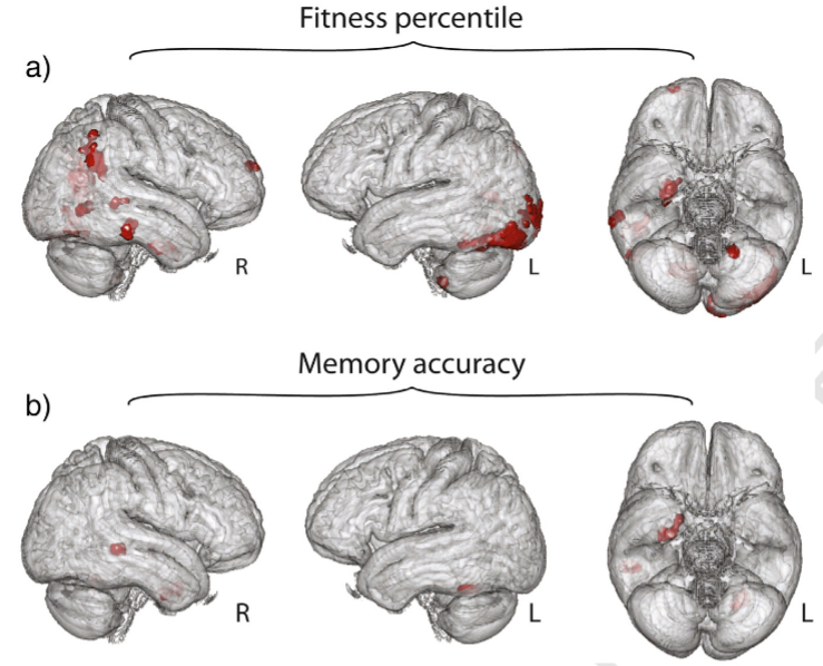
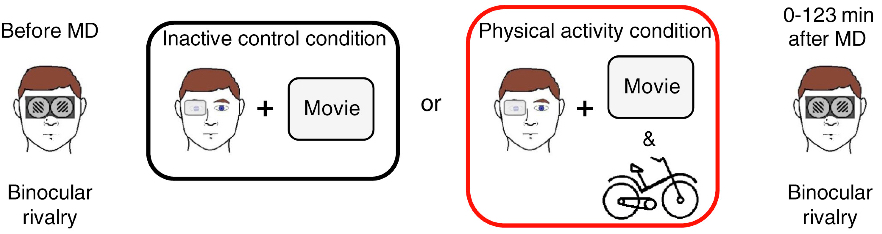
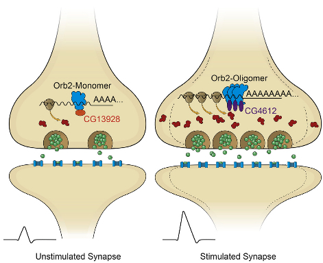
We also must keep in mind that studies reporting positive effects of brain games on cognition are more likely to be published than studies with null results –the so-called “file drawer effect”– such that even the available evidence is likely to draw an overly positive picture of the true state of affairs. Statistical methods such meta-analysis, which integrates the results of many studies in a given field of inquiry, allow estimation of effect magnitude as well as the likelihood of the file-drawer effect. While some meta-analyses report small positive effects of training on cognition, others note substantial disparities in methodological rigor among the studies that cast doubt on any firm conclusion. Further, the problems that haunt individual studies do not simply disappear when results from such studies are summarized in a meta-analysis. In particular, the practice of assessing specific tests rather than broader assays of ability is just as problematic on the level of meta-analytic integration as it is on the level of individual studies.

In summary, research on aging has shown that the human mind is malleable throughout life span. In developed countries around the world, later-born cohorts live longer and reach old age with higher levels of cognitive functioning than those who were born in earlier times. When researchers follow people across their adult lives, they find that those who live cognitively active, socially connected lives and maintain healthy lifestyles are less likely to suffer debilitating illness and early cognitive decline in their golden years than their sedentary, cognitively and socially disengaged counterparts. The goal of research on the effectiveness of computer-based cognitive exercise is to provide experimental evidence to support or qualify these observations. Some of the initial results are promising and make further research highly desirable. However, at present, these findings do not provide a sound basis for the claims made by commercial companies selling brain games. Many scientists cringe at exuberant advertisements claiming improvements in the speed and efficiency of cognitive processing and dramatic gains in “intelligence”, in particular when these appear in otherwise trusted news sources. In the judgment of the signatories below, exaggerated and misleading claims exploit the anxiety of adults facing old age for commercial purposes. Perhaps the most pernicious claim, devoid of any scientifically credible evidence, is that brain games prevent or reverse Alzheimer’s disease.

In closing, we offer five recommendations. Some of these recommendations reflect experimental findings in human populations, whereas others are based on a synthesis of correlational evidence in humans and mechanistic knowledge about risks and protective factors.

  • Much more research needs to be done before we understand whether and what types of challenges and engagements benefit cognitive functioning in everyday life. In the absence of clear evidence, the recommendation of the group, based largely on correlational findings, is that individuals lead physically active, intellectually challenging, and socially engaged lives, in ways that work for them. Before investing time and money on brain games, consider what economists call opportunity costs: If an hour spent doing solo software drills is an hour not spent hiking, learning Italian, making a new recipe, or playing with your grandchildren, it may not be worth it. But if it replaces time spent in a sedentary state, like watching television, the choice may make more sense for you.
  • Physical exercise is a moderately effective way to improve general health, including brain fitness. Scientists have found that regular aerobic exercise increases blood flow to the brain, and helps to support formation of new neural and vascular connections. Physical exercise has been shown to improve attention, reasoning, and components of memory. All said, one can expect small but noticeable gains in cognitive performance, or attenuation of loss, from taking up aerobic exercise training.
  • A single study, conducted by researchers with financial interests in the product, or one quote from a scientist advocating the product, is not enough to assume that a game has been rigorously examined. Findings need to be replicated at multiple sites, based on studies conducted by independent researchers who are funded by independent sources. Moreover, participants of training programs should show evidence of significant advantage over a comparison group that does not receive the treatment but is otherwise treated exactly the same as the trained group.
  • No studies have demonstrated that playing brain games cures or prevents Alzheimer’s disease or other forms of dementia.
  • Do not expect that cognitively challenging activities will work like one-shot treatments or vaccines; there is little evidence that you can do something once (or even for a concentrated period) and be inoculated against the effects of aging in an enduring way. In all likelihood, gains won’t last long after you stop the challenge.

In summary: We object to the claim that brain games offer consumers a scientifically grounded avenue to reduce or reverse cognitive decline when there is no compelling scientific evidence to date that they do. The promise of a magic bullet detracts from the best evidence to date, which is that cognitive health in old age reflects the long-term effects of healthy, engaged lifestyles. In the judgment of the signatories, exaggerated and misleading claims exploit the anxiety of older adults about impending cognitive decline. We encourage continued careful research and validation in this field.


Read Full Post »

Serum Folate and Homocysteine, Mood Disorders, and Aging

Larry H. Bernstein, MD, FCAP, Curator

LPBI


Dietary Folate and the Risk of Depression in Finnish MiddleAged Men

Tolmunen T, et al.
PSYCHOTHER AND PSYCHOSOM · OCT 2004; 73:334-339    DOI: http://dx.doi.org:/10.1159/000080385


Serum Folate, Vitamin B-12, and Homocysteine and Their Association With Depressive Symptoms Among U.S. Adults

M.A. BEYDOUN, M.R. SHROFF, H.A. BEYDOUN AND A.B. ZONDERMAN
PSYCHOSOM MED · NOV 2010;             DOI: http://dx.doi.org:/10.1097/PSY.0b013e3181f61863

Objective: To examine, in a nationally representative sample of U.S. adults, the associations of serum folate, vitamin B-12, and total homocysteine (tHcy) levels with depressive symptoms. Several nutritional and physiological factors have been linked to depression in adults, including low folate and vitamin B-12 and elevated tHcy levels.
Methods: Data on U.S. adults (age, 20–85 years; n 2524) from the National Health and Nutrition Examination Survey during the period 2005 to 2006 were used. Depressive symptoms were measured with the Patient Health Questionnaire (PHQ), and elevated symptoms were defined as a PHQ total score of 10. Serum folate, vitamin B-12, and tHcy were mainly expressed as tertiles. Multiple ordinary least square (OLS), logistic, and zero-inflated Poisson regression models were conducted in the main analysis.
Results: Overall, mean PHQ score was significantly higher among women compared with men. Elevated depressive symptoms (PHQ score of 10) were inversely associated with folate status, particularly among women (fully adjusted odds ratio [tertiles T3 versus T1] 0.37; 95% confidence interval, 0.17–0.86), but not significantly related to tHcy or vitamin B-12. No interaction was noted between the three exposures in affecting depressive symptoms. In older adults (50 years) and both sexes combined, tHcy was positively associated with elevated depressive symptoms (fully adjusted odds ratio [tertiles T2 versus T1] 3.01; 95% confidence interval, 1.01–9.03), although no significant dose-response relationship was found. Conclusions: Future interventions to improve mental health outcomes among U.S. adults should take into account dietary and other factors that would increase levels of serum folate.
Key words: depression, folate, vitamin B-12, homocysteine, adults.


Relationship of homocysteine, folic acid and vitamin B12 depression in a middle-aged community sample

P.S. SACHDEV, et al.   PSYCHOL MED · MAY 2005;   35, 529–538         http://dx.doi.org: /10.1017/S0033291704003721 

Background. Case control studies have supported a relationship between low folic acid and vitamin B12 and high homocysteine levels as possible predictors of depression. The results from epidemiological studies are mixed and largely from elderly populations.
Method. A random subsample of 412 persons aged 60–64 years from a larger community sample underwent psychiatric and physical assessments, and brain MRI scans. Subjects were assessed using the PRIME-MD Patient Health Questionnaire for syndromal depression and severity of depressive symptoms. Blood measures included serum folic acid, vitamin B12, homocysteine and creatinine levels, and total antioxidant capacity. MRI scans were quantified for brain atrophy, subcortical atrophy, and periventricular and deep white-matter hyperintensity on T2-weighted imaging.
Results. Being in the lowest quartile of homocysteine was associated with fewer depressive symptoms, after adjusting for sex, physical health, smoking, creatinine, folic acid and B12 levels. Being in the lowest quartile of folic acid was associated with increased depressive symptoms, after adjusting for confounding factors, but adjustment for homocysteine reduced the incidence rate ratio for folic acid to a marginal level. Vitamin B12 levels did not have a significant association with depressive symptoms. While white-matter hyperintensities had significant correlations with both homocysteine and depressive symptoms, the brain measures and total antioxidant capacity did not emerge as significant mediating variables. Conclusions. Low folic acid and high homocysteine, but not low vitamin B12 levels, are correlates of depressive symptoms in community-dwelling middle-aged individuals. The effects of folic acid and homocysteine are overlapping but distinct.


Association of folate intake with the occurrence of depressive episodes in middle-aged French men and women

P. Astorg, et al.    BRIT J  NUTR · AUG 2008; 100, 183–18       http://dx.doi.org:/10.1017/S0007114507873612

A low folate intake or a low folate status have been found to be associated with a higher frequency of depression in populations, but the existence and the direction of a causal link between folate intake or status and depression is still uncertain. The aim of this study was to seek the relation between the habitual folate intake in middle-aged men and women and the occurrence of depressive episodes. In a subsample of 1864 subjects (809 men and 1055 women) from the French SU.VI.MAX cohort, dietary habits have been measured at the beginning of the follow-up (six 24 h records) and declarations of antidepressant prescription, taken as markers of depressive episodes, have been recorded during the 8-year follow-up. No significant association was observed between folate intake and the risk of any depressive episode or of a single depressive episode during the follow-up, in both men and women. In contrast, the risk of experiencing recurrent depressive episodes (two or more) during the follow-up was strongly reduced in men with high folate intake (OR 0·25 (95% CI 0·06, 0·98) for the highest tertile v. the lowest, P for trend 0·046). This association was not observed in women. These results suggest that a low folate intake may increase the risk of recurrent depression in men.   Folate: Depression: Cohort studies


Homocysteine, vitamin B12, and folic acid levels in Alzheimer’s disease, mild cognitive impairment, and healthy elderly: baseline characteristics in subjects of the Australian Imaging Biomarker Lifestyle study.

Faux NG1, Ellis KA, Porter L, Fowler CJ,…, Ames D, Masters CL, Bush AI.
J Alzheimers Dis. 2011; 27(4):909-22.    http://dx.doi.org:/10.3233/JAD-2011-110752.

There is some debate regarding the differing levels of plasma homocysteine, vitamin B12 and serum folate between healthy controls (HC), mild cognitive impairment (MCI), and Alzheimer’s disease (AD). As part of the Australian Imaging Biomarker Lifestyle (AIBL) study of aging cohort, consisting of 1,112 participants (768 HC, 133 MCI patients, and 211 AD patients), plasma homocysteine, vitamin B12, and serum and red cell folate were measured at baseline to investigate their levels, their inter-associations, and their relationships with cognition. The results of this cross-sectional study showed that homocysteine levels were increased in female AD patients compared to female HC subjects (+16%, p-value < 0.001), but not in males. Red cell folate, but not serum folate, was decreased in AD patients compared to HC (-10%, p-value = 0.004). Composite z-scores of short- and long-term episodic memory, total episodic memory, and global cognition all showed significant negative correlations with homocysteine, in all clinical categories. Increasing red cell folate had a U-shaped association with homocysteine, so that high red cell folate levels were associated with worse long-term episodic memory, total episodic memory, and global cognition. These findings underscore the association of plasma homocysteine with cognitive deterioration, although not unique to AD, and identified an unexpected abnormality of red cell folate.


Homocysteine and folate as risk factors for dementia and Alzheimer disease1,2,3

Giovanni RavagliaPaola FortiFabiola Maioli, …., Nicoletta BrunettiElisa Porcellini, and Federico Licastro
Am J Clin Nutr Sept 2005; 82(3): 636-643


Background: In cross-sectional studies, elevated plasma total homocysteine (tHcy) concentrations have been associated with cognitive impairment and dementia. Incidence studies of this issue are few and have produced conflicting results.

Objective: We investigated the relation between high plasma tHcy concentrations and risk of dementia and Alzheimer disease (AD) in an elderly population.

Design: A dementia-free cohort of 816 subjects (434 women and 382 men; mean age: 74 y) from an Italian population-based study constituted our study sample. The relation of baseline plasma tHcy to the risk of newly diagnosed dementia and AD on follow-up was examined. A proportional hazards regression model was used to adjust for age, sex, education, apolipoprotein E genotype, vascular risk factors, and serum concentrations of folate and vitamin B-12.

Results: Over an average follow-up of 4 y, dementia developed in 112 subjects, including 70 who received a diagnosis of AD. In the subjects with hyperhomocysteinemia (plasma tHcy > 15 μmol/L), the hazard ratio for dementia was 2.08 (95% CI: 1.31, 3.30; P = 0.002). The corresponding hazard ratio for AD was 2.11 (95% CI: 1.19, 3.76; P = 0.011). Independently of hyperhomocysteinemia and other confounders, low folate concentrations (≤11.8 nmol/L) were also associated with an increased risk of both dementia (1.87; 95% CI: 1.21, 2.89; P = 0.005) and AD (1.98; 95% CI: 1.15, 3.40; P = 0.014), whereas the association was not significant for vitamin B-12.

Conclusions: Elevated plasma tHcy concentrations and low serum folate concentrations are independent predictors of the development of dementia and AD.


In Western societies, the prevalence and economic costs of Alzheimer disease (AD) are soaring in step with the increased number of elders in the population (1). Therefore, it is important to identify modifiable risk factors for this disease. The sulfur amino acid homocysteine is a unique candidate for this role because of its direct neurotoxicity (24) and its association with cerebrovascular disease (5), which is currently believed to play a significant role in AD etiology (6). Moreover, elevated concentrations of plasma total homocysteine (tHcy) are an indicator of inadequate folate and vitamin B-12 status (7) and can directly affect brain function via altered methylation reactions (8).

An association between AD and elevated tHcy concentrations has been reported in case-control (9, 10) and cross-sectional (11, 12) studies. Moreover, in nondemented elderly populations, plasma tHcy is inversely associated with poor performance at simultaneously performed tests of global cognitive function (1315) and specific cognitive skills (13, 16). However, cross-sectional studies cannot determine causality. Only 2 longitudinal studies investigated the relation between hyperhomocysteinemia and risk of incident AD, but their results were inconsistent; the Framingham Study reported a strong association (17), and the Washington Heights–Inwood Columbia Ageing Project (WHICAP) reported no association (18). Clarification of this issue is important because consistent evidence of a prospective association between homocysteine and AD would more strongly support the need for intervention trials testing the effectiveness of homocysteine-lowering vitamin therapy in preventing dementia.

Therefore, we examined baseline plasma tHcy in relation to risk of incident dementia and AD in the Conselice Study of Brain Aging (CSBA), an Italian population-based study of older persons.

Study population

The CSBA is a population-based survey, already described in detail elsewhere (19,20), the principal aim of which is to provide data about epidemiology and risk factors for dementia in the elderly. Its design includes both cross-sectional and longitudinal components. The study was approved by the Institutional Review Board of the Department of Internal Medicine, Cardioangiology, and Hepatology, University of Bologna, and written informed consent was obtained from all participants.

Briefly, in 1999–2000, 1016 (75%) of the 1353 individuals aged ≥65 y residing in the Italian municipality of Conselice (province of Ravenna, Emilia Romagna region) participated in the prevalence study. Data on cognitive status at the follow-up examination in 2003–2004 were collected for 861 of the 937 participants free of dementia at baseline. A flow chart detailing the derivation of the incidence sample used in this study is reported in Figure 1.

This prospective population-based study was the first to replicate previous findings from the Framingham Study (17), indicating that hyperhomocysteinemia doubles the risk of developing dementia and AD independently of several major confounders. Our results disagree with the negative findings recently reported in the WHICAP study (18). Possible explanations for this difference are the acknowledged insufficient statistical power of the WHICAP study, the rather homogeneously high tHcy concentrations of its sample—which did not permit enough variability to detect an association—and methodologic issues related to the prolonged time between blood sample collection and processing, which could have affected tHcy measurements.

Inconsistent results were also given by the only 2 studies that examined the association between homocysteine and cognitive decline at follow-up as measured with the MMSE (30, 31). These studies, however, differed in sample size and in which confounders were taken into account. Moreover, MMSE is a reliable global screening measure of cognitive function but was not developed to estimate changes in cognitive function or to diagnose dementia (32).

The substantial evidence that tHcy is an independent vascular risk factor (5) supports the role of hyperhomocysteinemia in AD. Subjects with vascular risk factors and cerebrovascular disease have an increased risk of AD (6), and hyperhomocysteinemia has been related to cerebral macro- and microangiopathy, endothelial dysfunction, impaired nitric oxide activity, and increased oxidative stress (3335). Moreover, as shown in cell cultures, homocysteine can directly cause brain damage through several mechanisms: increased glutamate excitoxicity via activation of N-methyl-D-aspartate receptors (2), enhancement of β-amyloid peptide generation (4), impairment of DNA repair, and sensitization of neurons to amyloid toxicity (3).

On the basis of cross-sectional observations, some authors have suggested that elevated plasma tHcy concentrations are not a causative factor in dementia and AD but are only a marker for concomitant vascular disease, independently of cognitive status (36, 37). Results from other cross-sectional investigations (9, 12, 38), as well as those from the present investigation and the Framingham Study (17), argue against this interpretation, but only intervention trials can give the ultimate proof of a causal relation between hyperhomocysteinemia and AD.

In contrast with both the Framingham (17) and WHICAP (18) studies, we also found that, independent of homocysteine and other confounders (including vitamin B-12), low serum folate is associated with an increased risk of incident dementia and AD. Mandatory folate fortification of food might partially explain the negative results of the US studies, whereas in Italy, where folate fortification is not practiced, relative folate deficiency may be endemic among the elderly population. Nondemented patients with poor cognitive performance and AD patients often exhibit poor folate status (reviewed in 8), but only one study specifically examined B vitamins in relation to incident dementia. In a selected sample of nondemented Swedish elderly participants in the Kungsholmen Study, low serum folate and vitamin B-12 were predictive of AD at 3 y of follow-up (39). The sample, however, was small (370 subjects), and a clear association was detected only when both vitamins were taken into account.

Biologic explanatory mechanisms relating folate deficiency to dementia include impaired methylation reactions in the central nervous system, with a consequent insufficient supply of methyl groups, which are required for the synthesis of myelin, neurotransmitters, membrane phospholipids, and DNA (8). However, because of the study design and the relatively short follow-up time, we cannot definitely establish whether the independent association between low folate and dementia risk indicates an actual effect of folate status on cognitive function or, on the contrary, that subtle functional alterations may affect the dietary intake of folate in the early preclinical stages of dementia.


Neurotoxicity associated with dual actions of homocysteine at the N-methyl-D-aspartate receptor

Stuart A. Lipton*Won-Ki KimYun-Beom Choi*,…, Derrick R. Arnelle§, and Jonathan S. Stamler
P
NAS 1997; 94(11):5923–5928    http://www.pnas.org/content/94/11/5923.abstract

Severely elevated levels of total homocysteine (approximately millimolar) in the blood typify the childhood disease homocystinuria, whereas modest levels (tens of micromolar) are commonly found in adults who are at increased risk for vascular disease and stroke. Activation of the coagulation system and adverse effects of homocysteine on the endothelium and vessel wall are believed to underlie disease pathogenesis. Here we show that homocysteine acts as an agonist at the glutamate binding site of the N-methyl-D-aspartate receptor, but also as a partial antagonist of the glycine coagonist site. With physiological levels of glycine, neurotoxic concentrations of homocysteine are on the order of millimolar. However, under pathological conditions in which glycine levels in the nervous system are elevated, such as stroke and head trauma, homocysteine’s neurotoxic (agonist) attributes at 10–100 μM levels outweigh its neuroprotective (antagonist) activity. Under these conditions neuronal damage derives from excessive Ca2+ influx and reactive oxygen generation. Accordingly, homocysteine neurotoxicity through overstimulation of N-methyl-D-aspartate receptors may contribute to the pathogenesis of both homocystinuria and modest hyperhomocysteinemia.


Vitamin B12 and folate in relation to the development of Alzheimer’s disease

H-X. Wang, Å. WahlinH. Basun, …, B. Winblad, and L. Fratiglioni
Neurology May 8, 2001; 56(9):1188-1194    http:/​/​dx.​doi.​org/​10.​1212/​WNL.​56.​9.​1188

Objective: To explore the associations of low serum levels of vitamin B12 and folate with AD occurrence.

Methods: A population-based longitudinal study in Sweden, the Kungsholmen Project. A random sample of 370 nondemented persons, aged 75 years and older and not treated with B12 and folate, was followed for 3 years to detect incident AD cases. Two cut-off points were used to define low levels of vitamin B12 (≤150 and ≤250 pmol/L) and folate (≤10 and ≤12 nmol/L), and all analyses were performed using both definitions. AD and other types of dementia were diagnosed by specialists according to DSM-III-R criteria.

Results: When using B12 ≤150pmol/L and folate ≤10 nmol/L to define low levels, compared with people with normal levels of both vitamins, subjects with low levels of B12or folate had twice higher risks of developing AD (relative risk [RR] = 2.1, 95% CI = 1.2 to 3.5). These associations were even stronger in subjects with good baseline cognition (RR = 3.1, 95% CI = 1.1 to 8.4). Similar relative risks of AD were found in subjects with low levels of B12or folate and among those with both vitamins at low levels. A comparable pattern was detected when low vitamin levels were defined as B12 ≤250 pmol/L and folate ≤12 nmol/L.

Conclusions: This study suggests that vitamin B12 and folate may be involved in the development of AD. A clear association was detected only when both vitamins were taken into account, especially among the cognitively intact subjects. No interaction was found between the two vitamins. Monitoring serum B12 and folate concentration in the elderly may be relevant for prevention of AD.


Assessing the association between homocysteine and cognition: reflections on Bradford Hill, meta-analyses, and causality

,
Hyperhomocysteinemia is a recognized risk factor for cognitive decline and incident dementia in older adults. Two recent reports addressed the cumulative epidemiological evidence for this association but expressed conflicting opinions. Here, the evidence is reviewed in relation to Sir Austin Bradford Hill’s criteria for assessing “causality,” and the latest meta-analysis of the effects of homocysteine-lowering on cognitive function is critically examined. The meta-analysis included 11 trials, collectively assessing 22 000 individuals, that examined the effects of B vitamin supplements (folic acid, vitamin B12, vitamin B6) on global or domain-specific cognitive decline. It concluded that homocysteine-lowering with B vitamin supplements has no significant effect on cognitive function. However, careful examination of the trials in the meta-analysis indicates that no conclusion can be made regarding the effects of homocysteine-lowering on cognitive decline, since the trials typically did not include individuals who were experiencing such decline. Further definitive trials in older adults experiencing cognitive decline are still urgently needed.
Mouse model for deficiency of methionine synthase reductase exhibits short-term memory impairment and disturbances in brain choline metabolism
, , , , , , ,
Biochem. J. 2014 461: 205212    http://dx.doi.org:/10.1042/BJ20131568
Hyperhomocysteinaemia can contribute to cognitive impairment and brain atrophy. MTRR (methionine synthase reductase) activates methionine synthase, which catalyses homocysteine remethylation to methionine. Severe MTRR deficiency results in homocystinuria with cognitive and motor impairments. An MTRR polymorphism may influence homocysteine levels and reproductive outcomes. The goal of the present study was to determine whether mild hyperhomocysteinaemia affects neurological function in a mouse model with Mtrr deficiency. Mtrr+/+, Mtrr+/gt and Mtrrgt/gtmice (3 months old) were assessed for short-term memory, brain volumes and hippocampal morphology. We also measured DNA methylation, apoptosis, neurogenesis, choline metabolites and expression of ChAT (choline acetyltransferase) and AChE (acetylcholinesterase) in the hippocampus. Mtrrgt/gt mice exhibited short-term memory impairment on two tasks. They had global DNA hypomethylation and decreased choline, betaine and acetylcholine levels. Expression of ChAT and AChE was increased and decreased respectively. At 3 weeks of age, they showed increased neurogenesis. In the cerebellum, mutant mice had DNA hypomethylation, decreased choline and increased expression of ChAT. Our work demonstrates that mild hyperhomocysteinaemia is associated with memory impairment. We propose a mechanism whereby a deficiency in methionine synthesis leads to hypomethylation and compensatory disturbances in choline metabolism in the hippocampus. This disturbance affects the levels of acetylcholine, a critical neurotransmitter in learning and memory.

Read Full Post »

Poverty and the American Dream

Larry H. Bernstein, MD, FCAP, Curator

LPBI


Brookings Institute: Poverty Report

http://www.brookings.edu/~/media/Research/Files/Reports/2015/12/aei-brookings-poverty-report/Chapter-1.pdf

Chapter 1:

Introduction

In 1931, the writer James Truslow Adams coined the term “The American Dream.” His definition holds up well today. The dream, he said, is of a land in which: life should be better and richer and fuller for everyone, with opportunity for each according to ability or achievement. It is a difficult dream for the European upper classes to interpret adequately, and too many of us ourselves have grown weary and mistrustful of it. It is not a dream of motor cars and high wages merely, but a dream of social order in which each man and each woman shall be able to attain to the fullest stature of which they are … capable, and be recognized by others for what they are, regardless of the fortuitous circumstances of birth or position.1

Today, many Americans fear that our country is no longer a land of opportunity. Although social mobility overall seems not to have decreased in recent decades,2 there is evidence that it is lower in America than in many other advanced economies.3 Scholars on both the left and the right are also increasingly worried that children growing up today in lower-income families have fewer social supports and pathways into the middle class than in past generations. As Robert Putnam showed in his recent book Our Kids, 4 children from wellto-do families today enjoy more material, emotional, and educational support than ever before, but children from low-income families often grow up in homes, schools, and communities that are in disarray. Charles Murray reached similar conclusions in Coming Apart. 5

The trends aren’t entirely bleak, and poor children today are better off in several ways than they were a few decades ago. They have better access to healthcare, fewer of them are born to teen mothers, their parents have more education, they are exposed to fewer environmental toxins and violence, and fewer live in foster care. We should celebrate these advances. But the circumstances and outcomes of upper-income children have improved even more rapidly, leading to ever-widening inequality in the human and financial resources that boost child development. And on a few important factors, such as family stability, the circumstances of poor children have gotten worse.

The reasons for the increasing gaps between childhoods in different social classes are many and intertwined, including: the loss of manufacturing jobs, stagnating wages for workers without a college degree, labor-saving technological changes, changing relationships between workers and management, the increasing importance of education and training in a post-industrial economy, a less energetic civil society, high rates of incarceration, weaker attachment to the labor force among less-educated men, and the rising prevalence of single-parent families among the less-educated.

The poor prospects for children born into poor families are an urgent national concern. This state of affairs contradicts our country’s founding ideals. It weakens the promise that inspired so many immigrants to uproot themselves from everything familiar to seek freedom, self-determination, and better lives for their children in America. It holds particularly grave implications for the well being of blacks and for the future of racial equality so courageously fought for over the course of generations.

At its best, the American credo of freedom and individual initiative has been uniquely able to unleash the energy and imagination of its citizens, inspiring them, as Adams put it, “to attain to the fullest stature of which they are capable.”6 For many American families—including many low-income families—that dream is still possible. But large numbers of children live in disadvantaged and often chaotic homes and communities, attend schools that don’t prepare them to navigate an increasingly complex economy, and have parents (often a single parent) who work in low-wage jobs with variable and uncertain hours. The massive waste and loss of this human potential costs the United States in economic terms, and it is a tragedy in human terms. Most Americans would agree that we can do better.

The political difficulty arises when we turn to solutions. Most new ideas for helping the poor are controversial and expensive, and when one political party offers a proposal, the other party usually disagrees with its premises or specifics. The parties often have deep philosophical differences, but research also shows that the mere fact that one party proposes an idea can motivate partisans on the other side to dismiss it.7 And yet, points of agreement are emerging that could serve as a foundation for consensus. Most Americans and their political representatives tend to agree on several key points.

  1. First, for able-bodied Americans, it is far better to earn money than to depend on public assistance, although economic conditions sometimes prevent people from becoming self-sufficient.
  2. Second, children are on average better off growing up with two parents committed to each other for the long term, an arrangement most likely to occur within the context of marriage.
  3. And third, our schools don’t adequately prepare the young for the economic and social environment in which they must make their way.

THE AEI-BROOKINGS WORKING GROUP

Our report has three distinctive features.

  1. the diversity of our perspectives and experiences.    We share an intense belief that poverty and opportunity are profoundly consequential and that our nation’s future prosperity and our common humanity compel us to work together to find credible strategies to reduce poverty and increase economic mobility.
  2. we consider three major domains of life simultaneously: family, work, and education.    they are highly interconnected. Improving family stability helps children succeed in school; improving the fit between schools and jobs helps teenagers transition into the labor force; when young people can find work that pays well, they create more stable families, and the cycle continues.
  3. it is grounded in values—the three broadly shared American values of opportunity, responsibility, and security. Focusing on these shared values has made it easier for us to work together and find many points of agreement.

OPPORTUNITY The concept of “opportunity” draws nearly universal support among Americans, and it’s the core concept of the American Dream. We endorse Truslow Adams’ definition of opportunity as the state of affairs when “each man and each woman shall be able to attain to the fullest stature of which they are capable,” regardless of the circumstances of their birth.8

RESPONSIBILITY America is a free society, but freedom comes with responsibilities. Responsibility is the state of being accountable for things over which one has control, or has a duty of care. Family life is a network of mutual responsibilities. So is work life. So is democratic citizenship.

The values of responsibility and opportunity are closely linked in the American mind. We can see the link in a line from President Clinton’s 1993 Labor Day speech that has had bipartisan resonance: We’ll think of the faith of our parents that was instilled in us here in America, the idea that if you work hard and play by the rules, you’ll be rewarded with a good life for yourself and a better chance for your children.10 The converse of this assertion is that if you fail to be responsible—if you don’t work hard or don’t play by the rules, then you aren’t entitled to a reward. These linked values of responsibility and opportunity were the linchpins of the bipartisan welfare reform law of 1996—whose official name included both “Personal Responsibility” and “Opportunity.”11

SECURITY Despite our best efforts to care for ourselves, we all know that life sometimes resembles a lottery.   The central idea of insurance is that we are all better off pooling some of the risks of life, and hoping that we never get to recover our insurance premiums.

Friedrich Hayek, an economist who was wary of collectivism in most forms and who is widely admired by conservatives, endorsed the value of security in 1944 in this famous passage from The Road to Serfdom: There is no reason why, in a society which has reached the general level of wealth ours has . . . should not be guaranteed to all . . . some minimum of food, shelter and clothing, sufficient to preserve health. Nor is there any reason why the state should not help to organize a comprehensive system of social insurance in providing for those common hazards of life against which few can make adequate provision.12

Several decades of research show that increasing security for children can better prepare them to break the cycle of poverty and grow up to be more responsible adults. A child’s brain is highly malleable. In the early years, when it is growing rapidly, the young brain responds to cues about the kind of environment that surrounds it. When children are raised in a chaotic and unpredictable environment, they become more attracted to immediate rewards, rather than larger but more distant rewards.13   Although children have great resilience and the capacity to overcome their early environment, some children—especially if they don’t have the benefit of interventions that reduce the stress to which they are exposed—are overwhelmed by early stress and trauma and suffer permanent damage.16

Conversely, when children are raised in more stable and predictable environments, they are more likely to learn that it pays to defer gratification and reap larger rewards in the future. Low stress, high predictability, and strong, stable relationships with caring adults all help children become measurably better at self-regulating, delaying gratification, and controlling their impulses.17 If we want adult citizens who can exercise responsibility, we should do as much as we can to improve the security of childhood, especially among the poor.

These three values guide the rest of our report. We offer a comprehensive plan for reducing poverty and promoting economic opportunity in the United States. In each chapter, we evaluate the best evidence about current approaches and then recommend policies that will increase opportunity, encourage people to take greater responsibility for their own lives, and increase security, especially among lower-income Americans and their children.

In the final chapter, we summarize our recommendations and suggest how the nation can pay for the policies we propose. We also lay out a path by which our recommendations might be carried out, evaluated, and improved, despite America’s political polarization. We have negotiated and compromised to create a plan that we believe is the best way forward. We are all enthusiastic about the final product because we believe it will reduce poverty and increase opportunity in America.


Read Full Post »

Long Term Memory and Prions

Larry H. Bernstein, MD, FCAP, Curator

LPBI


updated 12/12/2015


Possible biochemical mechanism underlying long-term memories identified

Why is a prion-like molecular state necessary for persistence of memory? Could a transient memory be made permanent with a “Limitless” NZT-type neurotropic drug — or permanently forgotten?

It’s a nagging question: why do some of our memories fade away, while others last forever? Now scientists at the Stowers Institute for Medical Research have identified a possible biochemical mechanism: a specific synaptic protein called Orb2 can either block or maintain neural synapses (connections between neurons), which create and maintain long-term memories.

So for a memory to persist, the synaptic connections must be kept strong. But how? The researchers previously identified a synaptic protein called CPEB that is responsible for maintaining the strength of such connections in the sea slug (a model organism used in memory research). Recently, they identified a similar protein, called Orb2, in the fruit fly.

Now, using a fruit fly model system, they found that the synaptic connections are kept strong by the transformation of Orb2 from one molecular state to another. And that transformation causes Orb2 molecules to solidify and strengthen the memory connections in the brain.

The authors conclude their paper, published in the current issue of the journal Cell, with several questions. How and what triggers this transformation, how long does it persist? Is the continued presence of a prion-like state necessary for the persistence of memory, and is it correlated with or predictive of long-lasting memory? And most interestingly: can a transient memory about to be forgotten be stabilized by artificial recruitment of the prion-like state (perhaps by a neurotropic compound)?

And what about that ironic link with prions, associated with neurodegenerative disorders? Are prions some twisted form of memory that could one day even have value? We’ll be keeping an eye on where this fascinating research leads.

Technical details: the memory switch

In their latest study, the researchers determined that Orb2 exists in two distinct physical states: monomeric (a single molecule that can bind to other molecules) and oligomeric (a molecular complex).

Like CPEB, oligomeric Orb2 is prion-like — that is, it’s a self-copying cluster. (But unlike prions, oligomeric Orb2 and CPEB are not toxic.) Monomeric Orb2 represses, and oligomeric Orb2 activates a crucial step in the complex cellular process that leads to protein synthesis.

During this crucial step, messenger RNA (mRNA), which is an RNA copy of a gene’s recipe for a protein, is translated by the cell’s ribosome into the sequence of amino acids that will make up a newly synthesized protein. The monomeric form of Orb2 binds to the target mRNA, keeping it in a repressed state.

The Stowers scientists also determined that prion-like Orb2 not only activates translation into amino acids but imparts its translational state to nearby monomer forms of Orb2. As a result, monomeric Orb2 is transformed into prion-like Orb2, so its role in translation switches from repression to activation.

Self-sustaining activation maintains synaptic activity

Stowers Associate Investigator Kausik Si, Ph.D. thinks this switch is the possible mechanism by which fleeting experiences create an enduring memory. “Because of the self-sustaining nature of the prion-like state, this creates a local and self-sustaining translation activation of Orb2-target mRNA, which maintains the changed state of synaptic activity over time,” says Si.

The discovery that the two distinct states of Orb2 have opposing roles in the translation process provides “for the first time a biochemical mechanism of synapse-specific persistent translation and long-lasting memory,” he states.

“To our knowledge, this is the first example of a prion-based protein switch that turns a repressor into an activator,” Si adds. “The recruitment of distinct protein complexes at the non-prion and prion-like forms to create altered activity states indicates the prion-like behavior is in essence a protein conformation-based switch.

“Through this switch, a protein can lose or gain a function that can be maintained over time in the absence of the original stimuli. Although such a possibility has been anticipated prior to this study, there was no direct evidence.”

The research builds upon previous studies by Si and Eric Kandel, M.D., of Columbia University and other scientists. These studies revealed that both short-term and long-term memories are created in synapses.


Abstract of Amyloidogenic Oligomerization Transforms Drosophila Orb2 from a Translation Repressor to an Activator

Memories are thought to be formed in response to transient experiences, in part through changes in local protein synthesis at synapses. In Drosophila, the amyloidogenic (prion-like) state of the RNA binding protein Orb2 has been implicated in long-term memory, but how conformational conversion of Orb2 promotes memory formation is unclear. Combining in vitro and in vivo studies, we find that the monomeric form of Orb2 represses translation and removes mRNA poly(A) tails, while the oligomeric form enhances translation and elongates the poly(A) tails and imparts its translational state to the monomer. The CG13928 protein, which binds only to monomeric Orb2, promotes deadenylation, whereas the putative poly(A) binding protein CG4612 promotes oligomeric Orb2-dependent translation. Our data support a model in which monomeric Orb2 keeps target mRNA in a translationally dormant state and experience-dependent conversion to the amyloidogenic state activates translation, resulting in persistent alteration of synaptic activity and stabilization of memory.


New Finding on Synapse Destruction May Open Path to Alzheimer’s Therapy

http://www.genengnews.com/gen-news-highlights/new-finding-on-synapse-destruction-may-open-path-to-alzheimer-s-therapy/81252029/

A team led by scientists at the University of New South Wales in Australia say they have discovered how connections between brain cells are destroyed in the early stages of Alzheimer’s disease. They believe their work opens up a new avenue for research on possible treatments for the degenerative brain condition.

“One of the first signs of Alzheimer’s disease is the loss of synapses—the structures that connect neurons in the brain,” noted study leader, Vladimir Sytnyk, Ph.D., of the UNSW School of Biotechnology and Biomolecular Sciences. “Synapses are required for all brain functions, and particularly for learning and forming memories. In Alzheimer’s disease, this loss of synapses occurs very early on, when people still only have mild cognitive impairment, and long before the nerve cells themselves die. We have identified a new molecular mechanism which directly contributes to this synapse loss, a discovery we hope could eventually lead to earlier diagnosis of the disease and new treatments.”

The team studied a specific protein in the brain, neural cell adhesion molecule 2 (NCAM2), one of a family of molecules that physically connects the membranes of synapses and help stabilize these long lasting synaptic contacts between neurons. The researchers paper (“Aβ-dependent reduction of NCAM2-mediated synaptic adhesion contributes to synapse loss in Alzheimer’s disease”) is published in Nature Communications.

Using post-mortem brain tissue from people with and without the condition, they discovered that synaptic NCAM2 levels in the part of the brain known as the hippocampus were low in those with Alzheimer’s disease. They also showed in mice studies and in the laboratory that NCAM2 was broken down by beta-amyloid, which is the main component of the plaques that build up in the brains of people with the disease.

“Our research shows the loss of synapses is linked to the loss of NCAM2 as a result of the toxic effects of beta-amyloid,” pointed out Dr. Sytnyk. “It opens up a new avenue for research on possible treatments that can prevent the destruction of NCAM2 in the brain.”


Aβ-dependent reduction of NCAM2-mediated synaptic adhesion contributes to synapse loss in Alzheimer’s disease

Iryna Leshchyns’kaHeng Tai LiewClaire ShepherdGlenda M. HallidayClaire H. StevensYazi D. KeLars M. Ittner & Vladimir Sytnyk
Nature Communications Nov 2015; 6(8836)        doi:10.1038/ncomms9836

Alzheimer’s disease (AD) is characterized by synapse loss due to mechanisms that remain poorly understood. We show that the neural cell adhesion molecule 2 (NCAM2) is enriched in synapses in the human hippocampus. This enrichment is abolished in the hippocampus of AD patients and in brains of mice overexpressing the human amyloid-β (Aβ) precursor protein carrying the pathogenic Swedish mutation. Aβ binds to NCAM2 at the cell surface of cultured hippocampal neurons and induces removal of NCAM2 from synapses. In AD hippocampus, cleavage of the membrane proximal external region of NCAM2 is increased and soluble extracellular fragments of NCAM2 (NCAM2-ED) accumulate. Knockdown of NCAM2 expression or incubation with NCAM2-ED induces disassembly of GluR1-containing glutamatergic synapses in cultured hippocampal neurons. Aβ-dependent disassembly of GluR1-containing synapses is inhibited in neurons overexpressing a cleavage-resistant mutant of NCAM2. Our data indicate that Aβ-dependent disruption of NCAM2 functions in AD hippocampus contributes to synapse loss.


Learning and memory processes depend on the number and correct functioning of synapses in the brain. Cell adhesion molecules are enriched in the pre- and postsynaptic membranes. These molecules physically connect synaptic membranes, providing mechanical stabilization of synaptic contacts1, 2, 3, are necessary for the formation of new synapses during neuronal development4, 5, and maintain and regulate synaptic plasticity in adults6, 7, 8, 9, 10.

Alzheimer’s disease (AD) is a neurodegenerative brain condition predominantly of the aging population. One of the earliest signs of AD is the loss of synapses11, which can at least partially be linked to the toxicity mediated by Aβ12, 13, 14, a peptide that accumulates in the brains of AD patients. The impact of AD on synaptic adhesion and the role of synaptic cell adhesion molecules in the progression of the disease remains poorly understood.

The neural cell adhesion molecule 2 (NCAM2), sometimes designated OCAM, belongs to the immunoglobulin superfamily of cell adhesion molecules. NCAM2 participates in homophilic trans-interactions15, 16. During human embryonic development, NCAM2 is expressed in several tissues, including lung, liver, and kidney with the highest expression in the brain17. The expression level of NCAM2 peaks around postnatal day 21 and remains high during adulthood15, suggesting that the protein is necessary both during development and in adult brains. Accordingly, studies with cultured neurons and in NCAM2 deficient mice show that NCAM2 is important for the development of the brain, and the olfactory system in particular18, 19.

The NCAM2 gene is located on chromosome 21 in humans and NCAM2 overexpression has been suggested to be one of the factors contributing to the symptoms of Down syndrome17, which presents with early-onset AD pathology. Single-nucleotide polymorphisms in the NCAM2 gene have been reported as a risk factor related to the progression of AD in the Japanese population20. A recent genome-wide association study has found an association between single-nucleotide polymorphisms in the NCAM2 gene and levels of Aβ in the cerebrospinal fluid in humans, suggesting that NCAM2 is involved in the pathogenic pathway to the senile plaques that concentrate in AD brains21. Since genetic association studies indicate a link between NCAM2 and AD, we have analysed whether AD pathology influences levels of NCAM2 in synapses. Our results indicate that the synaptic adhesion mediated by NCAM2 is highly susceptible to Aβ toxicity and that proteolytic fragments of NCAM2 generated in an Aβ-dependent manner can directly contribute to the induction of synapse disassembly.


Synaptic NCAM2 is reduced in the hippocampus in AD

To analyse whether functions of NCAM2 are affected in AD, frozen post-mortem brain tissue of AD patients and non-affected controls (n=10 each) was analysed by western blot with antibodies against NCAM2. The detailed demographic data for the subjects analysed are presented inSupplementary Table 1. Total levels of NCAM2 were slightly increased in the hippocampus, but not significantly affected in the cerebellum or superior temporal cortex in AD (Supplementary Fig. 1). In contrast, levels of VGLUT1, a presynaptic marker-protein of excitatory synapses, were reduced in AD hippocampus (Supplementary Fig. 1), indicating a loss of excitatory synapses. Levels of VGAT, a presynaptic marker-protein of inhibitory synapses, were not significantly affected in any brain region analysed (Supplementary Fig. 1).

Changes in the protein levels in brain homogenates do not necessarily reflect changes in the protein levels in synapses. To analyse whether the synaptic function of NCAM2 is affected in AD, we compared the enrichment of NCAM2 in synaptosomes isolated from the brain tissue of individuals with AD and non-affected controls by western blot analysis of synaptosomes and total homogenates of the brains used for synaptosome preparations. Equal total protein amounts from each probe were applied to the gels to compensate for any possible differences in the yield of synaptosomes because of the synapse loss observed in AD. Western blot analysis with antibodies against actin, VGLUT1, VGAT, synaptophysin (a general presynaptic marker-protein), and PSD95 (a postsynaptic marker-protein), showed that these proteins were enriched to similar levels in synaptosomes from AD and control brains, indicating similar purities of intact synaptosome isolations (Fig. 1a). Western blot analysis showed that in control individuals NCAM2 was highly enriched in synaptosomes from the hippocampus and to a lower degree in synaptosomes from the temporal cortex and cerebellum (Fig. 1a,b). This synaptic enrichment of NCAM2 was significantly reduced in synaptosomes from AD hippocampi (Fig. 1a,b). The synaptic enrichment of NCAM2 was slightly lower in the AD versus control cerebellum, however the difference was not statistically significant (Fig. 1a,b).


Figure 1: Synaptic accumulation of NCAM2 is reduced in the hippocampus of AD-affected individuals.

Figure 2: Cleavage of the membrane-adjacent extracellular fragment of NCAM2 is increased in AD brains.

Figure 3: The extracellular domain of NCAM2 binds to Aβ.

Cleavage of NCAM2aa682-701 is increased in AD brains

NCAM2 binds to Aβ in vitro

Figure 4: NCAM2 accumulates in excitatory synapses of cultured hippocampal neurons.

NCAM2 accumulates in excitatory synapses of cultured hippocampal neurons.

http://www.nature.com/ncomms/2015/151127/ncomms9836/images_article/ncomms9836-f4.jpg

(a) Low-magnification image of a cultured hippocampal neuron labelled by indirect immunofluorescence with antibodies against NCAM2, synaptophysin and MAP2. Note expression of NCAM2 along MAP2 positive dendrites. NCAM2 is also expressed in astrocytes (marked a) which are present in these cultures. Scale bar, 20μm. (b) High-magnification image of dendrites of neurons co-labelled with antibodies against NCAM2, synaptophysin and MAP2. Arrows show clusters of NCAM2 partially overlapping with synaptophysin accumulations. NCAM2-negative synapses are also observed (arrowheads). Scale bar, 10μm. (c) High-magnification image of a dendrite of a cultured hippocampal neuron labelled with antibodies against NCAM2, synaptophysin and PSD95. NCAM2 clusters partially overlap with accumulations of PSD95 and synaptophysin (arrows). Scale bar, 10μm. Three-dimensional analysis of the co-localization within the outlined area is on the right. Z-stack has been acquired with 0.15μm steps. The xz and yz sections along the dashed lines on the xy image are shown. Note co-localization of the NCAM2 cluster with synaptic markers. (d) Negative control, that is, labelling performed without primary antibodies, is shown. Scale bar, 10μm.


Figure 5: Aβ1–42 oligomers bind to NCAM2 at the cell surface of neurons.

Figure 6: Levels of NCAM2 are reduced in synaptosomes of cultured hippocampal neurons treated with Aβ1-42 oligomers.

Figure 7: NCAM2 co-localizes with Aβ1-42 in brains of APP23 transgenic mice.

Figure 8: NCAM2 binds to Aβ and its synaptic accumulation is reduced in the hippocampus of APP23 transgenic mice.

Aβ removes NCAM2 from synapses of hippocampal neurons

Western blot analysis showed that levels of soluble NCAM2 with the molecular weight of ~100kDa were significantly increased in culture medium from Aβ1-42-treated hippocampal neurons (Fig. 6b), further indicating that Aβ1-42 induces removal of NCAM2 off the neuronal cell surface. In contrast, levels of the soluble proteolytic products of CHL1, another synaptic cell adhesion molecule of the immunoglobulin superfamily26, 27, were not changed in the culture medium from Aβ1-42-treated hippocampal neurons (Fig. 6b). Incubation with Aβ1-42 did not increase levels of soluble NCAM2 in the culture medium from cortical neurons (Fig. 6b), suggesting that cortical neurons are more resistant to Aβ1-42-dependent NCAM2 proteolysis.

Aβ binds to and removes NCAM2 from synapses in APP23 mice

Disruption of NCAM2 adhesion promotes synapse disassembly

Figure 9: Disruption of NCAM2 functions at the neuronal cell surface promotes glutamatergic synapse disassembly.

Disruption of NCAM2 functions at the neuronal cell surface promotes glutamatergic synapse disassembly.

http://www.nature.com/ncomms/2015/151127/ncomms9836/images_article/ncomms9836-f9.jpg

(ae) Cultured hippocampal neurons were either mock-treated or incubated with the recombinant soluble extracellular domains of NCAM2 (NCAM2-ED), antibodies against the extracellular domain of NCAM2 (NCAM2mAb), or Aβ1-42 oligomers. In a,b, neurons were labelled with antibodies against the extracellular domain of GluR1 before permeabilization of membranes with detergent, and co-labelled with antibodies against synaptophysin after permeabilization of membranes with detergent. Representative images of dendrites are shown (a). Note co-localization of cell surface GluR1 accumulations with synaptophysin clusters in mock-treated neurons, and increased levels of non-synaptic cell surface GluR1 accumulations in neurons treated with NCAM2-ED, NCAM2mAb or Aβ1-42. Graphs (b) show the percentage of synaptic and non-synaptic GluR1 clusters relative to total number of GluR1 clusters along dendrites and numbers of synaptophysin accumulations per dendrite length (mean+s.e.m.). *P<0.0001 (analysis of variance with Dunnett’s multiple comparison test, n>80 dendrites from 20 neurons were analysed in each group). In c, neurons were labelled with antibodies against the extracellular domain of NR1 before permeabilization of membranes with detergent, and co-labelled with antibodies against synaptophysin after permeabilization of membranes with detergent. Graphs show the percentage of synaptic and non-synaptic NR1 clusters relative to total number of NR1 clusters along dendrites (mean+s.e.m.). *P<0.0001 (analysis of variance with Dunnett’s multiple comparison test, n>85 dendrites from 20 neurons were analysed). In d,e, neurons were co-labelled with fluorescent phalloidin and synaptophysin antibodies. Representative images of dendrites are shown in d. Note higher labelling intensity and co-localization with synaptophysin of the phalloidin-labelled polymerized actin accumulations in control neurons versus neurons treated with Aβ1-42, NCAM2-ED or NCAM2mAb. Note increased numbers of filopodia and lamellipodia in neurons treated with Aβ1-42, NCAM2-ED or NCAM2 mAb. Graphs (e) show ratio of the dendrite area-to-length and phalloidin labelling intensity of dendrites of neurons. Mean values+s.e.m. are shown. *P<0.0001 (analysis of variance with Dunnett’s multiple comparison test, n=50 dendrites from 20 neurons were analysed in each group). Scale bar, 10μm (in a,d).

Cleavage-resistant NCAM2 reduces Aβ-dependent synapse loss

Figure 10: Aβ1-42 reduces the number of GluR1-containing synapses in the NCAM2-dependent manner.

http://www.nature.com/ncomms/2015/151127/ncomms9836/images_article/ncomms9836-f10.jpg

(a) Representative images of dendrites of cultured hippocampal neurons transfected either with control negative miRNA (negative miR) or NCAM2miR and either mock-treated or incubated with Aβ1-42. Transfected neurons were identified by fluorescence of GFP, which is co-expressed together with miRNA. Neurons were co-labelled with antibodies against cell surface GluR1 and synaptophysin. Note that the number of synaptic GluR1 clusters is reduced and the number of non-synaptic GluR1 clusters is increased in neurons transfected with NCAM2miR. Scale bar, 10μm. (b,c) Graphs show mean+s.e.m. percentage of synaptic and non-synaptic GluR1 clusters relative to the total number of GluR1 clusters along dendrites (b) and numbers of synaptophysin accumulations per dendrite length normalized to the mean number in mock-treated neurons (c) for neurons described in (a). (df) Graphs show mean+s.e.m. percentage of synaptic and non-synaptic GluR1 clusters relative to the total number of GluR1 clusters along dendrites (d), number of synaptophysin accumulations per dendrite length normalized to the mean number in mock-treated neurons (e), and area/length ratio (f) in cultured hippocampal neurons transfected either with GFP alone or co-transfected with GFP and non-mutated NCAM2 (NCAM2WT) or NCAM2D693A mutant and either mock-treated or incubated with Aβ1-42. (g,h) Graphs show mean+s.e.m. percentage of non-synaptic GluR1 clusters relative to the total number of GluR1 clusters along dendrites (g) and area/length ratio (h) in cultured hippocampal neurons co-transfected with NCAM2 miR and either GFP, non-mutated NCAM2 (WT) or NCAM2D693A mutant (D693A) and either mock-treated or incubated with Aβ1-42. In bh, *P<0.01 (compared as indicated), ˆP<0.01 (compared with mock-treated neurons transfected with negative miR (b), GFP (df) or co-transfected with NCAM2miR and GFP (gh)), analysis of variance with Tukey’s multiple comparison test, n>50 dendrites from 20 neurons were analysed in each group.


Taken together, our results indicate that Aβ affects the numbers of GluR1-containing glutamatergic synapses in a NCAM2-dependent manner.

Alzheimer’s disease is characterized by loss of synapses, which is the strongest correlate of cognitive decline11, 29, 30, 31, 32 and possibly one of the earliest events in AD pathogenesis30, 33. Synapses are long lasting contacts between neurons, which are stabilized by a number of cell adhesion molecules that concentrate in pre- and postsynaptic membranes2, 5. Cell adhesion molecules play an essential role in maintaining synapse functionality and stability. Although cell adhesion molecules of many families are required for the synapse integrity8, 10, elimination of even one type of synaptic cell adhesion molecule is often sufficient to induce abnormalities in synapse ultrastructure and protein composition6, 7. In the present study, we show that levels of the synaptic cell adhesion molecule NCAM2 are markedly reduced in hippocampal synapses in AD brains and Aβ-forming APP23 mice. Our observations that disruption of NCAM2 interactions at the cell surface, knockdown of NCAM2 expression and Aβ exposure result in reduced numbers of glutamatergic synapses in hippocampal neurons suggest that abnormalities in NCAM2-mediated synaptic adhesion contribute to synapse loss in AD.

Although the mechanisms of synapse disassembly in AD remain poorly understood, previous studies indicated that synapse loss can be linked to Aβ-induced toxicity12, 34, 35. Our observations showing that synaptic levels of NCAM2 are similarly reduced in APP23 mice and in cultured hippocampal neurons from wild-type mice exposed to Aβ argue in favour of Aβ-dependent mechanisms in the disruption of NCAM2-mediated synaptic adhesion. We however do not exclude that other factors, such as disrupted trafficking of NCAM2 to synapses, may also contribute to the reduction of NCAM2 levels at synapses. Strikingly, the effects of Aβ on synaptic targeting of NCAM2 were particularly strong in hippocampal but not cortical or cerebellar neurons. The enhanced susceptibility of synaptic NCAM2 to Aβ-dependent proteolysis may therefore contribute to selective vulnerability of the hippocampus to AD.

Our observations that NCAM2 directly interacts with synthetic Aβ1-42, that Aβ1-42 forms a molecular complex with NCAM2 at the neuronal cell surface and that complexes of NCAM2 and oligomers of Aβ can be isolated from APP23 mouse brains, indicate that NCAM2 may function as a previously unrecognized receptor for Aβ at the neuronal cell surface. Previous studies have shown that Aβ can also bind to other cell adhesion molecules at the neuronal cell surface, among which are the prion protein36 and L137. In addition, a number of cell adhesion molecules have been shown to interact with APP, including the neural cell adhesion molecule 1 (NCAM1)38 and TAG1 (ref. 39). It remains to be investigated whether the NCAM2/Aβ complex comprises other adhesion molecules and cell surface proteins. Interestingly, NCAM1, a homologue of NCAM2, binds to prion protein40 and L1 (ref. 41). However, in spite of homology to NCAM2, NCAM1 binds to a region of APP which is different to the Aβ-containing region38.

…..

Taken together, we show that Aβ induces synaptic loss and proteolysis of NCAM2 in cell culture and APP transgenic mouse models, providing a mechanistic explanation for synaptic NCAM2 changes in AD brains. The detrimental effects of proteolyically cleaved extracellular NCAM2 on synapses may augment the Aβ toxicity in the pathogenesis of AD. The exact molecular mechanisms underlying Aβ-induced NCAM2 changes, and to which degree it contributes to onset and progression of disease remains to be established. Nevertheless, our data reveal a new role of NCAM2 in AD that warrants further investigation.

Read Full Post »

Autocrine selection of GLP-1 binding site

Larry H. Bernstein, MD, FCAP, Curator

LPBI

Update 12/15/2015

TSRI Team Finds Unique Anti-Diabetes Compound

Scientists from The Scripps Research Institute (TSRI) have deployed a powerful new drug discovery technique to identify an anti-diabetes compound with a novel mechanism of action

http://www.technologynetworks.com/HTS/news.aspx?ID=186055

The finding may lead to a new type of diabetes treatment. Just as importantly, it demonstrates the potential of the new technique, which enables researchers to quickly find drug candidates that activate cellular receptors in desired ways.

“In principle, we can apply this technique to hundreds of other receptors like the one we targeted in this study to find disease treatments that are more potent and have fewer side effects than existing therapies. It has been a very productive cross-campus collaboration, so we’re hoping to build on its success as we continue to collaborate on interrogating potential therapeutic targets,” said Patricia H. McDonald, an assistant professor at TSRI’s Jupiter, Florida campus and a senior investigator of the study.

McDonald’s laboratory collaborated on the study with the laboratory of Richard A. Lerner, the Lita Annenberg Hazen Professor of Immunochemistry at TSRI’s La Jolla campus, and with other TSRI groups. Lerner has pioneered techniques for generating and screening large libraries of antibodies or proteins to find new therapies.

In Search of a Better Activator

Three years ago, Lerner and colleagues devised a technique called autocrine selection, which enables scientists to screen very large libraries of molecules to find those that not only bind a given cellular receptor but also activate it to bring about a desired therapeutic effect. Since then, the Lerner laboratory and collaborating scientists have used the technique to find new molecules that block cold virus infection, boost red blood cell production and kill cancer cells, among other effects.

For the new study, Lerner and his laboratory used the technique to target a receptor linked to type 2 diabetes, a life-shortening disease estimated to affect 30 million people in the US alone.

The GLP-1 receptor, as it is known, is expressed by insulin-producing “beta cells” in the pancreas. Several drugs that activate this receptor—drugs called GLP-1 receptor agonists—are already approved for treating type 2 diabetes. In this case, the TSRI team’s aim was to find a molecule that activates the GLP-1 receptor in a unique way.

The GLP-1 receptor belongs to a large class of receptors known as G protein-coupled receptors (GPCRs). Scientists recently have come to understand that when a molecule activates a GPCR, it doesn’t necessarily trigger a single chain of biochemical signals within the cell. In fact, most GPCR agonists trigger signals via multiple distinct pathways—one being via a so-called G protein and another via a protein known as beta-arrestin. In some cases, a “biased agonist” that principally activates just one of these pathways would work better than one that activates both.

In this case, Lerner and his laboratory teamed up with McDonald, an expert on GPCRs and metabolic disease, to find a molecule that would preferentially activate the GLP-1 receptor’s G protein pathway.

To start, researchers in Lerner’s laboratory, including Hongkai Zhang, a senior staff scientist and co-first author of the study, generated a library of candidate molecules—based on a known GLP-1 receptor agonist, Exendin-4, a small protein (peptide) originally found in the venom of Gila monster lizards; a synthetic version of this protein is now used as a type 2 diabetes medication. Zhang created about one million new peptides by randomly varying one end of Exendin-4—the end that normally activates the G protein and beta arrestin pathways.

“The idea was that at least one of these many variants would induce a change in the shape of the GLP-1 receptor that would activate the G-protein pathway without activating the beta arrestin pathway,” Zhang said.

Using the autocrine selection system, Zhang and colleagues rapidly screened these variant peptides and eventually isolated one, P5, that potently and selectively activated the GLP-1 receptor’s G-protein pathway. An initial test in healthy mice showed that P5 worked well at boosting glucose tolerance—at about one-hundredth the dose of Exendin-4 needed for the same effect.

Protein expert Philip E. Dawson, an associate professor at TSRI’s La Jolla campus, synthesized sufficient quantities of P5, and McDonald and her laboratory performed more advanced tests in cultured cells and in mice.

A Different Mechanism

Exendin-4 and and other GLP-1 receptor agonists work in part by strongly stimulating pancreatic beta cells to produce more insulin—which signals muscle and fat cells to draw glucose from the blood, thus lowering blood glucose levels.

McDonald and her team found that although P5 equals or outperforms Exendin-4 in standard mouse models of diabetes, it stimulates insulin production only weakly.

“We didn’t expect that, but in fact, it was a nice finding because less reliance on stimulating insulin could mean less stress on the beta cells,” said Emmanuel Sturchler, staff scientist in the McDonald laboratory and co-first author of the study.

Investigating further, the team found that while the peptide doesn’t make mice fatter or heavier, it triggers the growth of new fat cells. In typical obesity-related diabetes, fat cells grow larger, not more numerous, and as they grow larger, they lose their ability to respond to insulin (insulin resistance). The proliferation of fat cells with P5 was accompanied by signs of increased insulin sensitivity in those cells, suggesting that the peptide works in part by alleviating insulin resistance.

Exendin-4 induces a feeling of satiety, causing mice (and people) to modestly lower food intake and thus lose weight. But the researchers found that P5 lacks this mechanism and appears to have no effect on appetite or weight.

“P5’s mechanisms of action turned out to be quite different from Exendin-4’s, and we think that this finding could lead to new therapeutics,” Sturchler said.

The team will now look for opportunities to develop P5 into a new diabetes drug. The researchers also see this as the first of many discoveries of GPCR-targeting compounds with unique and potentially valuable properties—as well as discoveries in basic GPCR biology.


New screening tech at Scripps spotlights diabetes drug candidates

Wednesday, December 9, 2015 | By John Carrol


The Scripps Research Institute has used a new drug screening platform to identify a drug which researchers believe has strong potential for treating diabetes.

Working with a technique dubbed autocrine selection, investigators are able to screen molecules in search of targets that can bind to and activate cellular receptors in order to achieve a sought-after drug effect.

In this latest study, published in Nature Communications, the Scripps team went after the GLP-1 receptor, which is already the target of a number of GLP-1 agonists. Scripps, though, wanted to activate the GLP-1 receptor’s G protein pathway.

Hongkai Zhang focused on the GLP-1 agonist Extendin-4, whipping up a million peptides that could alter the end of the protein that activates the G protein and beta arrestin pathways.

“The idea was that at least one of these many variants would induce a change in the shape of the GLP-1 receptor that would activate the G-protein pathway without activating the beta arrestin pathway,” Zhang said.

They then identified the one in a million that improved glucose tolerance at a radically reduced dose of Extendin-4, testing it on mice.

“P5’s mechanisms of action turned out to be quite different from Exendin-4’s, and we think that this finding could lead to new therapeutics,” said Emmanuel Sturchler, a staff scientist in the McDonald laboratory and co-first author of the study.

https://www.scripps.edu/news/press/2015/20151207lerner-mcdonald.html

Scientists from The Scripps Research Institute (TSRI) have deployed a powerful new drug discovery technique to identify an anti-diabetes compound with a novel mechanism of action.

The finding, which appeared online ahead of print in Nature Communications, may lead to a new type of diabetes treatment. Just as importantly, it demonstrates the potential of the new technique, which enables researchers to quickly find drug candidates that activate cellular receptors in desired ways.

“In principle, we can apply this technique to hundreds of other receptors like the one we targeted in this study to find disease treatments that are more potent and have fewer side effects than existing therapies. It has been a very productive cross-campus collaboration, so we’re hoping to build on its success as we continue to collaborate on interrogating potential therapeutic targets,” said Patricia H. McDonald, an assistant professor at TSRI’s Jupiter, Florida campus and a senior investigator of the study.


‘Fingerprints’ for Major Drug Development Targets

For the first time, scientists from the Florida campus of The Scripps Research Institute (TSRI) have created detailed “fingerprints” of a class of surface receptors that have proven highly useful for drug development.

http://www.technologynetworks.com/HTS/news.aspx?ID=185860

These detailed “fingerprints” show the surprising complexity of how these receptors activate their binding partners to produce a wide range of signaling actions.

The study focuses on interactions of G protein-coupled receptors (GPCRs) with their signaling mediators known as G proteins. GPCRs—currently accounting for about 40 percent of all prescription pharmaceuticals on the market—play key roles in many physiological functions because they transmit signals from outside the cell to the interior. When an outside substance binds to a GPCR, it activates a G protein inside the cell to release components and create a specific cellular response.

“Until now, it was generally believed that GPCRs are very selective, activating only a few G proteins they were designed to work with,” said TSRI Associate Professor Kirill Martemyanov, who led the study. “It turns out the reality is much more complex.”

Ikuo Masuho, a senior research associate in the Martemyanov lab, added, “Our imaging technology opens a unique avenue of developing drugs that would precisely control complex GPCR-G protein coupling, maximizing therapeutic potency by activating G proteins that contribute to therapeutic efficacy while inhibiting other G proteins that cause adverse side effects.”

The study found that individual GPCRs engage multiple G proteins with varying efficacy and rates, much like a dance where the most desirable partner, the GPCR, is surrounded by 14 suitors all vying for attention. The results, as in any dance, depend on which G proteins bind to the receptor—and for how long. The same receptor changes G protein partners—and the signaling outcome—depending on the action of the signal received from outside of the cell.

This finding was made possible by novel imaging technology used by the Martemyanov lab to monitor G protein activation in live cells. Using a pair of light-emitting proteins, one attached to the G protein, the other attached to what’s known as a reporter molecule, Martemyanov and his colleagues were able to measure simultaneously both the signal and activation rates of most G proteins present in the body.

“Our approach looks at 14 different types of G proteins at once—and we only have 16 in our bodies,” he said. “This is as close as it can get to what is actually happening in real time.”

In the accompanying commentary in Science Signaling, Alan Smrcka, a professor at University of Rochester Medical School and a prominent GPCR researcher, wrote, “[The findings] suggest the power of the GPCR fingerprinting approach, in that it could predict the G protein coupling specificity of a GPCR in a native system, which was previously undetected by conventional analysis. This could be very helpful for identifying previously unappreciated signaling pathways downstream of individual GPCRs that could be useful therapeutically or identified as potential side effects of GPCRs.”


Long-Acting Glucagon-Like Peptide 1 Receptor Agonists  

A review of their efficacy and tolerability

Alan J. Garber, MD, PHD

Diabetes Care May 2011; 34(Supplement 2): S279-S284    http://dx.doi.org/10.2337/dc11-s231

Targeting the incretin system has become an important therapeutic approach for treating type 2 diabetes. Two drug classes have been developed: glucagon-like peptide (GLP)-1 receptor agonists and dipeptidyl peptidase 4 (DPP-4) inhibitors. Clinical data have revealed that these therapies improve glycemic control while reducing body weight (GLP-1 receptor agonists, specifically) and systolic blood pressure (SBP) in patients with type 2 diabetes. Furthermore, incidence of hypoglycemia is relatively low with these treatments (except when used in combination with a sulfonylurea) because of their glucose-dependent mechanism of action. There are currently two GLP-1 receptor agonists available (exenatide and liraglutide), with several more currently being developed. This review considers the efficacy and safety of both the short- and long-acting GLP-1 receptor agonists. Head-to-head clinical trial data suggest that long-acting GLP-1 receptor agonists produce superior glycemic control when compared with their short-acting counterparts. Furthermore, these long-acting GLP-1 receptor agonists were generally well tolerated, with transient nausea being the most frequently reported adverse effect.

Careful consideration should be given to the selection of therapies for managing type 2 diabetes. In particular, antidiabetic agents that offer improved glycemic control without increasing cardiovascular risk factors or rates of hypoglycemia are warranted. At present, many available treatments for type 2 diabetes fail to maintain glycemic control in the longer term because of gradual disease progression as β-cell function declines. Where sulfonylureas or thiazolidinediones (common oral antidiabetic drugs) are used, the risk of hypoglycemia and weight gain can increase (1,2). The development of new therapies for the treatment of type 2 diabetes that, in addition to maintaining glycemic control, could reduce body weight and hypoglycemia risk (3,4), may help with patient management. Indeed, guidelines have been developed that support the consensus that blood pressure, weight reduction, and avoidance of hypoglycemic events should be targeted in type 2 diabetes management alongside glycemic targets. For example, the American Diabetes Association (ADA) defines multiple goals of therapy that include A1C <7.0% and SBP <130 mmHg and no weight gain (or, in the case of obese subjects, weight loss) (5). In particular, incretin-based therapies (GLP-1 receptor agonists, specifically) can help meet these new targets by offering weight reduction, blood pressure reduction, and reduced hypoglycemia in addition to glycemic control.

WHAT IS GLP-1?

The incretin effect, responsible for 50–70% of total insulin secretion after oral glucose administration, is defined as the difference in insulin secretory response from an oral glucose load compared with intravenous glucose administration (6) (Supplementary Fig. 1).

There are two naturally occurring incretin hormones that play a role in the maintenance of glycemic control: glucose-dependent insulinotropic polypeptide and GLP-1, both of which have a short half-life because of their rapid inactivation by DPP-4 (7). In patients with type 2 diabetes, the incretin effect is reduced or, in some cases, absent (8). In particular, the insulinoptropic action of glucose-dependent insulinotropic polypeptide is lost in patients with type 2 diabetes. However, it has been shown that, after administration of pharmacological levels of GLP-1, the insulin secretory function can be restored in this population (9), and thus GLP-1 has become an important target for research into new therapies for type 2 diabetes.

GLP-1 has multiple physiological effects that make it an attractive candidate for type 2 diabetes therapy. It increases insulin secretion while inhibiting glucagon release, but only when glucose levels are elevated (6,10), thus offering the potential to lower plasma glucose while reducing the likelihood of hypoglycemia. Furthermore, gastric emptying is delayed (10) and food intake is decreased after GLP-1 administration. Indeed, in a 6-week study investigating continuous GLP-1 infusion, patients with type 2 diabetes achieved a significant weight loss of 1.9 kg and a reduction in appetite from baseline compared with patients receiving placebo, where there was no significant change in weight or appetite (11). Preclinical studies reveal other potential benefits of GLP-1 receptor agonist treatment in individuals with type 2 diabetes, which include the promotion of β-cell proliferation (12) and reduced β-cell apoptosis (13). These preclinical results indicate that GLP-1 could be beneficial in treating patients with type 2 diabetes. However, because native GLP-1 is rapidly inactivated and degraded by the enzyme DPP-4 and has a very short half-life of 1.5 min (14), to achieve the clinical potential for native GLP-1, patients would require 24-h administration of native GLP-1 (15). Because this is impractical as a therapeutic option for type 2 diabetes, it was necessary to develop longer-acting derivatives of GLP-1.

DEVELOPMENT OF DPP-4–RESISTANT GLP-1 RECEPTOR AGONISTS

Two classes of incretin-based therapy have been developed to overcome the clinical limitations of native GLP-1: GLP-1 receptor agonists (e.g., liraglutide and exenatide), which exhibit increased resistance to DPP-4 degradation and thus provide pharmacological levels of GLP-1, and DPP-4 inhibitors (e.g., sitagliptin, vildagliptin, saxagliptin), which reduce endogenous GLP-1 degradation, thereby providing physiological levels of GLP-1. In this review, we focus on the GLP-1 receptor agonist class of incretin-based therapies. The efficacy and tolerability of the DPP-4 inhibitors have been reviewed elsewhere (16). Two GLP-1 receptor agonists are licensed at present in Europe, the U.S., and Japan: exenatide (Byetta, Eli Lilly) (17) and liraglutide (Victoza, Novo Nordisk) (18). For the purposes of this review, we refer to “short-acting” GLP-1 receptor agonists as those agents having duration of action of <24 h and “long-acting” as those agents with duration of action >24 h (Table 1).

….. more        http://care.diabetesjournals.org/content/34/Supplement_2/S279.full.pdf+html


Autocrine selection of a GLP-1R G-protein biased agonist with potent antidiabetic effects

Hongkai ZhangEmmanuel SturchlerJiang ZhuAinhoa NietoPhilip A. Cistrone,…., Patricia H. McDonald & Richard A. Lerner
Nature Communications Dec 2015; 6(8918)
       
     http://dx.doi.org:/10.1038/ncomms9918

Glucagon-like peptide-1 (GLP-1) receptor (GLP-1R) agonists have emerged as treatment options for type 2 diabetes mellitus (T2DM). GLP-1R signals through G-protein-dependent, and G-protein-independent pathways by engaging the scaffold protein β-arrestin; preferential signalling of ligands through one or the other of these branches is known as ‘ligand bias’. Here we report the discovery of the potent and selective GLP-1R G-protein-biased agonist, P5. We identified P5 in a high-throughput autocrine-based screening of large combinatorial peptide libraries, and show that P5 promotes G-protein signalling comparable to GLP-1 and Exendin-4, but exhibited a significantly reduced β-arrestin response. Preclinical studies using different mouse models of T2DM demonstrate that P5 is a weak insulin secretagogue. Nevertheless, chronic treatment of diabetic mice with P5 increased adipogenesis, reduced adipose tissue inflammation as well as hepatic steatosis and was more effective at correcting hyperglycemia and lowering hemoglobin A1clevels than Exendin-4, suggesting that GLP-1R G-protein-biased agonists may provide a novel therapeutic approach to T2DM.

Figure 1: Autocrine-based system for selection of agonists from large combinatorial peptide libraries

Autocrine-based system for selection of agonists from large combinatorial peptide libraries.

(a) Schematic representation of the peptide libraries. (b) Schematic representation of the membrane-tethered Exendin-4 (top) and FACS analysis of mCherry and GFP expression 2 days after transduction of HEK293-GLP-1R-GFP cells with the membrane-tethered Exendin-4 displaying different linker size (bottom). (c) Schematic representation of the autocrine-based selection of combinatorial peptide library. The lentivirus peptide libraries are preparred from lentiviral plasmids (step 1). The CRE-responsive GLP-1R reporter cell line is transduced with lentiviral library (step 2). GFP expressing cells are sorted (step 3) and peptide-encoding genes are amplified from genomic DNA of sorted cells to make the library for the next selection round (step 4). After iterative rounds of selection, enriched peptide sequences are analysed by deep sequencing (step 5). (d) Enrichment of GFP positive cells during three rounds of FACS selection. (e) N termini sequences of top 13 peptides (frequency>1.0% representation).


Type 2 diabetes mellitus (T2DM) is a complex metabolic disorder characterized by hyperglycaemia arising from a combination of insufficient insulin secretion together with the development of insulin resistance. The incretin, glucagon-like peptide-1 (GLP-1) is an endogenous peptide hormone secreted from intestinal endocrine cells in response to food intake1. GLP-1 lowers postprandial glucose excursion by potentiating glucose-stimulated insulin secretion from pancreatic β-cells and has also recently been shown to promote β-cell survival in rodents2. In addition, GLP-1 exerts extra-pancreatic actions such as promoting gastric emptying, weight loss and increasing insulin sensitivity in peripheral tissues3. Hence, incretin-based therapies represent a strategy for the treatment of T2DM.

GLP-1 exerts its action through the GLP-1 receptor (GLP-1R)4 expressed in the pancreas, other peripheral tissues, and the central nervous system. Activation of GLP-1R triggers Gαs-protein coupling leading to an elevation of cyclic AMP (cAMP), modulates intracellular calcium concentration5 and induces β-arrestin recruitment6, 7. Historically, β-arrestins were believed to serve an exclusive role in G-protein coupled receptor (GPCR) desensitization8. However, it has since been shown that β-arrestins can also function to activate signalling cascades9, 10. In this regard, in the pancreatic β-cell, elevation of both cAMP and cytosolic Ca2+ and β-arrestin signalling downstream of GLP-1R activation are critical events in promoting glucose-dependent insulin secretion.

Recently, the concept of ‘functional selectivity’ or ‘ligand bias’ has emerged whereby ligand binding promotes engagement of only a particular subset of the full GPCR signalling repertoire to the exclusion of others11. A better understanding of GLP-1R pleiotropic signalling and the underlying physiological consequences might provide new avenues for the development of drugs with novel modes of action that have the potential to provide greater therapeutic value while possibly avoiding unwanted side effects12, 13. Therefore we developed an autocrine-based system, to screen large and diverse, combinatorial peptide libraries containing up to 100 million different members with the aim of identifying potent, selective, G-protein-biased GLP-1R agonists. We identified one such ligand, designated P5 and have characterized its in vitro pharmacological phenotype, and explored its therapeutic potential.

P5 is a selective and potent G-protein-biased GLP-1R agonist

To assess potential signalling bias, the active peptides were further characterized in vitro using distinct assays that monitor receptor proximal signals. Cell-based assays for Gαs-protein (cAMP production), Gαq-protein (intracellular Ca2+ mobilization) and β-arrestin (1 and 2) signalling were used to determine the potency (EC50; effector concentration for half-maximum response) and maximal efficacy (Emax (%)) of peptides relative to the reference ligand Ex4 (Table 1). Peptides P1, P2, P5 and P10 all stimulated cAMP production. However, only P5 functioned as a full agonist (Emax=100%) displaying sub-nanomolar potency at both the human (hGLP-1R) and mouse receptor (mGLP-1R) (Fig. 2a,b; Table 1). The P5 EC50 was similar to the endogenous ligand GLP-1 but was slightly right shifted when compared with the reference peptide Ex4 (Fig. 2a,b; Table 1). Importantly, P5-induced cAMP production was inhibited by the selective GLP-1R antagonist Ex 9–39 in a concentration-dependent manner (Supplementary Fig. 1a,b). In addition, P5-induced cAMP production was negligible in HEK293 cells expressing the human glucagon receptor (Supplementary Fig. 1c). These data suggest that P5 selectively interacts with the GLP-1R.


In line with previous reports43, 44, 45 our data support the notion that non β-cell actions of GLP-1 agonists can improve glycaemic control. Importantly, GLP-1R is expressed in adipose tissue, in both the stromal vascular and the adipocyte fraction and its expression level has been found to correlate with the degree of insulin resistance46. In addition, the GLP-1 peptide has been reported to regulate adipogenesis in vitro47, 48. Given that P5, a G-protein-biased agonist with a severely blunted β-arrestin response has less propensity to induce GLP-1R desensitization, sustained activation of the receptor in adipose tissue may lead to the changes we observed in eWAT. Consistent with this notion, increased expression of adipogenic genes and a decrease in resistin expression was reported in β-arrestin 1 knockout mice49. Nevertheless, considering the multitude of metabolic pathways regulated by β-arrestin, further studies are warranted to determine the role of β-arrestin signalling downstream of GLP-1R activation in adipogenesis. Additionally, we found that chronic treatment with P5 increased circulating level of GIP to a greater extent than Ex4. Several studies demonstrated that GIP acts as an insulin sensitizer in adipocytes and disruption of the GIP/GIP-R axis has been reported in insulin-resistant states such as obesity50, 51. Interestingly, PPARγ activation was shown to increase GIP-R levels during adipocyte differentiation52. Thus, by increasing GIP and PPARγ levels, P5 chronic treatment may restore GIP/GIP-R signalling in adipocytes. Furthermore, previous studies have demonstrated that the simultaneous activation of the GLP-1R and the GIP-R results in enhanced glycaemic control, and lower HbA1c levels in human and rat, when compared with GLP-1R alone53, suggesting a GIP and GLP-1 synergism. Thus, the superior glycaemic control observed with the G-protein-biased agonist may result from P5-induced increases in GIP level and concomitant receptor activation. In addition, the GLP-1R can form homodimers as well as ligand-induced heterodimers with the GIP-R54. It is conceivable, that P5 may promote the formation of new and pharmacologically distinct homo/heterodimers displaying different signalling capacity. However, further studies are required to delineate more precisely the molecular and cellular mechanisms and the consequences of P5-induced increase in GIP levels.

In summary, high-throughput autocrine-based functional screening of combinatorial peptide libraries enabled the discovery of a high potency G-protein-biased GLP-1R agonist demonstrating new pharmacological virtues. In a series of translational preclinical studies we demonstrate that P5 is a weak insulin secretagogue yet displays superior antidiabetic effect (Fig. 7). Thus, GLP-1R G-protein-biased ligands may offer new and unappreciated advantages in the context of chronic treatment such as promoting adipocyte hyperplasia, restoring insulin responsiveness and long-term glycaemic control while preserving pancreatic β-cell function by minimizing the insulin secretory burden.


Figure 7: Schematic depicting the identification and characterization of a novel GLP-1R-biased agonist.

Schematic depicting the identification and characterization of a novel GLP-1R-biased agonist.

Using an autocrine-based system coupled to FACS, we screened large, diverse, combinatorial peptide libraries and identified P5, a potent and selective G-protein-biased GLP-1R agonist. P5 displayed a decreased insulinotropic effect, yet significantly improved glucose tolerance and insulin responsiveness by promoting white adipocyte tissue hyperplasia.


Exendin-4 Is a High Potency Agonist and Truncated Exendin-(9-39)- amide an Antagonist at the Glucagon-like Peptide 1-(7-36)-amide Receptor of Insulin-secreting ,&Cells*

Riidiger Goke, Hans-Christoph Fehmann, Thomas LinnS, Harald Schmidt, Michael Krause9, John EngT, and Burkhard GokeII
J Biol Chem  Sept 1993;268(26):19650-19655      http://www.jbc.org/content/268/26/19650.full.pdf

Exendin-4 purified from Heloderma suspecturn venom shows structural relationship to the important incretin hormone glucagon-like peptide 1-(7-36)- amide (GLP-1). We demonstrate that exendin-4 and truncated exendin-(9-39)-amide specifically interact with the GLP-1 receptor on insulinoma-derived cells and on lung membranes. Exendin-4 displaced “‘IGLP- 1, and unlabeled GLP- 1 displaced lZ6I-exendin-4 from the binding site at rat insulinoma-derived RINmSF cells. Exendin-4 had, like GLP-1, a pronounced effect on intracellular CAMP generation, which was reduced by exendin-(9-39)-amide. When combined, GLP-1 and exendin-4 showed additive action on CAMP. They each competed with the radiolabeled version of the other peptide in cross-linking experiments. The apparent molecular mass of the respective ligand-binding protein complex was 63,000 Da. Exendin-(9-39)-amide abolished the cross-linking of both peptides. Exendin-4, like GLP-1, stimulated dose dependently the glucose-induced insulin wcretion in isolated rat islets, and, in mouse insulinoma TC-1 cells, both peptides stimulated the proinsulin gene expression at the level of transcription. Exendin- (9-39)-amide reduced these effects. In conclusion, exendin-4 is an agonist and exendin-(9-39)-amide is a specific GLP- 1 receptor antagonist.


Glucagon-like peptide-1 receptor agonists for the treatment of type 2 diabetes mellitus

Kathleen Dungan, MDAnthony DeSantis, MD
http://www.uptodate.com/contents/glucagon-like-peptide-1-receptor-agonists-for-the-treatment-of-type-2-diabetes-mellitus

Despite advances in options for the treatment of diabetes, optimal glycemic control is often not achieved. Hypoglycemia and weight gain associated with many antidiabetic medications may interfere with the implementation and long-term application of “intensive” therapies [1]. Current treatments have centered on increasing insulin availability (either through direct insulin administration or through agents that promote insulin secretion), improving sensitivity to insulin, delaying the delivery and absorption of carbohydrate from the gastrointestinal tract, or increasing urinary glucose excretion.

Glucagon-like peptide-1 (GLP-1)-based therapies (eg, GLP-1 receptor agonists, dipeptidyl peptidase 4 [DPP-4] inhibitors) affect glucose control through several mechanisms, including enhancement of glucose-dependent insulin secretion, slowed gastric emptying, and reduction of postprandial glucagon and of food intake (table 1). These agents do not usually cause hypoglycemia in the absence of therapies that otherwise cause hypoglycemia.

This topic will review the mechanism of action and therapeutic utility of GLP-1 receptor agonists for the treatment of type 2 diabetes mellitus. DPP-4 inhibitors are discussed separately. A general discussion of the initial management of blood glucose and the management of persistent hyperglycemia in adults with type 2 diabetes is also presented separately. (See “Dipeptidyl peptidase 4 (DPP-4) inhibitors for the treatment of type 2 diabetes mellitus”.)

GLUCAGON-LIKE PEPTIDE-1

Glucose homeostasis is dependent upon a complex interplay of multiple hormones: insulin and amylin, produced by pancreatic beta cells; glucagon, produced by pancreatic alpha cells; and gastrointestinal peptides, including glucagon-like peptide-1 (GLP-1) and glucose-dependent insulinotropic polypeptide (GIP; gastric inhibitory polypeptide) (figure 1). Abnormal regulation of these substances may contribute to the clinical presentation of diabetes. The role of GLP-1 in glucose homeostasis is illustrative of the incretin effect, in which oral glucose has a greater stimulatory effect on insulin secretion than intravenous glucose [2]. This effect is mediated by several gastrointestinal peptides, particularly GLP-1, that are released in the setting of a meal and stimulate insulin synthesis and insulin secretion, which does not occur when carbohydrate is administered intravenously.

GLP-1 is produced from the proglucagon gene in L-cells of the small intestine and is secreted in response to nutrients (figure 1) [3]. GLP-1 binds to a specific GLP-1 receptor, which is expressed in various tissues including pancreatic beta cells, pancreatic ducts, gastric mucosa, kidney, lung, heart, skin, immune cells, and the hypothalamus [2,4]. GLP-1 exerts its main effect by stimulating glucose-dependent insulin release from the pancreatic islets [2]. It has also been shown to slow gastric emptying [5], inhibit inappropriate post-meal glucagon release [3,6], and reduce food intake (table 1) [3]. Owing in part to the effects of GLP-1 on slowed gastric emptying and appetite centers in the brain, therapy with GLP-1 and its receptor agonists is associated with weight loss, even among patients without significant nausea and vomiting.


Exendin-4, a glucagon-like peptide-1 receptor agonist, reduces Alzheimer disease-associated tau hyperphosphorylation in the hippocampus of rats with type 2 diabetes.
Impaired insulin signaling pathway in the brain in type 2 diabetes (T2D) is a risk factor for Alzheimer disease (AD). Glucagon-like peptide-1 (GLP-1) and its receptor agonist are widely used for treatment of T2D. Here we studied whether the effects of exendin-4 (EX-4), a long-lasting GLP-1 receptor agonist, could reduce the risk of AD in T2D.  RESULTS: The levels of phosphorylated tau protein at site Ser199/202 and Thr217 level in the hippocampus of T2D rats were found to be raised notably and evidently decreased after EX-4 intervention. In addition, brain insulin signaling pathway was ameliorated after EX-4 treatment, and this result was reflected by a decreased activity of PI3K/AKT and an increased activity of GSK-3β in the hippocampus of T2D rats as well as a rise in PI3K/AKT activity and a decline in GSK-3β activity after 4 weeks intervention of EX-4. CONCLUSIONS: These results demonstrate that multiple days with EX-4 appears to prevent the hyperphosphorylation of AD-associated tau protein due to increased insulin signaling pathway in the brain. These findings support the potential use of GLP-1 for the prevention and treatment of AD in individuals with T2D.

Read Full Post »

Role of Inflammation in Disease

Larry H. Bernstein, MD, FCAP, Curator

LPBI


Inflamed  

The debate over the latest cure-all craze.

BY

Medical Dispatch NOVEMBER 30, 2015 ISSUE     http://www.newyorker.com/magazine/2015/11/30/inflamed


The National Institutes of Health recently designated inflammation a priority.


The National Institutes of Health recently designated inflammation a priority.
CREDIT ILLUSTRATION BY CHAD HAGEN


Several years ago, I fell at the gym and ripped two tendons in my wrist. The pain was excruciating, and within minutes my hand had swollen grotesquely and become hot to the touch. I was reminded of a patient I’d seen early in medical school, whose bacterial infection extended from his knee to his toes. Latin was long absent from the teaching curriculum, but, as my instructor examined the leg, he cited the four classic symptoms of inflammation articulated by the Roman medical writer Celsus in the first century: rubor, redness; tumor, swelling; calor, heat; and dolor, pain. In Latin, inflammatio means “setting on fire,” and as I considered the searing pain in my injured hand I understood how the condition earned its name.

Inflammation occurs when the body rallies to defend itself against invading microbes or to heal damaged tissue. The walls of the capillaries dilate and grow more porous, enabling white blood cells to flood the damaged site. As blood flows in and fluid leaks out, the region swells, which can put pressure on surrounding nerves, causing pain; inflammatory molecules may also activate pain fibres. The heat most likely results from the increase in blood flow.
The key white blood cell in inflammation is called a macrophage, and for decades it has been a subject of study in my hematology laboratory and in many others. Macrophages were once cast as humble handmaidens of the immune system, responsible for recognizing microbes or debris and gobbling them up. But in recent years researchers have come to understand that macrophages are able to assemble, within themselves, specialized platforms that pump out the dozens of molecules that promote inflammation. These platforms, called inflammasomes, are like pop-up factories—quickly assembled when needed and quickly dismantled when the crisis has passed.

For centuries, scientists have debated whether inflammation is good or bad for us. Now we believe that it’s both: too little, and microbes fester and spread in the body, or wounds fail to heal; too much, and nearby healthy tissue can be degraded or destroyed. The fire of inflammation must be tightly controlled—turned on at the right moment and, just as critically, turned off. Lately, however, several lines of research have revealed that low-level inflammation can simmer quietly in the body, in the absence of overt trauma or infection, with profound implications for our health. Using advanced technologies, scientists have discovered that heart attacks, diabetes, and Alzheimer’s disease may be linked to smoldering inflammation, and some researchers have even speculated about its role in psychiatric conditions.

As a result, understanding and controlling inflammation has become a central goal of modern medical investigation. The internal research arm of the National Institutes of Health recently designated inflammation a priority, mobilizing several hundred scientists and hundreds of millions of dollars to better define its role in health and disease; in 2013, the journal Science devoted an entire issue to the subject. This explosion in activity has captured the public imagination. In best-selling books and on television and radio talk shows, threads of research are woven into cure-all tales in which inflammation is responsible for nearly every malady, and its defeat is the secret to health and longevity. New diets will counter the inflammation simmering in your gut and restore your mental equilibrium. Anti-inflammatory supplements will lift your depression and ameliorate autism. Certain drugs will tamp down the silent inflammation that degrades your tissues, improving your health and extending your life. Everything, and everyone, is inflamed.

Such claims aside, there is genuine evidence that inflammation plays a role in certain health conditions. In atherosclerosis, blood flow to the heart or the brain is blocked, resulting in a heart attack or a stroke. For a long time, atherosclerosis was thought to result mainly from eating fatty foods, which clogged the arteries. “Atherosclerosis was all about fats and grease,” Peter Libby, a professor at Harvard Medical School and a cardiologist at Brigham and Women’s Hospital, in Boston, told me recently. “Most physicians saw atherosclerosis as a straight plumbing problem.”

During his cardiology training, Libby studied immunology, and he became fascinated with the work of Rudolf Virchow, a nineteenth-century German pathologist. Virchow speculated that atherosclerosis might be an active process, caused by inflamed blood vessels, not one caused simply by the accumulation of fat. In the mid-nineteen-nineties, in studies with mice, Libby, working in parallel with other groups of scientists, found that low-density lipoproteins—LDLs, those particles of “bad” cholesterol—can work their way into the lining of arteries. There, they sometimes trigger an inflammatory response, which can cause blood clots that block the artery. Libby and others began to understand that atherosclerosis wasn’t a mere plumbing problem but also an immune disease—“our body’s defenses turned against ourselves,” he told me.

Paul Ridker, a cardiovascular expert and a colleague of Libby’s at Harvard and Brigham and Women’s, moved the research beyond the laboratory. He found that many patients who’d had heart attacks, despite lacking factors such as high blood pressure, high cholesterol, and a history of smoking, had an elevated level of C-reactive protein, a molecule produced in response to inflammation, in their blood. After demonstrating, in a separate study, that cholesterol-reducing statins could also reduce C-reactive-protein levels, Ridker launched the Jupiter trial, in which people with elevated levels of C-reactive protein but normal cholesterol levels were given a placebo or a statin medication. In 2008, the published results showed that the subjects who received the statin saw their levels of C-reactive protein drop and were less likely three and a half years later to suffer a heart attack. This suggested that elevated cholesterol isn’t the only factor at work in cardiovascular disease, and that in some cases statins, acting as anti-inflammatory agents, could be used to treat the condition.
The benefit was modest: the statin treatment reduced the risk of heart attack in only about one per cent of the patients. Still, that figure is statistically significant, and for one in every hundred patients—a hundred in every ten thousand—it’s meaningful. An independent safety-monitoring board ended the study early, saying that it was unethical to continue once it was clear that statins provided a benefit not available to the subjects on the placebo. (Critics argue that shortening the trial, which was funded by a drug company, exaggerated the potential benefits and underestimated long-term harm, but the researchers strongly disagree.) The N.I.H. and other scientific groups are funding new studies to further explore whether anti-inflammatory drugs—for example, low doses of immunomodulatory agents that are used for treating severe arthritis—can help prevent cardiovascular disease.
Another chronic condition that has been linked to inflammation is Type II diabetes. People with this condition can’t adequately use insulin, a molecule that enables the body’s cells to take glucose out of the bloodstream and derive energy from it. Their organs fail and glucose builds to dangerous levels in the blood. Recently, researchers have found macrophages in the pancreases of people with Type II diabetes. The macrophages release inflammatory molecules that are thought to impair insulin activity. One of these inflammatory molecules is called interleukin-1, and in 2007 the New England Journal of Medicine reported on a clinical trial in which an interleukin-1 blocker proved to be modestly effective at lowering blood-sugar levels in Type II diabetics. This suggests that, by blocking inflammation, it might be possible to restore insulin activity and alleviate some of the symptoms of diabetes.

Alzheimer’s disease, too, seems to show a link to inflammation. Alzheimer’s results from the buildup of amyloid and tau proteins in the brain; specialized cells called glial cells, which are related to macrophages, recognize these proteins as debris and release inflammatory molecules to get rid of them. This inflammation is thought to further impair the working of neurons, worsening Alzheimer’s. The connection is tantalizing, but it’s important to note that it doesn’t mean that inflammation causes Alzheimer’s. Nor is there strong evidence that inflammation contributes to other forms of dementia where the brain isn’t filled with protein debris. And in clinical trials anti-inflammatory drugs like naproxen and ibuprofen have failed to ameliorate or prevent Alzheimer’s.


On September 18, 2015, scientists at the N.I.H. convened a meeting to publicly present their research priorities, one of which is to decipher the consequences of inflammation. It’s increasingly apparent that inflammation plays some role in many health conditions, but scientists are far from grasping the nature of that relationship, the mechanisms involved, or the extent to which treating inflammation is helpful.

“We really don’t know how much inflammation contributes to diabetes, Alzheimer’s, depression, and other disorders,” Michael Gottesman, a director of research at the N.I.H., told me. “We know a lot about the mouse and its immune response. Much, much less is understood in humans. As we learn more, we see how much more we need to learn.” Gottesman pointed out that, of the thousand or so proteins circulating in our bloodstream, about a third are involved in inflammation and in our immune response, so simply detecting their presence doesn’t reveal much about their potential involvement in any particular disease. “Correlation is not causation,” he emphasized. “Because you find an inflammatory protein in a certain disorder, it doesn’t mean that it is causing that disorder.”
This lack of certainty hasn’t dampened the enthusiasm of a growing number of doctors who believe that inflammation is the source of a wide range of conditions, including dementia, depression, autism, A.D.H.D., and even aging. One of the most prominent such voices is that of Mark Hyman, whose books—including “The Blood Sugar Solution 10-Day Detox Diet”—are best-sellers. Hyman serves as a personal health adviser to Bill and Hillary Clinton and to the King and Queen of Jordan. Recently, he was recruited by the Cleveland Clinic with millions of dollars in funding to establish a center based on his ideas. Trained in family medicine, Hyman told me that he considers himself a new type of doctor. “I am a doctor who treats root causes and addresses the body as a dynamic system,” he wrote in an e-mail. “Being an inflammalogist is part of that.”

Studies with human subjects clearly indicate that, in cases where inflammation underlies a chronic condition, the inflammation is local: in the arteries (heart disease); or in the brain (Alzheimer’s); or in the pancreas (diabetes). And though there are associations between various forms of inflammatory disease—for example, people with psoriasis or periodontal disease have a somewhat higher risk of heart disease—it has not been proved that there is a causal connection. Hyman and other doctors, such as the neurologist David Perlmutter, promote a more radical idea: that certain foods and environmental toxins cause smoldering inflammation, which somehow spreads to other areas of the body, including the brain, degrading one’s health, mental acuity, and life span.

The notion of a gut-brain connection seems to derive from studies with mice, including one that showed that introducing a bacterium into a mouse’s gastrointestinal tract led to behavioral changes, such as a reluctance to navigate mazes. But there’s scant evidence that anything similar happens in people, or any rigorous study to show that “anti-inflammatory diets” reduce depression. Earlier this year, the journal Brain, Behavior, and Immunity published a meta-analysis of more than fifty clinical studies that found inflammatory molecules in patients with depression. The paper revealed that there was little consistency from study to study about which molecules correlated to the condition. Steven Hyman, a former director of the National Institute of Mental Health and now the head of the Stanley Center at the Broad Institute (and no relation to Mark Hyman), in Cambridge, Massachusetts, noted that depression is “one of those topics where exuberant theorization vastly outstrips the data.”

Nonetheless, Mark Hyman holds fast to his view. “Inflammation is the final common pathway for pretty much all chronic diseases,” he told me. His recommended solution is an “anti-inflammatory diet”—omitting sugar, caffeine, beans, dairy, gluten, and processed foods, as well as taking a variety of supplements, including probiotics, fish oil, Vitamins C and D, and curcumin, a key molecule in turmeric. Hyman introduced me to one of the patients he had treated with his anti-inflammatory diet and supplements, a forty-seven-year-old hedge-fund manager in Cambridge named Jim Silverman. Two decades ago, Silverman began noticing blood in his stool. A colonoscopy resulted in a diagnosis of ulcerative colitis. In the ensuing years, Silverman was treated by gastroenterologists with aspirin-based medication, anti-inflammatory suppositories, and even corticosteroids, but the problem persisted. Then, five years ago, on a flight home from a business conference, he happened to sit next to Hyman, who told him that he could cure colitis.
“I thought, What a bullshitter,” Silverman said. He travelled anyway to Hyman’s UltraWellness Center, in Lenox, Massachusetts, to consult with him. Hyman told him that dairy was inflaming his bowel. Silverman was skeptical, but he kept track of his diet and bleeding episodes, and ultimately concluded that restricting dairy products resulted in long periods without bleeding. He now thinks that he could be suffering from a dairy allergy. In addition to avoiding dairy products, he continues to follow the anti-inflammatory regimen of supplements prescribed by Hyman. “I’m just taking it because I’m doing well,” he said. “I have no idea if it’s doing anything, but I don’t want to rock the boat.”

I asked Gary Wu, a professor of gastroenterology at the Perelman School of Medicine, at the University of Pennsylvania, and one of the world’s experts on the gut microbiome, about the alleged value of treating inflammatory bowel disease by restricting specific foods. Recently, in the journal Gastroenterology, Wu and his colleagues published a comprehensive review of scientific studies on diet and inflammatory bowel disease. They found only two dietary interventions that had been proved to reduce inflammation: an “elemental diet,” which is a liquid mixture of amino acids, simple sugars, and triglycerides, and a slightly more complex liquid diet. The liquid mixtures are typically administered with a tube placed through the nose. “The diet is not palatable,” Wu said. “And you don’t eat during the day. There is no intake of whole foods at all.”

David Agus, a cancer specialist and a professor of medicine and engineering at the University of Southern California, is equally skeptical of Hyman’s claims for the anti-inflammation diet. Agus, who is perhaps best known for being the doctor on “CBS This Morning,” recently received a multimillion-dollar grant from the National Cancer Institute to study how inflammation may spur the growth of tumors. “This notion that foods cause inflammation and foods can block inflammation, there’s zero data that it changes clinical outcomes,” he told me. “If the idea gets people to eat fruits and vegetables, I love it, but it’s not real.” Agus noted that vitamins don’t counter inflammation, and that it’s been shown, in rigorous clinical trials, that they may increase one’s risk of developing cancer.
Still, Agus views inflammation as a component not only of cancer but also of chronic diseases like diabetes and dementia. Rather than special diets, he supports preventively taking approved anti-inflammatory medications, such as aspirin and statins, and scrupulously scheduling the standard vaccinations in order to prevent infections. In “The End of Illness,” Agus encourages the reader to “reduce your daily dose of inflammation” by, among other things, not wearing high heels, since these can inflame your feet and the inflammation could possibly affect your vital organs. When I pressed him on that suggestion, he told me, “What I meant is that if your feet hurt all day it’s probably not a good thing. The downside is you just wear a different pair of shoes. The upside is it gave you an understanding of inflammation and its role in disease.”

Mark Hyman, at times, acknowledges the possible limits of his paradigm. When I asked him about the alleged link among gut inflammation, diet, and psychological disorders, he conceded that some of his evidence was anecdotal, derived from his own clinical practice. He mentioned the case of a child with asthma, eczema, and A.D.H.D., whom he treated with “an elimination diet, taking him off processed foods, and giving him supplements.” The child’s allergic problems improved and his behavior was markedly better, Hyman said: “It was a light-bulb moment. I saw secondary effects on the brain that came out of treating physical problems.”

He also cited studies of patients with rheumatoid arthritis, a painful and debilitating auto-immune condition that inflames and erodes the joints, who became less depressed after being treated with inflammatory blockers. But had the anti-inflammatory treatment directly lifted their depression, or had their mood improved simply because they were more mobile and in less pain? I told Hyman that it was hard to connect the dots. “For sure,” he said.


Connecting the dots is a challenge even for scientists who are actively involved in inflammation research. One afternoon, I visited Ramnik Xavier, the chair of gastroenterology at Massachusetts General Hospital and an expert in Crohn’s disease and ulcerative colitis. The bowel is inflamed in both conditions: ulcerative colitis affects the colon, whereas Crohn’s disease can affect any part of the digestive system. But the nature of inflammation varies almost from person to person and involves interactions among DNA, many kinds of gastrointestinal cells, and the peculiarities of the gut microbiome. “Lots of cells, lots of genes, lots of bugs,” Xavier said.

Xavier, a compact man with a laconic manner and thick black hair marked by streaks of gray, initially studied the role of specialized white blood cells, known as T-cells and B-cells, in defending the body against the development of colitis. Eventually, with Mark Daly, a geneticist at the Broad Institute, Xavier began to search for genes that predispose people to inflammatory bowel disease and for genes that might protect them against it. The two scientists, as part of an international consortium, have identified at least a hundred and sixty areas of DNA that are associated with an increased risk of inflammatory bowel disease; Xavier’s lab has zeroed in on about two dozen genes within these regions of DNA.
One of the frustrations of treating inflammation is that our weapons against it are so imprecise. Drugs like naproxen and ibuprofen are the equivalent of peashooters. At the other extreme, cannon-like steroids shut down the immune system, raising the risk of infection, eroding the bones, predisposing the patient to diabetes, and causing mood swings. Even the peashooters can cause collateral damage: aspirin may help to protect against colon cancer, heart attack, and stroke, but it also raises the risk of gastrointestinal bleeding. Ibuprofen, naproxen, and similar drugs were labelled by the F.D.A. as increasing the risk of heart attack and stroke in people who’ve never suffered either condition, and clinical trials failed to show that they prevent or ameliorate dementia. (Although these drugs reduce inflammation, they may also alter the lining of blood vessels and increase the risk of clots.) Statins lower the chance of a heart attack, but there is growing concern not only about the side effect of muscle pain but also about increasing the likelihood of diabetes. And the absolute benefits of these preventive medications is slight, measured in single digits.

In the lab at the Broad Institute, Xavier and his team were trying to discover new treatments that can block inflammation in a targeted manner. The day I visited, they were assessing molecules associated with colitis, especially one called interleukin-10, or IL-10, which is known to decrease inflammation. In a cavernous room, I watched as a robotic arm moved among racks of plastic plates, each containing hundreds of small wells in which chemical compounds were being tested. Some people with Crohn’s disease have genetic mutations that disable the salubrious effects of IL-10. Xavier is trying to identify molecules that can compensate for this deficiency, in the hope that such molecules might eventually be turned into drugs to treat this subset of patients.

But other patients suffer from a different manifestation of Crohn’s—they can’t fully clear debris from cells in their gut, so it builds up, triggering inflammation. In a neighboring lab, members of Xavier’s research team were trying to develop drugs for that condition, too. A robotic arm was handling plates that contained genetically engineered cells and moving them under a fluorescent microscope. The images appeared on a computer screen—fields of cells studded with yellow and green dots, like the sky in van Gogh’s “Starry Night.”

On another visit, Xavier took me to his clinic at Mass General. Patients, ranging from the very young to the elderly, were reclining in Barcaloungers as nurses and physicians intravenously administered potent anti-inflammatory drugs. Later, I spoke by phone to one of Xavier’s patients, a forty-nine-year-old woman named Maria Ray, who received a diagnosis of colitis in 1998. She was treated with sulfa drugs and corticosteroids, which controlled the problem for several years, but in 2004, after a series of flare-ups, she underwent surgery to remove her colon. Soon after, she developed ulcers on her skin, arthritis of her knees and elbows, and inflammation in both eyes. Xavier prescribed other drugs, and for two years her condition improved, but lately her skin lesions and eye inflammation have returned. “We hoped surgery would cure her ulcerative colitis,” Xavier said. “But we don’t really understand why there is such an overactive immune system now inflaming these other parts of her body.”
At the very least, the fact that Ray has symptoms in many organs, despite the removal of her colon, complicates the simplistic view that treating the gut will suppress inflammation elsewhere. Moreover, there’s no evidence that patients with Crohn’s or colitis are more likely than average to develop dementia and other cognitive disorders. “What we see in mice is not always reproduced in people,” Xavier said.


Perhaps no aspect of inflammation is more compelling, or illusory, than the idea that it may be responsible for aging. An internist friend in Manhattan told me that healthy patients occasionally come in to her office carrying Mark Hyman’s books, eager to live longer by following his anti-inflammation life style. When I asked Hyman if he could introduce me to someone who follows his longevity regimen, he readily offered himself. “I’m aiming to live to a hundred and twenty,” he said.

The notion stems from grains of evidence, such as studies that have shown an increase in inflammation with age. The genesis of aging is still a mystery. It may occur for a host of reasons—a waning of the energy generated by the mitochondria within cells, the tendency of DNA to grow fragile and more mutation-prone over time—and it’s much too simplistic to attribute the process to inflammation alone. Luigi Ferrucci, the scientific director of the National Institute on Aging, conducted some of the early research on inflammation and aging, and for a while, he told me, he believed the avenue held promise. On the morning we spoke, he had just finished his daily six-mile run. Sixty-one years old, born in Livorno, on the coast of Tuscany, Ferrucci is an animated man with a stubbly beard who favors crew-neck sweaters. In the past four decades, he has studied thousands of people in order to identify the biological processes that result in aging. He measured scores of molecules in the blood, hoping to find clues that would lead him to the cause of aging’s hallmarks, particularly sarcopenia, or loss of muscle mass, and cognitive decline.

His most illuminating studies involved people in late middle age who showed no sign of heart disease, diabetes, dementia, or other conditions that might be associated with inflammation. He found that a single inflammatory molecule, called interleukin-6, was the most powerful predictor of who would eventually become disabled. Healthy patients with high levels of the IL-6 molecule aged more quickly and grew sicker than those without the inflammatory molecule. “I thought I had discovered the cause of aging and was going to win the Nobel Prize,” Ferrucci said, laughing.
But then he found other subjects with no evidence of inflammation, and without elevated levels of IL-6 or other inflammatory molecules, whose bodies nevertheless began to decline. “We are looking at the layer, not at the core of the problem,” he said. “Inflammation may accelerate aging in some people—but it is a manifestation of something that is occurring underneath.” He reiterated the point that correlation is not causation. “If you have the curiosity of the scientist, you can’t stop there, because you want to know why,” he said. “You want to break the toy so you can see how it’s working inside.”

Toward that end, Ferrucci recently organized a large team of collaborators and launched a new clinical study, GESTALT, which stands for Genetic and Epigenetic Signatures of Translational Aging Laboratory Testing. Groups of healthy people will be studied intensively as they age, with detailed analyses of their DNA, RNA, proteins, metabolic capacity, and other sophisticated parameters, every two years for at least a decade. “Then we can say what mechanisms account for increased inflammation with aging, and the loss of muscle mass, or the loss of memory, or the loss of energy capacity or fitness,” Ferrucci said. “These have never really been addressed on a deep level in humans.”

In the meantime, he sticks to a Mediterranean diet, mainly out of fealty to his heritage. (Ferrucci is known among his N.I.H. colleagues as a gourmet Italian cook.) The media recently gave much attention to a study, published in 2013 in the New England Journal of Medicine, on the benefits of a Mediterranean diet in preventing heart attack or stroke. But, as Ferrucci noted, the benefits weren’t clearly related to inflammation and they accrued to a very small percentage of the subjects on the diet. “Believe me, if there were a diet that prevented aging, I would be on it,” he said.

We’d all like a simple solution for complex medical problems. We’re desperate to feel in command of our lives, particularly as we age and see friends and family afflicted by Alzheimer’s, stroke, and heart failure. “My patients, understandably, are very focussed on the foods they eat, wanting control, hoping they won’t have to take immune-suppressive treatments,” Gary Wu, the University of Pennsylvania gastroenterologist, told me.

Some years ago, I became obsessed with a restrictive diet—no bread, cheese, ice cream, cookies—in an attempt to lower my cholesterol levels. (My father died of a heart attack in his fifties, and I was haunted by his fate.) After nearly six months, I’d lost some fifteen pounds, but my cholesterol level had hardly budged, and I’d become so vigilant about everything I ate that I stopped enjoying meals. Gradually, I resumed a balanced and more reasonable diet and regained an appreciation for one of life’s fundamental pleasures.

Scientists may yet discover that inflammation contributes to disease in unexpected ways. But it’s important to remember, too, that inflammation serves a vital role in the body. “We are playing with one of the primary mechanisms selected by nature to maintain the integrity of our body against the thousand environmental attacks that we receive every day,” Ferrucci said. “Inflammation is part of our maintenance and repair system. Without it, we can’t heal.”

Read Full Post »

« Newer Posts - Older Posts »