Healthcare analytics, AI solutions for biological big data, providing an AI platform for the biotech, life sciences, medical and pharmaceutical industries, as well as for related technological approaches, i.e., curation and text analysis with machine learning and other activities related to AI applications to these industries.
Group of Researchers @ University of California, Riverside, the University of Chicago, the U.S. Department of Energy’s Argonne National Laboratory, and Northwestern University solve COVID-19 Structure and Map Potential Therapeutics
Reporters: Stephen J Williams, PhD and Aviva Lev-Ari, PhD, RN
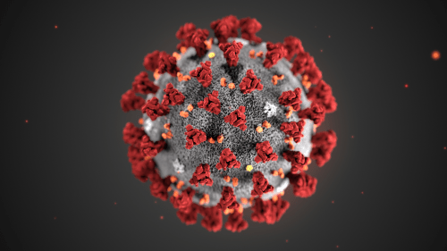
This illustration, created at the Centers for Disease Control and Prevention (CDC), reveals ultrastructural morphology exhibited by coronaviruses. Note the spikes that adorn the outer surface of the virus, which impart the look of a corona surrounding the virion, when viewed electron microscopically. A novel coronavirus virus was identified as the cause of an outbreak of respiratory illness first detected in Wuhan, China in 2019.
Image of newly mapped coronavirus protein, called Nsp15, which helps the virus replicate.
How UC is responding to the coronavirus (COVID-19)
The University of California is vigilantly monitoring and responding to new information about the coronavirus (COVID-19) outbreak, which has been declared a global health emergency.
The 3-D structure of a potential drug target in a newly mapped protein of COVID-19, or coronavirus, has been solved by a team of researchers from the University of California, Riverside, the University of Chicago, the U.S. Department of Energy’s Argonne National Laboratory, and Northwestern University.
The scientists said their findings suggest drugs previously developed to treat the earlier SARS outbreak could now be developed as effective drugs against COVID-19.
The initial genome analysis and design of constructs for protein synthesis were performed by the bioinformatic group of Adam Godzik, a professor of biomedical sciences at the UC Riverside School of Medicine.
The protein Nsp15 from Severe Acute Respiratory Syndrome Coronavirus 2, or SARS-CoV-2, is 89% identical to the protein from the earlier outbreak of SARS-CoV. SARS-CoV-2 is responsible for the current outbreak of COVID-19. Studies published in 2010 on SARS-CoV revealed inhibition of Nsp15 can slow viral replication.This suggests drugs designed to target Nsp15 could be developed as effective drugs against COVID-19.
Adam Godzik, UC Riverside professor of biomedical sciences Credit: Sanford Burnham Prebys Medical Discovery Institute
“While the SARS-CoV-19 virus is very similar to the SARS virus that caused epidemics in 2003, new structures shed light on the small, but potentially important differences between the two viruses that contribute to the different patterns in the spread and severity of the diseases they cause,” Godzik said.
The structure of Nsp15, which will be released to the scientific community on March 4, was solved by the group of Andrzej Joachimiak, a distinguished fellow at the Argonne National Laboratory, University of Chicago Professor, and Director of the Structural Biology Center at Argonne’s Advanced Photon Source, a Department of Energy Office of Science user facility.
“Nsp15 is conserved among coronaviruses and is essential in their lifecycle and virulence,” Joachimiak said. “Initially, Nsp15 was thought to directly participate in viral replication, but more recently, it was proposed to help the virus replicate possibly by interfering with the host’s immune response.”
Mapping a 3D protein structure of the virus, also called solving the structure, allows scientists to figure out how to interfere in the pathogen’s replication in human cells.
“The Nsp15 protein has been investigated in SARS as a novel target for new drug development, but that never went very far because the SARS epidemic went away, and all new drug development ended,” said Karla Satchell, a professor of microbiology-immunology at Northwestern, who leads the international team of scientists investigating the structure of the SARS CoV-2 virus to understand how to stop it from replicating. “Some inhibitors were identified but never developed into drugs. The inhibitors that were developed for SARS now could be tested against this protein.”
Rapid upsurge and proliferation of SARS-CoV-2 raised questions about how this virus could become so much more transmissible as compared to the SARS and MERS coronaviruses. The scientists are mapping the proteins to address this issue.
Over the past two months, COVID-19 infected more than 80,000 people and caused at least 2,700 deaths. Although currently mainly concentrated in China, the virus is spreading worldwide and has been found in 46 countries. Millions of people are being quarantined, and the epidemic has impacted the world economy. There is no existing drug for this disease, but various treatment options, such as utilizing medicines effective in other viral ailments, are being attempted.
Godzik, Satchell, and Joachimiak — along with the entire center team — will map the structure of some of the 28 proteins in the virus in order to see where drugs can throw a chemical monkey wrench into its machinery. The proteins are folded globular structures with precisely defined functions and their “active sites” can be targeted with chemical compounds.
The first step is to clone and express the genes of the virus proteins and grow them as protein crystals in miniature ice cube-like trays. The consortium includes nine labs across eight institutions that will participate in this effort.
Above is a modified version of the Northwestern University news release written by Marla Paul.
Gene panels for breast cancer recurrence risk have arrived. In fact, they’ve been around since the mid-2000s. And now, like guests at a wedding reception, it’s a matter of figuring out where to seat them.
Like it or not, tests such as Oncotype DX (Genomic Health Inc.), MammaPrint (Agendia), and Mammostrat (Clarient)—to name just a few—are making their presence felt.
Clinicians favor these tests for a simple reason: the results help them decide if patients with breast cancer need chemotherapy. More broadly, the tests reflect a shift in thinking among physicians, one that emphasizes molecular profiling of tumors. They’ve arrived on the scene when physicians are also starting to question the value of lymph node status to help determine treatment.George W. Sledge, MD, finds these changes remarkable. Not all that long ago, he might have pink-slipped a test that would help parse treatment decisions. When the NIH held its consensus development conference on adjuvant therapy with breast cancer in 2000, he recalls, the agreement was, basically, that everyone with a tumor greater than one centimeter ought to be treated with chemotherapy. “There’s no question that resulted in us hugely overtreating patients,” he says. “So I think a test that reduces the quantity of human suffering by half in that group is a useful test,” says Dr. Sledge, professor of medicine and pathology, Indiana University, Indianapolis, and immediate past president of the American Society of Clinical Oncology.
In clinical practice, these tests are functioning like traffic managers. “We now see fewer patients getting chemotherapy who would have gotten it before,” says Thomas Julian, MD, professor of surgery, Drexel University College of Medicine, Philadelphia, and director of breast surgical oncology for the West Penn Allegheny Health Care System, Pittsburgh. “We’re also seeing a few who are getting chemo who might not have gotten it before. So it’s changed in both directions,” says Dr. Julian, who is also senior surgical director for medical affairs for the National Surgical Adjuvant Breast and Bowel Project.
Oncotype DX is a real-time RT-PCR assay measuring RNA expression in 16 cancer-related genes and five reference genes, using paraffin-embedded tissue. Results are given as a recurrence score between zero and 100, which are translated as low risk (a score of 18 or lower), medium risk (19 to 30), or high risk (31 or above). The MammaPrint microarray assay measures expression of 70 genes in fresh tissue; it categorizes patients as either high risk, with a so-called poor signature, or low risk (a so-called good signature) for recurrence. There is no intermediate category. Mammostrat is an immunohistochemistry test measuring five markers: p53, HTF9C, CEACAM5, NDRG1, and SLC7A5. The results are combined into a quantitative risk index: low, moderate, and high. For now, only MammaPrint has FDA clearance.
The test is not useful in patients whose tumors are HER2 positive. The test nearly always will show such patients to be at high risk; moreover, the paradigm for treating such patients is with chemotherapy and trastuzumab (Herceptin).It is used for patients who are lymph node negative, ER positive, and HER2 negative, with “moderate-size tumors—say, tumors that are over a centimeter but less than four or five centimeters. Another consideration is tumor size. The test is most useful for tumors of around five millimeters or greater in size.For patients with very, very small tumors—one, two, three millimeters—there’s no need for the test. Elizabeth Hammond, MD, agrees these tests are useful, although she suspects they may best prove their mettle in second- or third-generation assays. It’s simple biology: phenotypic expression of a genetic alteration of ER or HER2 status is the result of cell-signaling pathway changes. “Looking at multiple expressions of that problem with a gene panel, either by RT-PCR or some other method, will in the long run give us better information.
Comment: In 1982, labs were running RIA assays for Estrogen Receptor. It was known for some time that breast cancer is estrogen-dependent. This was a major discovery by a surgeon at University of Chicago, that led to oophorectomy with resection of the lesion. The assay was quite elaborate and required a “scatchard plot”. The assay was no longer used when a good histochemical stain became widely used with a progesterone receptor a few years later. We went into the 1990’s knowing that if the patient is pre-menopausal, positive ER+/PR+ is likely, and the cancer is aggressive. If the patient was postmenopausal, the test is more likely ER/PR negative. This gives us a perspective on how far we have come.
Image via CrunchBase
English: Validation chart for Agendia’s MammaPrint Assay, part of the Symphony Breast Cancer Suite (Photo credit: Wikipedia)
Ovarian and breast cancer patients in a pedigree chart of a family (Photo credit: Wikipedia)