Feeds:
Posts
Comments

Posts Tagged ‘regenerative medicine’

Transplantation of modified human adipose derived stromal cells expressing VEGF165

Author: Larry H. Bernstein, MD, FCAP and Curator: Aviva Lev-Ari, PhD, RN

http://pharmaceuticalintelligence.com/2013-11-03/larryhbern/Transplantation of modified human adipose derived stromal cells expressing VEGF165 

This contribution to the series on stem cells and regenerative medicine deals with transplantation of modified human adipose tissue to repair  ischemic damaged skeletal muscle by apparently increase neovascularization, essentrial for laying down the circulation that feeds the tissue.

Transplantation of modified human adipose derived stromal cells expressing VEGF165 results in more efficient angiogenic response in ischemic skeletal muscle

Evgeny K Shevchenko1*Pavel I Makarevich12Zoya I Tsokolaeva1,Maria A Boldyreva1Veronika Yu Sysoeva2Vsevolod A Tkachuk23 andYelena V Parfyonova12
1Laboratory of angiogenesis, Russian Cardiology Research and Production Complex;  2Lomonosov Moscow State University; 3Laboratory of molecular endocrinology, Russian Cardiology Research and Production Complex, Moscow,  Russia.
Journal of Translational Medicine 2013, 11:138.   http://www.translational-medicine.com/content/11/1/138   http://dx.doi.org/10.1186/1479-5876-11-138
This is an Open Access article distributed under the terms of the Creative Commons Attribution License   http://creativecommons.org/licenses/by/2.0

Abstract

Background

Modified cell-based angiogenic therapy has become a promising novel strategy for ischemic heart and limb diseases. Most studies focused on myoblast, endothelial cell progenitors or bone marrow mesenchymal stromal cells transplantation. Yet adipose-derived stromal cells (in contrast to bone marrow) are abundantly available and can be easily harvested during surgery or liposuction. Due to high paracrine activity and availability ADSCs appear to be a preferable cell type for cardiovascular therapy. Still neither genetic modification of human ADSC nor in vivo therapeutic potential of modified ADSC have been thoroughly studied. Presented work is sought to evaluate angiogenic efficacy of modified ADSCs transplantation to ischemic tissue.

Materials and methods

Human ADSCs were transduced using recombinant adeno-associated virus (rAAV) serotype 2 encoding human VEGF165. The influence of genetic modification on functional properties of ADSCs and their angiogenic potential in animal models were studied.

Results

We obtained AAV-modified ADSC with substantially increased secretion of VEGF (VEGF-ADSCs). Transduced ADSCs retained their adipogenic and osteogenic differentiation capacities and adhesion properties.

  • The level of angiopoetin-1 mRNA was significantly increased in VEGF-ADSC compared to unmodified cells yet
    • expression of FGF-2, HGF and urokinase did not change.

Using matrigel implant model in mice it was shown that

  • VEGF-ADSC substantially stimulated implant vascularization with paralleling increase of capillaries and arterioles.

In murine hind limb ischemia test we found

  • significant reperfusion and revascularization after intramuscular transplantation of VEGF-ADSC compared to controls with no evidence of angioma formation.

Conclusions

Transplantation of AAV-VEGF- gene modified hADSC resulted in stronger therapeutic effects in the ischemic skeletal muscle and may be a promising clinical treatment for therapeutic angiogenesis.

Keywords:

Therapeutic angiogenesis; Cell therapy; Gene modified cells; Adipose stromal cells; Vascular endothelial growth factor; Adeno-associated virus; Ischemia

Background

Despite advances in revascularization techniques, the treatment of ischemic heart and limb diseases remains a worldwide problem. Therapeutic angiogenesis represents alternative new strategy for ischemia resolution that utilizes regenerative capacity of human body and

  • stimulates natural process of
  1. vessel growth,
  2. remodeling and
  3. tissue revascularization [1].

Commonly adopted approaches for therapeutic angiogenesis include

  • direct introduction of recombinant growth factors and gene therapy.

Yet clinical trials have shown several drawbacks of these modalities. Thus low efficacy of recombinant protein administration is explained by

  • dissemination after injection and
  • rapid degradation of therapeutic agent, which
  • requires multiple and long-term infusions thus
    • leading to tremendous expenses [2,3].

Delivery of cDNA coding angiogenic factors via different expression mammalian vectors (plasmids, recombinant viruses) was found more feasible and allowed to achieve great improvement in some cases yet

  • efficacy was still not high enough especially in double blind placebo controlled trials [4].

Many authors discussed possible reasons of gene therapy low efficacy and most of them are univocal to emphasize transfection efficacy and transient transgene expression after plasmid delivery. This can be circumvented by administration of viral vectors but their use is limited due to possible danger of insertional mutagenesis and immune reactions [5,6].

Recently, autologous transplantation of bone marrow stromal cells or endothelial progenitor cells has been shown to enhance angiogenesis and peripheral blood flow [79]. However,

  • the regenerative capacity of these cells decreases with age and
  • in patients with co-morbidities such as diabetes mellitus which reduces efficacy of autologous cell administration, and
  • limited cell viability after transplantation into ischemic tissues also restricts their angiogenic potential [1012].

It was shown in several experimental studies that this problem could be circumvented by gene modified cell therapy strategy utilizing stem or progenitor cells overexpressing angiogenic proteins [13,14]. To develop a feasible and potent gene modified cell therapy for ischemic diseases

  1. the cells should be both effective and accessible in large numbers as well as
  2. the chosen viral vector should be both safe and effective in terms of gene delivery.

The majority of experimental studies have evaluated gene modified bone marrow stromal cells or endothelial progenitor cells for ischemia treatment [1517]. However, cells extracted from bone marrow or peripheral blood after mobilization are available in limited numbers and as for bone marrow cells painful aspiration procedure is required.

In contrast to bone marrow or myoblasts, stromal fraction of adipose tissue contains an abundant population of multipotent stem cells that can be easily harvested in high numbers by minimally invasive surgical techniques [1821]. These adipose –derived stromal cells (ADSCs)

  • share common properties with bone marrow stromal cells and represent a very convenient object for therapeutic use.

However the best development of ADSC for angiogenic therapy still needs to be determined.

As for genetic modification of cells the choice of safe and effective gene transfer vector as well as the appropriate transgene determines the quality and safety of the cell product affecting the efficacy of modified cell based therapy. Recombinant adeno-associated viruses (rAAV) are one of the most promising and versatile tools in this field due to

  1. low immunogenicity and
  2. high transduction potency in vitro

in many types of both – dividing and non-dividing mammalian cells. Besides that until now no human disease caused by AAV has been identified [22].

In this study we genetically modified human ADSCs with a key regulator of angiogenesis – VEGF165 [23] via rAAV-transduction and then evaluated effects of rAAV-transduction and VEGF165 overexpression on human ADSC

  1. growth,
  2. differentiation capacity,
  3. adhesion and
  4. angiogenic factor expression as well as
  5. revascularization and
  6. functional improvement

after intramuscular injection in a mice hind limb model.

Methods

Cell culture

(refer to doi:10.1186/1479-5876-11-138)

DNA constructs production of rAAV particles and cell transduction

(refer to doi:10.1186/1479-5876-11-138)

Western blotting and ELISA

(refer to doi:10.1186/1479-5876-11-138)

ADSC proliferation activity assay

To assess population doubling time (PDT) of gene modified (transduced with rAAV at passage 1) or untreated ADSC (passage 2) seeded on 6-well plates (2 × 104 cells/well). After a 9 day incubation average cell numbers for three wells were obtained using a hemocytometer chamber. PDT was calculated as follows:

 PDT=(log2)*t/(log(Nt/N0))

 where t is period of incubation (hours), Nt – endpoint amount of cells, N0 – initial number of cells.

ADSC cell cycle stage analysis by flow cytometry

(refer to doi:10.1186/1479-5876-11-138)

ModFit LT 3.2 software (Verity Software House, USA) was used for analysis of cell distribution over cell cycle stages according to intensity of propidium iodide fluorescence in a wavelength range of 600–625 nm (excitation wavelength – 488 nm). Results are presented as a percentage of cells in S + G2/M stages.

Apoptosis assay

Analysis of spontaneous apoptosis frequency in ADSC culture was performed using Annexin-V FITC Apoptosis Kit (Invitrogen, USA) according to manufacturer’s protocol.

Adipogenic, osteogenic and endothelial differentiation of ADSC

To confirm adipogenesis intracellular lipid droplets were detected using Oil red O staining reagent (Millipore, USA) 2 weeks after induction. To confirm osteogenesis Alizarine Red C staining was used to detect extracellular matrix mineralization 2 and 3 weeks post induction. Endothelial cells were stained for CD31 and VEGFR2 surface antigens and cell counts were obtained using flow cytometry.

Cell attachment assay

(refer to doi:10.1186/1479-5876-11-138)

Flow cytometry

Antigen expression analysis was performed on cell sorter MoFlo (DakoCytomation, Denmark) or flow cytometry scanner BD FACS CantoTM II (BD Pharmingen, USA). 10 000 events were acquired and analyzed for antigen expression.

Quantitative polymerase chain reaction

Quantitative polymerase chain reaction (qPCR) was performed using primers specific for human VEGF165, ANGPT1, HGF, FGF2 and PLAU mRNAs.

Animals

8–10 week-old male BALB/c NUDE mice

Matrigel plug assay

(Refer to doi:10.1186/1479-5876-11-138)

Hind limb ischemia model

Ten week-old male BALB/с NUDE mice were anaesthetized by intraperitoneal injection of 0.3 ml of 2.5% avertin. Femoral artery was separated in its distal part and ligated proximal to its popliteal bifurcation (keeping v. femoralis and n. ischiadicus intact). ADSC, GFP-ADSC or VEGF-ADSC (5×10⁵ cells per animal) were resuspended in 150 μl of PBS, and injected in 3 equally divided doses tom. tibialis anterior, m. gastrocnemius and m. biceps femoris to generate three experimental animal groups: “GFP-ADSC”, “VEGF-ADSC”, “ADSC” (14 animals per group). PBS (150 μl) was injected in negative control “PBS” group. Blood flow was subsequently measured by laser Doppler imaging.

Laser doppler imaging

(Refer to doi:10.1186/1479-5876-11-138)

Muscle explants

M. tibialis anterior explant culture was prepared on matrigel according to Jang et al. [26] protocol and cultured in M199 medium (Gibco, USA), containing 2% FBS. At day 3 and 7 medium was collected for determination of human VEGF165 concentration by ELISA.

Specimen preparation and histological analysis

At designated period (day 20 for muscles, day 14 for matrigel plugs) animals were sacrificed by lethal isoflurane dose followed by cervical dislocation. Afterwards m. tibialis anterior or matrigel implants respectively were harvested,

For muscle necrosis analysis we used routine hematoxylin-eosin staining of formalin-fixed muscle sections. Necrotic tissue was defined by loss of fiber morphology, cytoplasm disruption, inflammatory cells infiltration and fibrosis.

Statistical analysis

Results were analyzed in Statsoft Statistica 6.0 (Statsoft, USA).

Results

Effective transduction of human ADSC by adeno-associated virus serotype 2

Low-passage human ADSC obtained from different donors were transduced using rAAV encoding GFP to assess gene delivery efficacy. Transduced to total cells ratio was counted by flow cytometry. GFP-positive ADSC (GFP-ADSC) were detected as early as day 2 after viral infection. Maximum number of positive cells (65.6±3%) and highest GFP-fluorescence intensity was reached by day 4–5 (Figure 1). GFP signal was detectable for at least 30 days. At day 15 and 30 flow cytometry showed that 45±2% and 25±1.5% of ADSC were GFP-positive respectively.

Figure 1. Human ADSC transduction by recombinant adeno-associated virus

Figure 1. Human ADSC transduction by recombinant adeno-associated virus

A. GFP-positive cell count by FACS in GFP-ADSC culture at day 4 after transduction by rAAV. B.Representative image of GFP-positive human ADSC (green) transduced by rAAV, 100 × magnification.

Increase of VEGF expression and secretion after rAAV transduction of human ADSC

To obtain gene modified ADSC we constructed rAAV vector encoding human VEGF165. In ADSC transduced by rAAV-VEGF (VEGF-ADSC) VEGF165 mRNA level increased 80±15-fold compared to basal expression in unmodified ADSC or GFP-ADSC (Figure 2A). Protein production was analyzed by Western blotting and ELISA. Data presented at Figure 2B, C shows that in VEGF-ADSC secretion of VEGF increased 45-50-fold (4.5±1.8 ng/ml/105 cells) compared to unmodified cells (0.1±0.02 ng/ml/105 cells) or GFP-ADSC (0.09±0.02 ng/ml/105cells). VEGF concentration in conditioned medium decreased over time during VEGF-ADSC cultivation but remained 30-fold higher (2.9±1.1 ng/ml/105 cells) than in controls (0.09 ± 0.02 ng/ml/105 cells) at day 30 post transduction. Material from a total of 10 donors was used to obtain mean values of VEGF expression increase.

 Figure 2. Validation of VEGF165 expression in AAV-modified VEGF-ADSC.
Figure 2. Validation of VEGF165 expression in AAV-modified VEGF-ADSC.

A. VEGFA expression level in human ADSC 10 days after AAV transduction determined by quantitative PCR. B, C. Analysis of VEGF secretion by GFP-ADSC, VEGF-ADSC and unmodified cells using ELISA (B) and immunoblotting (C). In immunosorbent assay protein content was determined in conditioned media samples obtained at days 7 and 30 post genetic modification of ADSC.

rAAV-mediated modification of human ADSC suppresses their proliferation activity yet does not influence apoptosis

We found that proliferation rate of VEGF-ADSC and GFP-ADSC was reduced compared to unmodified cells (Figure 3A). ADSC population doubling time was 61.3±7 h, while for GFP-ADSC and VEGF-ADSC it was 116.9±11 and 145.4±12 h respectively (n=5, p<0.01 vs unmodified cells). At the same time spontaneous apoptosis rate in all three cell cultures was comparable and comprised about 2±0.5% of total cell population.

Figure 3. . Proliferation of gene modified ADSC.

Figure 3. Proliferation of gene modified ADSC.
Population doubling time in GFP-ADSC, VEGF-ADSC and ADSC cultures. Data of five serial runs. B. Cells distribution in S-G2 cell cycle stages according to cytometry analysis of GFP-ADSC, VEGF-ADSC and ADSC. Data of three serial runs.

Analysis of cell cycle stages distribution in ADSC, GFP-ADSC and VEGF-ADSC cultures (Figure 3B) showed that number of cells in S-G2 stages was more than 1.5-fold lower in modified cells: GFP-ADSC (16±4% cells) and VEGF-ADSC (13±6% cells) compared to unmodified ADSC (25±3% cells; n=3; p<0.05 vs unmodified cells).

ADSC adhesion does not change after genetic modification

Interactions with extracellular matrix proteins play important role in incorporation and integration to recipient’s tissue, cell viability and their functional properties upon transplantation. ADSC did adhere on main extracellular protein collagen type 1 as well as vitronectin and fibronectin while almost none of cells attached to laminin-coated plastic. We did not observe statistically significant differences in adhesion properties between ADSC, GFP-ADSC and VEGF-ADSC cultures (Figure 4).

Figure 4. Data from comparative study of ADSC, GFP-ADSC and VEGF-ADSC adhesion on culture plates

 Figure 4. Data from comparative study of ADSC, GFP-ADSC and VEGF-ADSC adhesion on culture plates coated by collagen 1, vitronectin, fibronectin or laminin (n=4).

Modified ADSC retain their adipogenic, osteogenic and endothelial differentiation potential in vitro

To analyze potential influence of viral transduction and transgene overexpression on differentiation capacity of gene modified cells we performed experiments on adipogenic and osteogenic differentiation of ADSC.

Microscopic analysis of gene modified and untreated ADSC stained with Oil Red O reagent after 14 days of incubation in adipogenic media showed >30% of differentiated (visualized by intracellular lipid droplets accumulation) cells (Figure 5). Oil Red O+ cell count did not reveal statistically significant differences in both GFP-ADSC (33.7±8.1%) and VEGF-ADSC (34.1±11.5%) as well as unmodified ADSC (34.3±11.7%). Similar results were obtained in osteogenic differentiation assay of ADSC. It was confirmed by Alizarin Red C staining that detects extracellular matrix mineralization. At 14 days of incubation in osteogenic media we detected dye-positive cells in ADSC, GFP-ADSC, VEGF-ADSC culture. At day 21 it was followed by dramatic increase of extracellular matrix calcification in both – modified and untreated cells without significant differences (Figure 5).

Figure 5. Adipogenic and osteogenic differentiation of gene modified ADSC

Figure 5. Adipogenic and osteogenic differentiation of gene modified ADSC

Representative images of ADSC and VEGF-ADSC cultures stained by Oil Red O (lipid droplets detection, kjadipogenic differentiation, 100 × magnification) and Alizarine Red C (matrix mineralization, osteogenic differentiation, 100 × magnification for “day 14” and 50 × magnification for “day 21”) reagents after incubation in specific differentiation medium, n=3.

Taking into account mitogenic activity of VEGF we analyzed possible effect of genetic modification and VEGF overexpression on endothelial cell fraction in VEGF-ADSC. Using flow cytometry we determined amount of cells that carry CD31 and VEGFR2 endothelial markers in ADSC, GFP-ADSC and VEGF-ADSC (rAAV-modified at passage 1) cultures at passage 2. Less than 1.5% of CD31, VEGFR2-positive cells were detected in all three populations. Subsequently modified and untreated ADSC at passage 2 that reached >90% confluency were subject to incubation in EGM-2 medium to stimulate endothelial differentiation. After 14 days of cultivation in EGM-2 repeated analysis of CD31 and VEGFR2 expression showed that percentage of endothelial marker-positive cells did not change and remained about 1% in all assayed cultures.

Level of angiopoietin-1 mRNA increases in VEGF-ADSC

Using qPCR we studied potential impact of genetic modification and augmented VEGF secretion on expression activity of hepatocyte growth factor (HGF), fibroblast growth factor-2 (FGF2), angiopoietin-1 (ANGPT-1) and urokinase (PLAU) genes in VEGF-ADSC. As shown in Figure 6 we did not find any changes in FGF2 and HGF expression in GFP-ADSC and VEGF-ADSC compared to ADSC. We found a 3-fold increase in urokinase expression in VEGF-ADSC yet it was not statistically significant. At the same time increase of ANGPT-1 expression in VEGF-ADSC was significant and 5.3±0.6-fold higher than in unmodified cells or GFP-ADSC (n=6, p<0.05).

Figure 6. Comparison of ANGPT1, FGF2, PLAU and HGF genes expression

Figure 6. Comparison of ANGPT1, FGF2, PLAU and HGF genes expression by quantitative PCR in GFP-ADSC, VEGF-ADSC and unmodified ADSC. Charts represent relative expression for assayed genes from a total of 6 runs.

Analysis of VEGF and PDGF receptors expression on ADSC surface

Analysis of VEGF receptors expression on human ADSC was carried out to assess possible autocrine action of VEGF on VEGF-ADSC functional properties. Flow cytometry of ADSC and VEGF-ADSC (at passage 1–2) from different donors stained for VEGF receptor 1 and 2 showed <1% of positive cells (Figure 7). Taking into account observation of Ball et al. which indicated platelet-derived growth factor receptors (PDGFRα and PDGFRβ) as facultative receptors for VEGF165 [27] we analyzed the presence of cells which expressed PDGFRβ in human ADSC culture. Using specific monoclonal antibodies and subsequent flow cytometry we found that >90% of human ADSC were positive for PDGFRβ (Figure 7).

Figure 7. Analysis of VEGF and PDGF receptors expression on ADSC surface.

Figure 7. Analysis of VEGF and PDGF receptors expression on ADSC surface. VEGFR1, VEGFR2 or PDGFRβ-positive cell count by flow cytometry in ADSC culture.

Increased vascularisation of matrigel implants after VEGF-ADSC transplantation

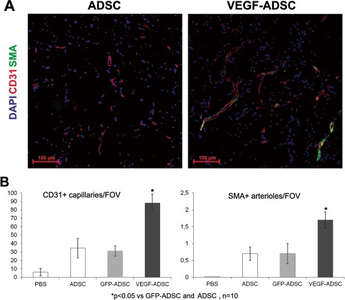
We used matrigel plug assay to determine angiogenic properties of gene modified ADSC in vivo. At day 14 matrigel implants were harvested and subject to histological analysis (Figure 8). In negative control group we found only small sporadic capillaries (<1 capillary per FOV) were detected while in “ADSC”, “GFP-ADSC” and “VEGF-ADSC” groups formation of vessel network was more evident. Vessel counts revealed a 2.7-fold increase of CD31-positive vessels in group “VEGF-ADSC” (88.1±10.4 vessels per FOV) compared to “GFP-ADSC” (31.3±6.2 vessels per FOV) and “ADSC” (34.5±11.6 per FOV). Number of smooth muscle actin (SMA)-positive vessels was also 2.5-fold higher in “VEGF-ADSC” (1.7±0.24 vessels per FOV) than in “GFP-ADSC” (0.7±0.3 vessels per FOV) and “ADSC” (0.7±0.2 vessels per FOV). Thus capillaries/SMA+vessels ratio did not vary among experimental groups.

Figure 8. Effect of VEGF-ADSC or ADSC on vascularization of matrigel implants in nude mice.

Figure 8. Effect of VEGF-ADSC or ADSC on vascularization of matrigel implants in nude mice.
A.Representative images of matrigel sections from “VEGF-ADSC” and “ADSC” groups stained by antibodies against murine CD31 and SMA, 100× magnification. B. Capillaries and arterioles count in matrigel implants.

Blood flow recovery after VEGF-ADSC transplantation into ischemic murine limb

Perfusion assessment in hind limb ischemia model showed maximum blood flow recovery in “VEGF-ADSC” group (Figure 9). By day 20 spontaneous reperfusion of ischemic limb in «PBS» group was feeble and did not exceed 30%. In contrast we observed evident augmentation of blood supply in three experimental groups that received cell injections. At the end of experiment perfusion in “ADSC” and “GFP-ADSC” groups reached 50% and 55% respectively. Blood flow recovery after VEGF-ADSC transplantation was much more effective. At day 12 perfusion in group “VEGF-ADSC” significantly exceeded values in “ADSC” and “GFP-ADSC” by 15-20% and towards the end of experiment (day 20) it reached 80-90%. Thus transplantation of ADSC overexpressing VEGF was more effective than of untreated or GFP-ADSC.

Figure 9. Reperfusion of murine ischemic limb after ADSC administration.

Figure 9. Reperfusion of murine ischemic limb after ADSC administration.
A. Representative laser-doppler scans of subcutaneous blood flow in mice from “ADSC” and “VEGF-ADSC” groups obtained at days 4 and 20 after ischemia induction and cell transplantation. B. Dynamics of blood flow recovery in ischemic limbs within 20 days after intramuscular injection of ADSC, GFP-ADSC, VEGF-ADSC or PBS.

Transplantation of VEGF-ADSC reduces necrosis and stimulates stable vessel formation in ischemic muscle

Histological analysis of hematoxylin-eosin stained m. tibialis anterior specimens obtained at day 20 after and cell transplantation showed significant decrease in necrotic tissue span in «VEGF-ADSC» group (31.3±7%) compared to «ADSC» and «GFP-ADSC» groups (54.3±8.4% and 55.63±6.8%). Animals that received PBS injection as a negative control were characterized by the highest muscle necrosis span that reached 84±6.7% (Figure 10).

Figure 10. Morphometric analysis of tissue necrosis in ischemic muscle from study group animals.

Figure 10. Morphometric analysis of tissue necrosis in ischemic muscle from study group animals.
A. Images of hematoxylin-eosin stained m. tibialis anterior sections. Necrotic tissue is marked by black line. (N* – necrotic tissue, B* – border zone, H* – healthy or regenerating tissue). B. Representative images of muscle tissue from different zones of section. Labels: star – vasa in normal muscle tissue with; black dot – inflammatory demarcation zone between anucleic disrupted tissue and regenerating muscle fibers; triangle – regenerating round-shaped muscle fibers with multiple centrally located nuclei. C. Statistical data of necrotic tissue area in “PBS”, “ADSC”, “GFP-ADSC” and “VEGF-ADSC” groups. Measurements made in 4–5 animals per group.

To assess vascular density muscle tissue sections were stained by specific antibodies against mouse CD31 and SMA (Figure 11). Vessel count showed that in “ADSC” and “GFP-ADSC” groups capillary and arteriolar densities were similar reaching 129±11 and 125±14 capillaries/FOV, 1.35±0.12 and 1.37±0.09 arterioles/FOV respectively. In specimens from animals that received VEGF-ADSC capillary density was 189±19 per FOV (p<0.05) with arteriolar density of 3.1±0.2 per FOV (p<0.01). Furthermore, we found that arterioles/CD31+ vessels ratio was similar in all experimental groups and slightly higher in group “VEGF-ADSC” (1% vs 1.6%). In addition morphometric analysis of muscle tissue from group “VEGF-ADSC” did not reveal angioma or abnormal vessel formation.

Figure 11. Vascularization of murine ischemic muscles after ADSC administration.

Figure 11. Vascularization of murine ischemic muscles after ADSC administration.
A. Representative images ofm. tibialis anterior sections from “VEGF-ADSC” and “ADSC” groups stained by antibodies against murine CD31 and SMA, 100× magnification. B. Capillaries and arterioles count in m. tibialis anterior sections. Counts made in 5–6 animals per group.

ADSC retain viability and transgene expression after transplantation into ischemic muscle

To evaluate viability of transplanted ADSC after injection into ischemic tissue m. tibialis anterior specimens from “GFP-ADSC” group were harvested at day 7 after induction of ischemia and cell transplantation. Frozen muscle sections were analyzed using fluorescence microscopy that allowed to detect GFP-positive cells distributed throughout muscle (Figure 12A).

1479-5876-11-138-12  Figure 12. Human ADSC viability and VEGF expression

Figure 12. Human ADSC viability and VEGF expression after transplantation to ischemic murine muscle. 

A. Representative image of m. tibialis anterior section from “GFP-ADSC” group obtained at day 7 after ischemia induction and GFP-ADSC injection, 50× magnification. GFP-positive cells are distributed in tissue around injection site.B. Analysis of VEGF165 content by ELISA in explants culture medium from “ADSC”, “GFP-ADSC”, “VEGF-ADSC” groups obtained at days 3 and 20 after cell trasplantation.

Data from experimental studies indicates that prolonged expression of therapeutic transgene is essential for effective stimulation of angiogenesis and ischemic tissue recovery. Muscle explant model was carried out to confirm the presence of viable and functionally active human ADSC overexpressing VEGF in ischemic muscle at hind limb ischemia experiment endpoint. M. tibialis anteriorwere harvested from “ADSC”, “GFP-ADSC” and “VEGF-ADSC” group animals at day 3 and 20 after cell transplantation and cultured as explant in matrigel. In culture medium samples collected after 3 days of “VEGF-ADSC” explant incubation (obtained at day 3 after cell transplantation) human VEGF165 concentration determined by ELISA reached 2.86±0.21 ng/ml (Figure 12B). Protein concentration was expectedly lower (0.145±0.015 ng/ml) in conditioned medium from muscle explants harvested at day 20. In addition comparison of VEGF concentration in culture medium samples collected at day 3 and 7 post incubation of explant culture revealed accumulation of VEGF. It indirectly confirms presence of functionally active human VEGF-ADSC in ischemic muscle up to 20 days post transplantation. In contrast to “VEGF-ASDC” human VEGF165 concentration in explant cultures from “GFP-ADSC” and “ADSC” groups was below limit of detection.

 Discussion

Gene modified cell-based therapy for ischemic disorders: myocardium infarction and limb ischemia is a rapidly evolving trend in experimental and regenerative medicine. Promoting angiogenesis in ischemic tissues via paracrine action of transplanted modified cells is an emerging alternative modality for patients who are unsuitable for surgical and interventional revascularization. Still choice of

  1. appropriate cell type,
  2. angiogenic factor and
  3. gene delivery tool

are crucial issues for efficacy and safety of the method.

Regarding type of cells there are certain issues concerning their derivation and preparation prior to grafting. Thus, embryonic stem cells application is doubtful due to

  1. ethical reasons,
  2. potential risks of teratogenesis and
  3. immune response to their differentiated progenies [28].

Use of endothelial progenitor cells from peripheral blood and bone marrow are limited by

  • expensive procedures of isolation and difficulties in obtaining sufficient amount of cells.

Regarding the latter point it is known that prolonged incubation of cells in vitro prior to transplantation is associated with

  1. potential risks of malignancy,
  2. proliferation decrease and
  3. commitment to terminal differentiation.

Use of skeletal myoblasts or bone marrow derived mesenchymal stromal cells (BMMSC) is associated with painful isolation procedure of muscle biopsy and suprailiac puncture respectively.

ADSC used in our study share a lot of similar properties and characteristics with BMMSC, while they are easier to obtain in sufficient quantity using minimally invasive liposuction procedure. Various data suggests that up to 1.5 × 10adipose stromal cells can be isolated from 1 ml of adipose tissue [29,30]. This allows to reduce the time of cell propagation in vitro prior to transplantation. As for therapeutic angiogenesis,

human ADSC produce a wide spectrum of biologically active molecules – angiogenic growth factors, cytokines, proteases etc. [31,32].

Multiple experimental studies accumulate data on relatively high therapeutic potential of ADSC for tissue regeneration and stimulation of angiogenesis [21,33,34]. However well-known reduction of cell regenerative potential with age and among patients with severe co-morbidities is also relevant for ADSC. Donor age-associated decrease of proliferation activity and differentiation capabilities was shown for human ADSC [35,36]. Angiogenic potential of ADSC also decreases with ageing and is characterized by reduced secretion of

  • VEGF,
  • HGF,
  • angiopoietin-1 and other angiogenic factors [37].

Thus, attempts to improve regenerative potential of ADSC are reasonable.

We have shown high efficacy of rAAV-mediated genetic modification of human ADSC. Using rAAV encoding VEGF165 we obtained human ADSC with increased level of VEGF165 secretion which retained for at least 30 days. VEGF-A and particularly its most abundant 165-amino acid isoform triggers multiple reactions promoting new vessel formation and growth [23] that supported our choice of therapeutic gene in presented study. Observed gradual decrease of transgene expression can be attributed to proliferation activity of ADSC together with known episomal subsistence of rAAV [38]. Moreover cellular mechanism of addressed

  • methylation can be activated after transduction leading to
  • suppression of cytomegalovirus promoter which triggers
  • transgene expression in our vector [39].

Potential influence of genetic modification and transgene expression on cell behavior and functional activity is frequently kept out of consideration while this issue is of great importance, especially for potential clinical application. We examined possible effects of rAAV-transduction and VEGF overexpression on functional properties of ADSC which included

  • proliferation,
  • spontaneous apoptosis,
  • adhesion and
  • differentiation capability.

We observed a decline in ADSC proliferation after modification by rAAV that was evident by

  • increase of population doubling time as well as
  • decrease in number of cells in S–G2 stages of cell cycle.

At the same time spontaneous apoptosis rate did not exceed 2% in modified and unmodified cells. These results contribute to previously published data that showed transient cell cycle arrest after AAV transduction of embryonic fibroblasts and BMMSC [40]. This effect was observed whenever

  • wild-type,
  • recombinant or
  • genome-empty AAV particles were used.

It was suggested that changes in expression profile and decreased proliferation were related to initial stage of virus entry and caused by capsid proteins interaction with cellular signaling pathways [40]. Growth inhibitory effect was transient and

  • proliferation restored to normal level over time of cell passaging [41].

It appears that proliferation decline of rAAV-modified ADSC occurs by a common mechanism.

ADSC are known to be able to differentiate into

  • adipocytes, chondrocytes, osteoblasts, myocytes, neural cells, cardiomyocytes, endothelial and liver cells
  • when cultured in special induction medium [42,43].

Analyzing data from our differentiation experiments we concluded that rAAV-mediated genetic modification of human ADSC and VEGF overexpression did not alter their adipogenic and osteogenic differentiation properties.

There are several observations indicating ability of ADSC for endothelial differentiation [44,45] as well as evidence for presence of small amount of endothelial cells in ADSC population at early passages [18,19]. In our experiments we did not find an increase in amount of cells positive for endothelial markers CD31 and VEGFR2 in VEGF-ADSC compared to unmodified ADSC population. This suggests that VEGF overexpression

  • neither induces endothelial differentiation of modified ADSC
  • or stimulates proliferation of preexisting endothelial cells in ADSC culture.

Adhesion tests conducted in our study were based on a fact that

  • interaction with extracellular matrix proteins is a key factor
  • that contributes to cell viability and integration into host tissue after transplantation [46].

We found that both modified and untreated ADSC showed very common adhesion on collagen type 1, vitronectin and fibronectin. Thus we can suggest that

  • rAAV-mediated genetic modification did not alter expression of adhesion molecules on cell surface of ADSC.

Our results showing low ADSC adhesion on laminin are not surprising taking into account published observations which indicate diminished or lack of α6, α7 and ß1 integrins expression in ADSC-components of α6/ß1 and α7/ß1 receptors for laminin [47,48].

Since VEGF can regulate multiple signaling pathways [23] we next determined whether expression of HGF, FGF2, urokinase and angiopoietin-1 might be altered in VEGF-ADSC. HGF and FGF2 are mitogens and chemoattractants for both endothelial and mural cells and directly participate in angio- and arteriogenesis [4]. Angiopoietin-1 is characterized as a stabilizing factor that provides formation of functionally mature vessel network [49]. Urokinase plasminogen activator is a key regulator of extracellular proteolysis which is

  • responsible for cleavage activation of growth factors and migration of endothelial cells during vessel growth [50,51].

We found almost 3-fold yet not statistically significant increase of urokinase expression while expression of HGF and FGF2 did not change. Another interesting finding is a 5-fold increase of angiopoietin-1 expression in VEGF–ADSC compared to GFP–ADSC or unmodified cells. We assumed that up-regulation of angiopoietin-1 expression occurs due to autocrine action of VEGF165 produced by VEGF-ADSC. However according to our data supported by other studies [30,52] cultured human ADSC population contains <1% of cells that express receptors to VEGF165 – VEGFR1 and VEGFR2. At the same time we found that

  • >90% of ADSC carry receptor to platelet-derived growth factor – PDGFRβ.
There is a published observation that
  • PDGFRα and PDGFRβ can act as a facultative receptor for VEGF [27].
  • it is also known that PDGFR activation leads to increase of angiopoietin-1 expression [53].

Considering that more than 90% of human ADSC are PDGFRβ-positive

  • we can speculate that increased expression of angiopoetin-1 in VEGF-ADSC could be attributed to PDGFRβ-mediated autocrine action of VEGF.
In our study we evaluated therapeutic potential of gene modified human ADSC in terms of their ability to induce angiogenesis in ischemic muscle tissue. It was found that matrigel implants after transplantation of VEGF-ADSC had higher vascular density than after delivery of untreated cells or ADSC transduced by a reporter gene. Along with

  • capillary formation we also found
  • proportional increase in amount of mature blood vessels characterized by smooth-muscle wall.

This can occur due to the fact that cells transplanted in matrigel produce other angiogenic factors besides VEGF that can promote vessel maturation and stabilization.

Key angiogenic property of cell therapies in experimental study is ability to induce reperfusion of ischemic tissue in appropriate animal models. We used hind limb ischemia model to show that

  • VEGF-ADSC transplantation led to significantly higher perfusion restoration than
  • after untreated of GFP-transduced cell administration.

It was also found that intramuscular injection of VEGF-ADSC had a tissue-protective effect and led to vivid decrement of necrosis span. VEGF is known to be significant antiapoptotic factor that can enhance cell survival. We suggest that

  • increased VEGF content during the first days after onset of acute ischemia and cells administration leads to promotion of cell survival and thus to reduction of necrotic disruption in muscle tissue.

We should also point that during the experiment we did not observe any blood flow decrease after cell administration or rapid “plateau” formation like it was previously described for plasmid-mediated gene delivery due to short-term transgene expression [4]. This can be explained by

  • presence of viable and functionally active ADSC that produced VEGF throughout the experiment.

In our muscle explant experiments we showed that VEGF-ADSC retain functional activity even at long terms after injection (up to 27 days) and produce VEGF in detectable quantities. Thus we can confidently attribute

  • tissue protection and restoration of blood flow in mice that received VEGF-ADSC to increased long-term VEGF production by modified cells.

As for decrease of human VEGF content in murine tissue by day 20 we suggest that cells undergo apoptosis over time. Besides that methylation of CMV promoter which drives VEGF expression in our vector could take place. Taking into account that Nude mice were used we find it hard to assume possible rejection of transplanted cells as far as this animal strain lacks T-cells immunity which plays a crucial role in graft rejection. Still, it seems that produced amount of VEGF is sufficient to trigger angiogenesis and relief tissue ischemia via restoration of blood flow.

Histological analysis of ischemic muscle injected with modified VEGF-ADSC revealed that

  • capillary density was significantly higher than in specimens from animals that received untreated cells or GFP-ADSC.

We noticed that this increase was not only due to higher capillary count, but also to SMA-positive blood vessels of arteriolar type. Furthermore arteriole/capillary ratio was constant throughout experimental groups that indicated formation of a stable mature vascular network. Thus, despite high level of VEGF produced by modified ADSC we did not observe any evidence for abnormal tumour-like vascular structures in muscle as it was previously shown e.g. in studies of adenovirus-mediated delivery of VEGF gene [54]. In contrast to matrigel implants experiment in case of skeletal muscle we do not state that increase of vascular density in experimental groups was only due to de novo formed vessels. Besides promoting endothelial cell proliferation VEGF also prevents endothelial apoptosis leading to survival of preexisting vessels. There was surely a vast amount of persisted capillaries in the muscles due to VEGF anti-apoptotic effect of VEGF.

It is often speculated that low efficacy reported in clinical trials using gene delivery of VEGF alone can be explained by its high mitogenic activity which is not supported by vessel stabilizing stimuli and consequently ends up with dissociation of formed capillaries [55]. This led to a concept of combined gene delivery

  • indicating that combinations of angiogenic and vascular stabilizing factors should be used to treat ischemic tissues [5558].

Cell therapy for ischemic disorders has a valuable advantage since transplanted cells produce a whole “cocktail” of biologically active molecules which render combined effect in impaired tissue. We suggest that stable vessel formation observed in our study is

  • mediated by aforementioned ADSC ability to produce a wide spectrum of angiogenic factors
  • including ones responsible for vessel stabilization and maturation: angiopoietin-1, TGF-β, PDGF,
  • which can act synergistically with increased production of VEGF165 by modified cells.

Besides that, genetic modification can alter cell’s expression profile. Observed increase in expression of angiopoietin-1 in VEGF-ADSC can further contribute to

  • formation of mature vascular network that also
  • supports therapeutic effect of transplanted cells.
Increased concentration of VEGF in ischemic tissue plays a substantial role in vessel stabilization and therapeutic effect if maintained over a significant period of time, which was achieved in our study and exceeded a substantial term of 3 weeks.

Conclusions

Thus we can conclude that human ADSC with their accessibility and angiogenic paracrine activity is an appropriate and preferable type of cells for therapeutic angiogenesis. Obtained results indicate that relatively safe rAAV holds great potential for gene transfer into human ADSC. Taken together, we suggest that

  • the use of AAV-modified ADSC overexpressing VEGF165 is a feasible and effective approach for stimulation of stable vascular network formation in ischemic muscle and

can be implied for therapeutic angiogenesis or tissue-engineered transplants. Further study and improvements in vector design, regulated transgene expression, cell preparation and propagation conditions are still to be completed to allow clinical application of modified cell-based therapeuticals.

Abbreviations

ADSC: Adipose derived stromal cells; BMMSC: Bone marrow derived mesenchymal stem cells; CGS: Cell growth supplement; DMEM: Dulbecco’s modified Eagle’s medium; ELISA: Enzyme-linked immunosorbent assay; FBS: Fetal bovine serum; FGF2: Fibroblast growth factor 2; FOV: Field of view; GFP: Green fluorescent protein; HEK293T: Human embryonic kidney 293 T; HGF: Hepatocyte growth factor; PDGF: Platelet derived growth factor; PDT: Population doubling time; PBS: Phosphate buffer saline; rAAV: Recombinant adeno-associated virus; SMA: Smooth muscle actin; VEGF: Vascular endothelial growth factor

Read Full Post »

Notable Contributions to Regenerative Cardiology by Richard T. Lee – Part I


Author and Curator: Larry H Bernstein, MD, FCAP

and

Article Commissioner: Aviva Lev-Ari, PhD, RD


Introduction

This presentation is a two part discussion of selected articles of a large body of research from Dr. Richard T. Lee, at Harvard Medical School’s Lee Laboratory and Brigham & Womens Hospital.  This work is innovative in the field of stem cell research and myocardial regeneration.  It devolves the complex cellular processes that are involved in the management of a cell transforming from a progenitor to a functional cardiomyocyte.  The cell engineering involves investigating interactions between a cell placed into the layer derived from the interstitial layer between viable cardiomyocytes.  This is only possible from a through actionable knowledge of the mechanism involved in the transformation process, which has occupied the Lee Laboratory for many years.  Part II will cover the cellular mechanisms underlying the conceptual approach to cardiac myocyte regeneration.

The Lee Laboratory uses emerging biotechnologies to discover and design new approaches to cardiovascular diseases. A central theme of the laboratory is that merging bioengineering and molecular biology approaches can yield novel approaches. Thus, the Lee Laboratory works at this interface using a broad variety of techniques in genomics, imaging, nanotechnology, physiology, cell biology, and molecular biology. The approach is to understand problems and design solutions in the laboratory and then demonstrate the effectiveness of these solutions in vivo. Ongoing projects in the laboratory include studies of cardiac regeneration, diabetic vascular disease, wound healing, heart failure, and cardiac hypertrophy.

Richard T. Lee is Professor of Medicine at Harvard Medical School and lecturer in Biological Engineering at the Massachusetts Institute of Technology. He is a 1979 graduate of Harvard College in Biochemical Sciences and received his M.D. from Cornell University Medical College in 1983.  He went on to complete his residency in 1986 and cardiology fellowship in 1989, both at Brigham and Women’s Hospital in Boston, and he obtained post-doctoral training at MIT in Bioengineering.

Dr. Lee is certified by the American Board of Internal Medicine in cardiovascular disease and is a Fellow of the American College of Cardiology. He is Leader of the Cardiovascular Program of the Harvard Stem Cell Institute.  He is a member of the Editorial Boards of the journals Circulation Research, Journal of Clinical Investigation, and Circulation, and has published over 180 peer-reviewed articles based on his research, which combines approaches in biotechnology and molecular biology to discover new avenues to manage and treat heart disease.

Regeneration of the heart

Matthew L. Steinhauser, Richard T. Lee
Division of Cardiovascular Medicine, Department of Medicine, Brigham and Women’s Hospital, Harvard Medical School, and (2)             Harvard Stem Cell Institute, Cambridge, MA
EMBO Mol Med 2011; 3: 701–712   http://dx.doi.org/10.1002/emmm.201100175

The death of cardiac myocytes diminishes the heart’s pump function and is a major cause of heart failure. With the exception of heart transplantation and implantation of mechanical ventricular assist devices, current therapies do not address the central problem of decreased pumping capacity owing to a depleted pool of cardiac myocytes. The field is evolving in two important directions. First, although endogenous mammalian cardiac regeneration clearly seems to decline rapidly after birth, it may still persist in adulthood. The careful elucidation of the cellular and molecular mechanisms of endogenous heart regeneration may therefore provide an opportunity for developing therapeutic interventions that amplify this process. Second, recent breakthroughs have enabled reprogramming of cells that were apparently terminally differentiated, either by dedifferentiation into pluripotent stem cells or by trans-differentiation into cardiac myocytes.
The longstanding paradigm held that the mammalian heart is a terminally differentiated organ, incapable of replenishing any myocyte attrition. During the past decade, however, studies revealed not only that mammalian cardiac myocytes retain some capacity for division (Beltrami et al, 2001), but also identified endogenous cardiac progenitor cells in the heart (Beltrami et al, 2003) or bone marrow (Orlic et al, 2001). These cells retain some potential for differentiation into the cellular components of the heart, including endothelial cells, smooth muscle cells and cardiac myocytes.
If progenitor cells residing in the adult are capable of producing new heart cells, the therapeutic delivery of such progenitors might facilitate the generation of de novo functional myocardium. In this context, cell-based therapies for the heart have been rapidly translated into the clinic to treat heart disease, but randomized clinical trials with bone marrow progenitors have shown at best modest improvements in ventricular function (Martin-Rendon et al, 2008). In short, the promise of complete cardiac regeneration has not yet been realized.  Therefore, it is worth revisiting both the foundations of cardiac regeneration and highlight recent advances that may portend future directions in the field.
We will first define the problem, that is elucidating the scope of endogenous mammalian regeneration and, by extension, the scale of the regenerative deficit. We will then summarize current regenerative approaches, including both cell-based therapies and pharmacoregenerative strategies. In this context, we will summarize the many challenges that stand in the way of cardiac regeneration, both endogenous repair processes and exogenous regenerative therapies.
The regenerative deficit of the mammalian heart is obvious when compared with organisms such as zebrafish and newts, which demonstrate a remarkable survival capacity after removal of up to 20% of the heart by transection of the ventricular apex. Pre-existing cardiac myocytes adjacent to the site appear to undergo a process of dedifferentiation, characterized by dissolution of sarcomeric structures. This is followed by incorporation of deoxyribonucleic acid (DNA) synthesis markers (e.g. nucleotide analogues) consistent with proliferation. Ultimately, newly generated cardiac myocytes are functionally integrated with the preexisting myocardium. The heart is left with little residual evidence of the injury, thus providing a natural example of complete myocardial regeneration.

Evidence for heart regeneration in mammals

During embryonic development and the early post-natal period, mice also demonstrate a remarkable regenerative capacity. Embryos heterozygous for a cardiac myocyte-specific null mutation in the x-linked holocytochrome c synthase (Hccs) gene demonstrate cardiac myocyte replacement during foetal development (Drenckhahn et al, 2008): when one of two X-chromosomes is randomly inactivated in each female somatic cell, approximately 50% of the cardiac myocytes are rendered Hccs-null and hence dysfunctional. Proliferative functional Hccs-expressing cardiac myocytes compensate for dysfunctional Hccs-null myocytes, such that, at birth, 90% of the heart is derived from myocytes containing one functional Hccs allele. However, after the first week in post-natal mice, injured myocardium is largely replaced by fibrosis and scarring.  Distinguishing whether the adult mammalian heart is incapable of cardiac myocyte replacement or whether it retains a low-level capacity for repair is therefore fundamentally important. This is the basis for an evolving view of a more plastic mammalian heart.
Arguments against the age old view of the terminally differentiated quiescent cardiac myocyte:

  1. evidence supporting cardiac myocyte plasticity relied on mathematical modelling of the myocyte population based on cytometric indices. (the measured average volume increase of cardiac myocytes was calculated to fall short of the increase predicted by the observed volumetric changes in the whole heart
  • changes in heart volume could not be explained by hypertrophy alone, and that cardiac myocyte hyperplasia contributed to changes in heart mass, but the conclusions relied on a number of assumptions about myocyte size and DNA content.
  • detecting cell cycle markers such as Ki67 or the incorporation of nucleotide analogues (e.g. iododeoxyuridine or 3H-thymidine) into newly synthesized DNA further support the notion that the mammalian heart may generate new myocytes
  • human cardiac myocytes can reenter the cell cycle, but the described rates of this phenomenon differ by more than one order of magnitude
  • experiments, made possible by nuclear arms testing in the middle of the 20th century, provide the most convincing evidence for post-natal human cardiac myocyte turnover.
  • the period of nuclear testing serves as a historical DNA labelling pulse, and the period after the test ban treaty serves as a chase.
  • the genomic DNA of cells generated during either the pulse or the chase reflect the earth’s atmospheric 14C concentration at that point in time, which allows investigators to date the age of cardiac myocytes by measuring the concentration of 14C in their nuclei
  1. Listed are a number of problems in detecting the generation of cardiac myocytes:
  • small errors may magnify projections of absolute yearly or lifetime myocyte turnover
  • mis-identification of cellular components by light microscopy
  • autofluorescence of myocardium, which complicates any method that relies on the detection of a fluorescent signal
  • confounders could also affect the 14C dating method, because it requires the isolation of cardiac myocyte nuclei by digestion and flow cytometric sorting

The heart also presents a unique challenge compared to other organs owing to the propensity of cardiac myocytes to synthesize DNA during S-phase without completing either mitosis and/or cytokinesis (Fig 1).

Figure 1. The majority of post-natal human DNA synthesis in the heart does not lead to new myocyte formation.

Cardiac myocytes can complete S-phase, followed by mitosis and cytokinesis (centre) resulting in myocyte doubling. Cardiac myocytes can also complete mitosis without cytokinesis (left), resulting in a binucleated cell. Cardiac myocytes can also undergo chromosomal replication without completing either mitosis or cytokinesis (right), resulting in polyploidy nuclei. By the completion of post-natal development, the majority of human myocyte nuclei contain ~4n chromosomal copies.

During early post-natal development, for example, the majority of rodent cardiac myocytes and an estimated 25–57% of human cardiac myocytes become binucleated. By adulthood, most cardiac myocyte nuclei have also become polyploid with at least one or two additional rounds of chromosomal replication.

The ploidy state of cardiac myocytes may increase with myocardial hypertrophy or injury, which could be mistaken for myocyte division. Conversely, hearts that have been unloaded by implantation of a ventricular assist device may have a lower percentage of polyploid myocytes, because more 2n cardiac myocytes are being generated. These aspects of cardiac myocyte biology inevitably represent potential confounders that must be considered in any quantification of cardiac myocyte formation.

Defining the cellular source of new cardiac myocytes

The majority of reports suggest some endogenous capacity for cardiac myocyte renewal, which has generated a broad focus on finding the cellular source of newly generated cardiac myocytes.  Newly generated adult mammalian cardiac myocytes may arise from an endogenous pool of progenitor cells after injury. The Lee Laboratory developed a genetic lineage mapping approach to quantify progenitor-dependent cardiac myocyte turnover (Fig 2) (Hsieh et al, 2007). In the bitransgenic MerCreMer/ZEG inducible cardiac myocyte reporter mouse, mature cardiac myocytes undergo an irreversible genetic switch from constitutive 3-galactosidase expression to green fluorescent protein (GFP) expression upon tamoxifen pulse. During a chase period, we evaluated the effect of myocardial injury on the proportion of GFP+ or 3-gal+ cardiac myocytes. Pressure overload or myocardial infarction resulted in a significant reduction in the percentage of GFP+ cardiac myocytes and a corresponding increase in the percentage of B-gal+ cardiac myocytes, consistent with repletion of the myocyte pool by B-gal— expressing progenitors. This approach cannot directly identify the molecular identity or anatomic location of the progenitor pool.
One approach to characterizing the molecular phenotype of cardiac progenitors is to study cardiac embryologic develop-ment, guided by the assumption that developmental paradigms are recapitulated during post-natal repair. When examined through a developmental lens, an increasingly detailed picture emerges of the soluble and transcriptional signals that guide the cardiogenic programme from gastrulation (formation of distinct germ layers) through the ultimate maturation of cardiac myocytes. The induction of mesoderm posterior (MESP)-1 expression by brachyury-expressing primitive mesodermal cells is a proximal require¬ment for the ultimate production of differentiated heart cells. As the developing embryo grows beyond the germ layer phase, its developing heart receives cells from distinct anatomic progeni¬tor sources: the 1st and 2nd heart fields provide the majority of the myocardium, with some contribution from epicardial progenitors.
Also, Certain fields may be preferentially marked by specific transcription factors;

  • the first heart field by T-box transcription factor 5 (Tbx5)
  • the second heart field by
    • Lim-homeodomain protein Islet1 (Isl1)
    • and epicardial progenitors by Wilms tumour-1 (WT1) or
    • T-box transcription factor 18 (Tbx18)
    • identified by embryonic lineage tracing or analysis of gene silencing include
      • homeobox protein nkx2.5
      • myocyte enhancer factor 2C (Mef2c)
      • GATA4
      • there is no consensus yet about the molecular identity of post-natal mammalian cardiac progenitor cells or ‘adult cardiac stem cells’

Figure 2. Lineage-mapping in the adult heart.

Left: Theoretical progenitor lineage-mapping is depicted. Progenitors would be selectively marked by fluorescent protein expression. After injury, the appearance of fluorescently labelled cardiac myocytes would support the concept that these progenitors were contributing to new myocyte formation. Right: Differentiated cell (cardiac myocyte) lineage-mapping. Upon treatment of the MerCreMer-ZEG mouse with OH-tamoxifen, approximately 80% of the cardiac myocytes undergo a permanent switch from I3-galactosidase to GFP expression. The dilution of the GFPþ cardiac myocyte pool after injury is consistent with repletion by I3-galþ progenitors.

A number of laboratories have identified cell populations within the post-natal mouse, which fulfil some criteria of cardiac progenitors:

  • expression of a developmentally important gene (isl-1(Laugwitz et al, 2005))
  • specific cell surface receptor profile (c-kit (Beltrami et al, 2003)
  • or sca-1 (Oh et al, 2003))
  • capacity to actively exclude Hoechst dye (so-called side population cells (Martin et al, 2004)) or based on the outgrowth of typical spherical colonies in tissue culture 

In general, the label of ‘cardiac stem cell’ results from the observation of self-propagation and cardiac myocyte transdifferentiation when exposed to cardiogenic conditions in vitro or when delivered in vivo after injury. However, the field will benefit from careful in vivo lineage tracing studies—without ex vivo culture steps—to study if and how a given cell type contributes to cardiac myocyte replenishment during either normal homeostasis or after injury (Fig 2). The lack of such publications to date owes in part to the lack of specificity of many stem cell markers (Fig 3).

Figure 3. Possible recapitulation of developmental paradigms by endogenous post-natal cardiac stem cells.

Between mesodermal development and the emergence of cardiac myocytes, cardiovascular progenitors express a number of markers that have also been detected in the various post-natal cardiac stem cell (CSC) preparations. Expression as measured by messenger RNA (mRNA) or protein expression is denoted with (þ). Absent expression is denoted by (-). Blank1/4 untested.

Moving towards a regenerative therapy

The therapeutic challenge is considerable: a typical large myocardial infarction that leads to heart failure will kill around 1 billion cardiac myocytes,  roughly a quarter of the heart’s myocytes. A possible therapeutic approach would coax an endogenous stem cell population or an exogenously delivered cell-based therapy to replace lost cardiac myocytes in a coordinated fashion. Amongst the myriad of potential cell-based therapies, no clear winning strategy has so far emerged (Segers & Lee, 2008).

Bone marrow derived progenitors

Conflicting studies sparked excitement and also uncertainty about a possible adult cardiogenic progenitor originating outside of the heart. A post-mortem examination of male heart transplant patients who had received female donor hearts found that approximately 10% of -sarcomeric actin-positive cardiac myocytes had Y-chromosomes, and two cases in which a bone marrow cell population with a higher density of the cell surface receptor c-kit, showed repopulation of murine cardiac myocytes after experimental myocardial infarction. A number of studies that followed failed to demonstrate similar rates of chimerism in transplanted hearts or potency of bone marrow stem cell.  However, some therapeutic effect was observed even in studies with no detectable transdifferentiation.

Figure 4. The challenge of regenerating the heart.

Both exogenously delivered cell therapies and progenitors in the endogenous niche encounter a similar hostile environment after myocardial injury, often including inadequate blood supply (ischemia), inflammation and fibrosis/scarring. Regenerative pathways may be activated by as yet unknown paracrine pathways, responsible for recruiting progenitors from the niche, stimulating proliferation and coaxing differentiation.  Cell-based therapy using autologous bone marrow
progenitors was rapidly translated into the clinic to treat human ischemic heart disease. A number of randomized trials, using bone marrow mononuclear cells have been performed and most studies demonstrated modest cell therapy-mediated improvements in ventricular function.

Pluripotent stem cells

Embryonic stem (ES) cells represent the prototypical stem cells with the hallmarks of clonogenicity, self-renewal and pluripotency. The potency of these cells also represents a real safety concern, given their tendency to form teratomas. One approach to overcoming this prohibitive safety problem has been to generate pluripotent-derived progenitors that have already committed to a cardiogenic pathway. As a proof-of-principle example of such a strategy, cells with an expression profile of Oct4, stage-specific embryonic antigen 1 (SSEA-1) and MESP1 demonstrated some regenerative potency when delivered therapeutically in a primate infarct model, without detectable teratoma formation. One could envision a similar strategy using cardiogenic intermediates that express any of the previously mentioned transcription factors associated with cardiac progenitors or cell surface markers such as the receptor for vascular endothelial growth factor (Flk1/KDR). Yet, such a strategy should still demonstrate both substantial preclinical efficacy without tumorigenicity before human translation. If such criteria are met, ES-derived therapies have the potential of providing ‘off-the-shelf’ cardiac myocytes to treat acute myocardial infarctions or chronic heart failure.
A second approach, which may also obviate the risk of teratomas, is to generate a pure population of ES-derived cardiac myocytes for therapeutic delivery either as a cell suspension or after ex vivo tissue engineering. There has already been enormous progress during the past decade in defining the factors and transcription signals to differentiate cardiac myocytes from ES-cells. As discussed in greater detail, cardiac myocyte development is dictated by the time and dose-dependent exposure to a series of growth factors from the wingless-type MMTV integration site (Wnt), fibroblast growth factor (FGF), bone morphogenetic protein (BMP) and nodal families. Several laboratories have successfully generated ES-derived preparations with more than 50% of functional cardiac myocytes.  The most realistic future for such technical advances may be as an unlimited source of cardiac myocytes for engineering myocardial grafts.
The generation of induced pluripotent stem (iPS) cells may overcome two important limitations of ES cells: ethical concerns about harvesting ES cells from embryos and graft rejection

  • iPS cells can be custom-engineered from a patient’s stromal cells for autologous transplantation.
  • immunogenecity in syngeneically transplanted iPS cells, suggests that the immune system cannot yet be discounted in the development of iPS-based therapies

The initial protocols for iPS cell generation involved retro-viral-mediated expression of four stem-cell genes.
But virally reprogrammed cells may harbour an associated risk of neoplastic conversion. Alternative reprogramming strategies, such as the use of small molecules (Shi et al, 2008) or non-viral gene modifying approaches (Warren et al, 2010) will probably be a necessary component of any future therapeutic strategies. However, the most important lesson from these landmark studies may be the remarkable plasticity of even the most terminally differentiated cells when exposed to the right combination of signals.

Tissue engineering

Historically, the greatest challenge in tissue engineering has been an adequate supply of oxygen and nutrients for metabolically active tissues such as the heart. One approach has been to engineer thin cardiac sheets, which can then be stacked for in vivo delivery. Although these layered sheets demonstrate some degree of electromechanical coordination and neovascularization in vivo, it is not clear yet if such an approach can be optimized to yield full-thickness myocardium with an adequate blood supply. The addition of non-myocyte cellular components, such as fibroblasts and endothelial cells, leads to the formation of primitive vascular structures within engineered grafts, but the electro-mechanical properties are not sufficient for normal functionality.

Circumventing cell-based therapy with pharmacoregeneration?

A short-term goal may be to exploit paracrine signalling to amplify the existent endogenous regenerative response. Recent cell transplantation experiments conducted in our laboratory, using an inducible cre-based genetic lineage mapping approach, tested the hypothesis that cell-based therapies might exert proregenerative effects via a paracrine mechanism (Loffredo et al, 2011) (Fig 5).  Consistent with some prior studies, we found no evidence for transdifferentiation of exogenously delivered bone marrow cells into cardiac myocytes. However, we did find increased generation of cardiac myocytes from endogenous progenitors in mice, which were administered c-kit+ bone marrow cells but not mesenchymal stem cells. This finding suggests paracrine signalling between exogenously delivered cells and endogenous resident progenitors. It provides a rationale for therapeutic interventions aimed at activating progenitors or recruiting them from their niche to the injury site.

Figure 5. Proposed of action for cell-based therapies.

In theory, exogenously delivered cells may directly differentiate into endothelial cells, smooth muscle cells and cardiac myocytes. They may also release paracrine factors which may result in non-regenerative effects, such as immunomodulation, angiogenesis or cardioprotection. Recent work from our laboratory suggests that a dominant mechanism achieved with bone marrow progenitor therapy may be via the activation of endogenous progenitor recruitment (Loffredo et al, 2011).

Controlling the mitotic activity of mononucleated cardiac myocytes may provide an alternative approach to replenishing cardiac myocytes. A major concern with systemic growth factor therapy, however, is the potential for mitogenic effects that may impact other organs. Thus, the future of pharmacologic regeneration may lie in the local delivery of engineered proteins and small molecules that target 

Future directions

In this review, we have described the current status of research on cardiac regeneration, highlighting important recent discoveries and ongoing controversies. The initial hope that a cell progenitor would emerge with the capacity to regenerate the injured mammalian heart in the same manner that bone marrow may be reconstituted has not been realized.
Cardiac myocyte regeneration may lie in the local delivery of engineered proteins and small molecules that target specific survival, growth and differentiation pathways.

Pending issues

Dissect the mechanistic differences between adult mammals with limited regenerative capacity and organisms, such as neonatal mice, zebrafish and newts, that demonstrate unambiguous cardiac myocyte regeneration. Understanding these differences may reveal new pathways that can be therapeutically targeted to achieve more robust regeneration.

Complete molecular and functional characterization of endogenous cardiac myocyte progenitors. Multiple laboratories have isolated progenitors from the heart with different molecular characteristics. What are the in vivo functional roles of these progenitors? Do the observed molecular differences between these isolated cells represent functionally distinct cell types?

Identify paracrine signalling pathways responsible for activation and recruitment of endogenous cardiac myocyte progenitors. This may facilitate a pharmacoregenerative therapy, in which treatment with a protein or small molecule would hold the promise of amplifying endogenous regeneration.

Refine reprogramming strategies to more efficiently produce mature cardiac myocytes, both in vitro and in vivo. The ultimate bioengineering goal is to produce a pure population of mature, fully functional cardiac myocytes for ex vivo tissue engineering (or) to control the proliferation and differentiation of endogenous cell populations or exogenously delivered cell therapies such that scar tissue is replaced by myocardium. These different strategies are unified by an underlying requirement to understand the fundamental pathways involved in cardiac myocyte differentiation and maturation.

There is reason for optimism for a regenerative medicine approach to heart failure, given the intense research efforts and the capacity of higher organisms, including the neonatal mouse, to regenerate myocardium. Perhaps the most important issue in this field is identifying which findings are consistently supported by multiple experimental approaches. Ultimately, the findings that are easily reproduced by most or all laboratories will most likely benefit patients.

Selected references

Hsieh et al, 2007.  Hsieh PC, Segers VF, Davis ME, MacGillivray C, Gannon J, Molkentin JD, Robbins J, Lee RT (2007) Evidence from a genetic fate-mapping study that stem cells refresh adult mammalian cardiomyocytes after injury. Nat Med 13: 970¬974
Laugwitz et al, 2005.  Laugwitz KL, Moretti A, Lam J, Gruber P, Chen Y, Woodard S, Lin LZ, Cai CL, Lu MM, Reth M et al (2005) Postnatal isl1þ cardioblasts enter fully differentiated cardiomyocyte lineages. Nature 433: 647-653
Beltrami et al, 2003.  Beltrami AP, Barlucchi L, Torella D, Baker M, Limana F, Chimenti S, Kasahara H, Rota M, Musso E, Urbanek K et al (2003) Adult cardiac stem cells are multipotent and support myocardial regeneration. Cell 114: 763¬776
Oh et al, 2003. Oh H, Bradfute SB, Gallardo TD, Nakamura T, Gaussin V, Mishina Y, Pocius J, Michael LH, Behringer RR, Garry DJ et al (2003) Cardiac progenitor cells from adult myocardium: homing, differentiation, and fusion after infarction. Proc Natl Acad Sci USA 100: 12313-12318
Segers & Lee, 2008.  Segers VF, Lee RT (2008) Stem-cell therapy for cardiac disease. Nature 451:937-942
Loffredo et al, 2011.  Loffredo FS, Steinhauser ML, Gannon J, Lee RT (2011) Bone marrow-derived cell therapy stimulates endogenous cardiomyocyte progenitors and promotes cardiac repair. Cell Stem Cell 8: 389-398.
Shi et al, 2008.  Shi Y, Desponts C, Do JT, Hahm HS, Scholer HR, Ding S (2008) Induction of pluripotent stem cells from mouse embryonic fibroblasts by Oct4 and Klf4 with small-molecule compounds. Cell Stem Cell 3: 568-574.
Warren et al, 2010.  Warren L, Manos PD, Ahfeldt T, Loh YH, Li H, Lau F, Ebina W, Mandal PK, Smith ZD, Meissner A et al (2010) Highly efficient reprogramming to pluripotency and directed differentiation of human cells with synthetic modified mRNA. Cell Stem Cell 7: 618-630.

Mammalian Heart Renewal by Preexisting Cardiomyocytes

SE Senyo, ML Steinhauser, CL Pizzimenti, VK. Yang, Lei Cai, Mei Wang, …,and Richard T. Lee
Cardiovascular and Genetics Divisions, Brigham and Women’s Hospital and Harvard Medical School,
INSERM, Orsay (Fr), Institut Curie, Laboratoire de Microscopie Ionique, Orsay (Fr), National Resource for Imaging Mass Spectrometry, Harvard Stem Cell Institute
Nature. 2013 January 17; 493(7432): 433–436.  http://dx.doi.org/10.1038/nature11682

Although recent studies have revealed that heart cells are generated in adult mammals, the frequency and source of new heart cells is unclear. Some studies suggest a high rate of stem cell activity with differentiation of progenitors to cardiomyocytes. Other studies suggest that new cardiomyocytes are born at a very low rate, and that they may be derived from division of pre-existing cardiomyocytes. Thus, the origin of cardiomyocytes in adult mammals remains unknown. Here we combined two different pulse-chase approaches — genetic fate-mapping with stable isotope labeling and Multi-isotope Imaging Mass Spectrometry (MIMS). We show that genesis of cardiomyocytes occurs at a low rate by division of pre-existing cardiomyocytes during normal aging, a process that increases by four-fold adjacent to areas of myocardial injury. Cell cycle activity during normal aging and after injury led to polyploidy and multinucleation, but also to new diploid, mononucleated cardiomyocytes. These data reveal pre-existing cardiomyocytes as the dominant source of cardiomyocyte replacement in normal mammalian myocardial homeostasis as well as after myocardial injury.

Despite intensive research, fundamental aspects of the mammalian heart’s capacity for self-renewal are actively debated. Estimates of cardiomyocyte turnover range from less than 1% per year to more than 40% per year. Turnover has been reported to either decrease or increase with age, while the source of new cardiomyocytes has been attributed to both division of existing myocytes and to progenitors residing within the heart or in exogenous niches such as bone marrow. Controversy persists regarding the plasticity of the adult heart in part due to methodological challenges associated with studying slowly replenished tissues. Toxicity attributed to radiolabeled thymidine and halogenated nucleotide analogues limits the duration of labeling and may produce direct biological effects. The challenge of measuring cardiomyocyte turnover is further compounded by the faster rate of turnover of cardiac stromal cells relative to cardiomyocytes.

We used Multi-isotope Imaging Mass Spectrometry (MIMS) to study cardiomyocyte turnover and to determine whether new cardiomyocytes are derived from preexisting myocytes or from a progenitor pool (Fig 1a). MIMS uses ion microscopy and mass spectrometry to generate high resolution quantitative mass images and localize stable isotope reporters in domains smaller than one micron cubed15,16,17. MIMS generates 14N quantitative mass images by measuring the atomic composition of the sample surface with a lateral resolution of under 50nm and a depth resolution of a few atomic layers. Cardiomyocyte cell borders and intracellular organelles were easily resolved (Fig 1b). Regions of interest can be analyzed at higher resolution, demonstrating cardiomyocyte-specific subcellular ultrastructure, including sarcomeres (Fig 1c, Supplemental Fig 1a). In all subsequent analyses, cardiomyocyte nuclei were identified by their location within sarcomere-containing cells, distinguishing them from adjacent stromal cells.
An immense advantage of MIMS is the detection of nonradioactive stable isotope tracers. As an integral part of animate and inanimate matter, they do not alter biochemical reactions and are not harmful to the organism18. MIMS localizes stable isotope tracers by simultaneously quantifying multiple masses from each analyzed domain; this enables the generation of a quantitative ratio image of two stable isotopes of the same element15. The incorporation of a tracer tagged with the rare stable isotope of nitrogen (15N) is detectable with high precision by an increase in 15N:14N above the natural ratio (0.37%). Nuclear incorporation of 15N-thymidine is evident in cells having divided during a one-week labeling period, as observed in the small intestinal epithelium, which turns over completely in 3–5 days16 (Fig 1d); in contrast, 15N-thymidine labeled cells are rarely observed in the heart (Fig 1e) after 1 week of labeling. In subsequent studies, small intestine was used as a positive control to confirm label delivery.
To evaluate for an age-related change in cell cycle activity, we administered 15N-thymidine for 8 weeks to three age groups of C57BL6 mice starting at day-4 (neonate), at 10-weeks (young adult) and at 22-months (old adult) (Supplemental Fig 2). We then performed MIMS analysis (Fig 2a, b, Supplemental Fig 3). In the neonatal group, 56% (±3% s.e.m., n=3 mice) of cardiomyocytes demonstrated 15N nuclear labeling, consistent with the well-accepted occurrence of cardiomyocyte DNA synthesis during post-natal development19. We observed a marked decline in the frequency of 15N-labeled cardiomyocyte nuclei (15N+CM) in the young adult (neonate= 1.00%15N+CM/day ±0.05 s.e.m. vs young adult=0.015% 15N+CM/ day ±0.003 s.e.m., n=3 mice/group, p<0.001) (Fig 2a, c; Supplemental Fig 3). We found a further reduction in cardiomyocyte DNA synthesis in old mice (young adult=0.015%15N+CM/day ±0.003 s.e.m. vs. old adult=0.007 %15N+CM/day ±0.002 s.e.m., n=3/group, p<0.05) (Fig 2c). The observed pattern of nuclear 15N-labeling in cardiomyocytes is consistent with the known chromatin distribution pattern in cardiomyocytes20 (Supplemental Fig 1b) and was measured at levels that could not be explained by DNA repair (Supplemental Fig 4). Extrapolating DNA synthesis measured in cardiomyocytes over 8 weeks yields a yearly rate of 5.5% in the young adult and 2.6% in the old mice. Given that cardiomyocytes are known to undergo DNA replication without completing the cell cycle19,21,22, these calculations represent the upper limit of cardiomyocyte generation under normal homeostatic conditions, indicating a low rate of cardiogenesis.
To test whether cell cycle activity occurred in preexisting cardiomyocytes or was dependent on a progenitor pool, we performed 15N-thymidine labeling of double-transgenic MerCreMer/ZEG mice, previously developed for genetic lineage mapping (Fig 3a)23,24. MerCreMer/ZEG cardiomyocytes irreversibly express green fluorescent protein (GFP) after treatment with 4OH-tamoxifen, allowing pulse labeling of existing cardiomyocytes with a reproducible efficiency of approximately 80%. Although some have reported rare GFP expression by non-cardiomyocytes with this approach25, we did not detect GFP expression in interstitial cells isolated from MerCreMer/ZEG hearts nor did we detect GFP expression by Sca1 or ckit-expressing progenitors in histological sections (Supplemental Fig 5). Thus, during a chase period, cardiomyocytes generated from progenitors should be GFP−, whereas cardiomyocytes arising from preexisting cardiomyocytes should express GFP at a frequency similar to the background rate induced by 4OH-tamoxifen. We administered 4OH-tamoxifen for two weeks to 8 wk-old mice (n=4); during a subsequent 10-week chase, mice received 15N-thymidine via osmotic minipump.

We next used MIMS and genetic fate mapping to study myocardial injury. Cardiomyocyte GFP labeling was induced in MerCreMer/ZEG mice with 4OH-tamoxifen. Mice then underwent experimental myocardial infarction or sham surgery followed by continuous labeling with 15N-thymidine for 8wks. The frequency of 15N-labeled cardiomyocytes in sham-operated mice was similar to prior experiments in unoperated mice (yearly projected rates: sham=6.8%; unoperated=4.4%), but increased significantly adjacent to infarcted myocardium (total 15N+ cardiomyocyte nuclei: MI=23.0% vs sham=1.1%, Fig 4a–b, Supplemental Fig 8). We examined GFP expression, nucleation and ploidy status of 15N-labeled cardiomyocytes and surrounding unlabeled cardiomyocytes. We found a significant dilution of the GFP+ cardiomyocyte pool at the border region as previously shown23,24 (67% vs. 79%, p<0.05, Table 2, Supplemental Fig 9); however, 15N+ myocytes demonstrated a similar frequency of GFP expression compared to unlabeled myocytes (71% vs. 67%, Fisher’s exact=n.s.), suggesting that DNA synthesis was primarily occurring in pre-existing cardiomyocytes. Of 15N-labeled cardiomyocytes, approximately 14% were mononucleated and diploid consistent with division of pre-existing cardiomyocytes (Supplemental Fig 6, 7). We observed higher DNA content (>2N) in the remaining cardiomyocytes as expected with compensatory hypertrophy after injury. Thus, in the 8wks after myocardial infarction, approximately 3.2% of the cardiomyocytes adjacent to the infarct had unambiguously undergone division (total 15N+ × mononucleated diploid fraction = 23% × 0.14 = 3.2%). The low rate of cardiomyocyte cell cycle completion is further supported by the absence of detectable Aurora B Kinase, a transiently expressed cytokinesis marker, which was detected in rapidly proliferating small intestinal cells but not in cardiomyocytes (Supplemental Fig 10). We also considered the possibility that a subset of 15N+ myocytes that were multinucleated and/or polyploid resulted from division followed by additional rounds of DNA synthesis without division. However, quantitative analysis of the 15N+ population did not identify a subpopulation that had accumulated additional 15N-label as would be expected in such a scenario (Supplemental Fig 11). Together, these data suggest that adult cardiomyocytes retain some capacity to reenter the cell cycle, but that the majority of DNA synthesis after injury occurs in preexisting cardiomyocytes without completion of cell division.
If dilution of the GFP+ cardiomyocyte pool cannot be attributed to division and differentiation of endogenous progenitors, do these data exclude a role for progenitors in the adult mammalian heart? These data could be explained by preferential loss of GFP+ cardiomyocytes after injury, a process that we have previously considered but for which we have not found supporting evidence23. Such an explanation excludes a role for endogenous progenitors in cardiac repair and would be consistent with data emerging from lower vertebrates8,26 and the neonatal mouse27 in which preexisting cardiomyocytes are the cellular source for cardiomyocyte repletion. A second possibility to explain the dilution of the GFP+ cardiomyocyte pool is that injury stimulates progenitor differentiation without division; inevitably, this would lead to exhaustion of the progenitor pool, which if true could explain the limited regenerative potential of the adult mammalian heart.

In summary, this study demonstrates birth of cardiomyocytes from preexisting cardiomyocytes at a projected rate of approximately 0.76%/year (15N+ annual rate × mononucleated diploid fraction = 4.4% × 0.17) in the young adult mouse under normal homeostatic conditions, a rate that declines with age but increases by approximately four-fold after myocardial injury in the border region. This study shows that cardiac progenitors do not play a significant role in myocardial homeostasis in mammals and suggests that their role after injury is also limited.

Engineering insulin-like growth factor-1 for local delivery

T Tokunou, R Miller, P Patwari, ME Davis, VFM Segers, AJ Grodzinsky, and RT Lee
Cardiovascular Division, Department of Medicine, Brigham and Women’s Hospital, Harvard Medical School, Boston, MA and Biological Engineering, MIT, Cambridge, MA
FASEB J. 2008 June ; 22(6): 1886–1893.   http://dx.doi.org/10.1096/fj.07-100925

Insulin-like growth factor-1 (IGF-1) is a small protein that promotes cell survival and growth, often acting over long distances. Although for decades IGF-1 has been considered to have therapeutic potential, systemic side effects of IGF-1 are significant, and local delivery of IGF-1 for tissue repair has been a long-standing challenge. In this study, we designed and purified a novel protein, heparin-binding IGF-1 (Xp-HB-IGF-1), which is a fusion protein of native IGF-1 with the heparin-binding domain of heparin-binding epidermal growth factor-like growth factor. Xp-HB-IGF-1 bound selectively to heparin as well as the cell surfaces of 3T3 fibroblasts, neonatal cardiac myocytes and differentiating ES cells. Xp-HB-IGF-1 activated the IGF-1 receptor and Akt with identical kinetics and dose response, indicating no compromise of biological activity due to the heparin-binding domain. Because cartilage is a proteoglycan-rich environment and IGF-1 is a known stimulus for chondrocyte biosynthesis, we then studied the effectiveness of Xp-HB-IGF-1 in cartilage. Xp-HB-IGF-1 was selectively retained by cartilage explants and led to sustained chondrocyte proteoglycan biosynthesis compared to IGF-1. These data show that the strategy of engineering a “long-distance” growth factor like IGF-1 for local delivery may be useful for tissue repair and minimizing systemic effects.

INSULIN-LIKE GROWTH FACTOR-1 (IGF-1) is a growth factor well known as an important mediator of cell growth and differentiation. IGF-1 stimulates several signaling pathways through the tyrosine kinase IGF-1 receptor, including phosphatidylinositol (PI) 3-kinase and mitogen-activated protein kinases (MAPKs). PI3-kinase has many downstream targets, including the kinase Akt, and activation of Akt promotes survival, proliferation, and growth.
IGF-1 has been extensively studied for its therapeutic potential in tissue repair and regeneration. IGF-1 is a small and highly diffusible protein that can act over long distances. However, systemic administration of IGF-1 has significant side effects as well as the potential to promote diabetic retinopathy and cancer. Therefore, local delivery of IGF-1 has been a longstanding challenge. Here, we describe the design of a new protein, formed by fusion of IGF-1 with the heparin-binding (HB) domain of heparin-binding epidermal growth factor-like growth factor (HB-EGF). HB-EGF binds selectively to glycosaminoglycans through its highly positively charged heparin-binding domain.

Thus, we hypothesized that engineering IGF-1 to bind to glycosaminoglycans could provide selective delivery of IGF-1 to cell surfaces or to specific tissues. We demonstrate that this heparin-binding IGF-1 (Xp-HB-IGF-1) can bind to cell surfaces as well as the proteoglycan-rich tissue of cartilage; furthermore, Xp-HB-IGF-1 prolongs the stimulation of chondrocyte biosynthesis, demonstrating its potential for tissue specific repair.

Purification of Xp-HB-IGF-1

Figure 1A—C shows the constructs for Xp-HB-IGF-1 and the control Xp-IGF-1 fusion proteins.

IGF-1 has 3 disulfide bonds and includes 70 amino acids. The IGF-1 fusion proteins both contain polyhistidine tags for protein purification and Xpress tags for protein detection. The expected molecular masses of Xp-HB-IGF-1 and Xp-IGF-1 are 14,018 and 11,548 Da, respectively. Xp-HB-IGF-1 has the HB domain on the N terminus of IGF-1. The HB domain has 21 amino acids and includes 12 positively charged amino acids. Final purification of the new fusion proteins after refolding was performed with RP-HPLC (Fig. 1D, E). Identification of the correctly folded protein was performed as described previously and confirmed with bioactivity assays. These 3 IGF-1s (Xp-HB-IGF-1, Xp-IGF-1, and unmodified IGF-1) yielded similar intensities.

Xp-HB-IGF-1 binds to heparin and cell surfaces

1. Xp-HB-IGF-1 binds selectively to heparin compared with Xp-IGF-1 (Fig. 2A).
2. Xp-HB-IGF-1 bound to 3T3 fibroblast cells when treated with 10 and 100 nM concentrations.
3. Xp-HB-IGF-1 binds with neonatal cardiac myocytes, with clear selective binding of Xp-HB-IGF-1 (Fig 2C)
4. These results are consistent with binding of this HB domain to heparan sulfate in the submicromolar range
5. Xp-HB-IGF-1 was readily detected on the surfaces of ES cells in embryoid bodies — which contain multiple cell types.
6. There is more Xpress epitope tag in Xp-HB-IGF-1 group than the Xp-IGF-1 group, suggesting that Xp-HB-IGF-1 binds with heparan sulfate on the cell surface.

Xp-HB-IGF-1 bioactivity

Bioassays for IGF-1 receptor phosphorylation and Akt activation were performed. Control IGF-1, Xp-HB-IGF-1, and Xp-IGF-1 all activated the IGF-1 receptor of neonatal cardiac myocytes dose-dependently and induced Akt phosphorylation identically (Fig. 3A), and they  activated Akt with a similar time course (Fig. 3B), indicating — addition of the heparin-binding domain does not interfere with the bioactivity of IGF-1.

  1. Xp-HB-IGF-1 transport in cartilage
  2. Cartilage is a proteoglycan-rich tissue, and chondrocytes respond to IGF-1 with increased extracellular matrix synthesis (19). Because prolonged local stimulation of IGF-1 signaling could thus be beneficial for cartilage repair, we studied the ability of Xp-HB-IGF-1 to bind to cartilage.
  3. Xp-HB-IGF-1 is selectively retained by cartilage, while Xp-IGF-1 is rapidly lost.
  4. Xp-HB-IGF-1 can bind to cartilage after chondroitin sulfate digestion

To explore the possibility of nonspecific binding of Xp-HB-IGF-1 to glycosaminoglycans other than heparan sulfate, we studied the binding of Xp-HB-IGF-1 after chondroitinase ABC digestion.
Xp-HB-IGF-1 retention is not mediated by the pool of chondroitin sulfated proteoglycans in the cartilage matrix.

  1. Xp-HB-IGF-1 increases chondrocyte biosynthesis
  2. Xp-HB-IGF-1, which is selectively retained in the cartilage, stimulates chondrocyte biosynthesis over a more sustained period.

DISCUSSION

In this study, we describe a novel IGF-1 protein, Xp-HB-IGF-1, which binds to proteoglycan-rich tissue and cell surfaces but has the same bioactivity as IGF-1. Our data indicate that Xp-HB-IGF-1 can activate the IGF-1 receptor and Akt and thus that the heparin-binding domain does not interfere with interactions of IGF-1 and its receptor. IGF-1 has four domains: B domain (aa 1–29), C domain (aa 30 – 41), A domain (aa 42–62) and D domain (aa 63–70), with the C domain playing the most important role in binding to the IGF-1 receptor. Replacement of the entire C domain causes a 30-fold decrease in affinity for the IGF-1 receptor. Thus, the addition of the heparin-binding domain to the N terminus of IGF-1 was not anticipated to interfere with interactions with the IGF-1 C domain.
Both extracellular matrix and cell surfaces are rich in proteoglycans and can serve as reservoirs for proteoglycan-binding growth factors. A classic example is the fibroblast growth factor-2 (FGF-2) system, where a low-affinity, high-capacity pool of proteoglycan receptors serves as a reservoir of FGF-2 for its high-affinity receptor. Our experiments suggest that Xp-HB-IGF-1 could function in some circumstances in a similar manner, since Xp-HB-IGF-1 is selectively retained on cell surfaces. Many growth factors are known to interact with heparan sulfate, including HB-EGF (10-12), FGF-2 (26), vascular endothelial growth factor-A (VEGF-A), transforming growth factor beta (TGF-β) (28), platelet-derived growth factors (PDGFs), and hepatocyte growth factor (HGF). However, other proteins such as nerve growth factor (NGF), which induces differentiation and reduces apoptosis of neurons, does not have the heparin-binding domain. Thus, the strategy of engineering growth factors for selective matrix or cell surface binding could be used for other growth factors.
IGF-1 can also bind with extracellular matrix via IGF binding proteins (IGFBPs); in the circulation, at least 99% of IGF-1 is bound to IGFBPs (IGFBP-1 to −6). Further experiments are necessary to determine whether addition of a heparin-binding domain to IGF-1 changes interactions with IGFBPs and whether this changes its biological activity.
IGF-1 can promote the synthesis of cartilage extracellular matrix and inhibit cartilage degradation (19); however, a practical mode of IGF-1 delivery to cartilage has yet to be developed (33). Heparan sulfate proteoglycans are prevalent in the pericellular matrix of cartilage, particularly as chains on perlecan and syndecan-2, and are known to bind other ligands such as FGF-2 (34). Our experiments suggest that Xp-HB-IGF-1 protein can bind with matrix and increase local, long-term bioavailability to chondrocytes and thus may improve cartilage repair.

Selected References

Hameed M, Orrell RW, Cobbold M, Goldspink G, Harridge SD. Expression of IGF-I splice variants in young and old human skeletal muscle after high resistance exercise. J. Physiol 2003;547:247–254. [PubMed: 12562960]
Shavlakadze T, Winn N, Rosenthal N, Grounds MD. Reconciling data from transgenic mice that overexpress IGF-I specifically in skeletal muscle. Growth Horm. IGF Res 2005;15:4–18. [PubMed: 15701567]
Milner SJ, Francis GL, Wallace JC, Magee BA, Ballard FJ. Mutations in the B-domain of insulin-like growth factor-I influence the oxidative folding to yield products with modified biological properties. Biochem. J 1995;308(Pt 3):865–871. [PubMed: 8948444]
Milner SJ, Carver JA, Ballard FJ, Francis GL. Probing the disulfide folding pathway of insulin-like growth factor-I. Biotechnol. Bioeng 1999;62:693–703. [PubMed: 9951525]
Bonassar LJ, Grodzinsky AJ, Srinivasan A, Davila SG, Trippel SB. Mechanical and physicochemical regulation of the action of insulin-like growth factor-I on articular cartilage. Arch. Biochem. Biophys 2000;379:57–63. [PubMed: 10864441]
Denley A, Cosgrove LJ, Booker GW, Wallace JC, Forbes BE. Molecular interactions of the IGF system. Cytokine Growth Factor Rev 2005;16:421–439. [PubMed: 15936977]
Musaro A, Dobrowolny G, Rosenthal N. The neuroprotective effects of a locally acting IGF-1 isoform. Exp. Gerontol 2007;42:76–80. [PubMed: 16782294]
Farndale RW, Buttle DJ, Barrett AJ. Improved quantitation and discrimination of sulphated glycosaminoglycans by use of dimethylmethylene blue. Biochim. Biophys. Acta 1986;883:173–177. [PubMed: 3091074]
Yayon A, Klagsbrun M, Esko JD, Leder P, Ornitz DM. Cell surface, heparin-like molecules are required for binding of basic fibroblast growth factor to its high affinity receptor. Cell 1991;64:841– 848. [PubMed: 1847668]
Martin P. Wound healing—aiming for perfect skin regeneration. Science 1997;276:75–81. [PubMed: 9082989]

Figure 1.  Construction and purification of a new Xp-HB-IGF-1 fusion protein.

Figure 1.  Construction and purification of a new Xp-HB-IGF-1 fusion protein.

A) The heparin binding domain of HB-EGF was inserted N-terminal to IGF-1 to generate the fusion protein. The construct included the hexahistidine and Xpress tags from the pTrcHis vector for purification and detection. B) The resulting amino acid sequence of HB-IGF-1. C) Schematic for the structure of HB-IGF-1. Red circles: positively charged amino acids; blue circles: negatively charged amino acids; yellow circles: cysteines. The arrow shows the HB domain. In this figure the epitope tags are not shown. D, E) Representative reverse-phase high-performance liquid chromatography (RP-HPLC) elution profiles with single peaks containing correctly folded protein. Readings of optical density at 214 nm are in blue; readings at 280 nm are in red; elution is by acetonitrile (ACN) gradient. F) After RP-HPLC, Coomassie blue staining and Western blot analysis demonstrate isolation of single bands containing Xpress-tagged protein. The right panel shows that the Western blot analysis of IGF-1, and the two engineered IGF-1 proteins yield similar results using an anti-IGF-1 antibody.

Protein Therapeutics for Cardiac Regeneration after Myocardial Infarction

Vincent F.M. Segers and Richard T. Lee
Provasculon Inc, 14 Cambridge Center, and Harvard Stem Cell Institute and the Cardiovascular Division, Department of Medicine, Brigham and Women’s Hospital, Harvard Medical School, Cambridge, MA
J Cardiovasc Transl Res. 2010 October ; 3(5): 469–477.   http://dx.doi./10.1007/s12265-010-9207-5.

Although most medicines have historically been small molecules, many newly approved drugs are derived from proteins. Protein therapies have been developed for treatment of diseases in almost every organ system, including the heart. Great excitement has now arisen in the field of regenerative medicine, particularly for cardiac regeneration after myocardial infarction. Every year, millions of people suffer from acute myocardial infarction, but the adult mammalian myocardium has limited regeneration potential. Regeneration of the heart after myocardium infarction is therefore an exciting target for protein therapeutics.  

In this review, we discuss different classes of proteins that have therapeutic potential to regenerate the heart after myocardial infarction. Protein candidates have been described that induce angiogenesis, including fibroblast growth factors and vascular endothelial growth factors, although thus far clinical development has been disappointing. Chemotactic factors that attract stem cells, e.g. hepatocyte growth factor and stromal cell derived factor-1, may also be useful. Finally, neuregulins and periostin are proteins that induce cell cycle reentry of cardiomyocytes, and growth factors like IGF-1 can induce growth and differentiation of stem cells. As our knowledge of the biology of regenerative processes and the role of specific proteins in these processes increases, the use of proteins as regenerative drugs could develop as a cardiac therapy.
Keywords: protein therapeutics; myocardial infarction; regeneration; heart failure

The current standard of care for MI is early reperfusion of the occluded vessel with angioplasty or thrombolysis to reverse ischemia and increase the number of surviving myocytes. Efforts to decrease delays between onset of symptoms and reperfusion have resulted in decreased morbidity and mortality, but the maximal benefit of early reperfusion has reached a point close to practical limits. Besides early reperfusion therapy, ACE inhibitors and beta-blockers are used to prevent remodeling after MI and progression to heart failure. Both ACE inhibitors and beta-blockers improve long term survival but no therapies besides cardiac transplantation are currently available that restore cardiac function.
In the last decade, a large number of pre-clinical and clinical studies have been published on the potential use of stem cells for cardiac regeneration after MI. Different stem cell types have been shown to improve cardiac function in animal studies and can induce a small but potentially significant increase in ejection fraction in clinical studies. Stem cell therapy is a promising treatment option for heart failure, but numerous technical challenges and gaps in our understanding of stem cell behavior may limit translation to the clinic.
With the advent of biotechnology, protein and peptide drugs are becoming increasingly important in modern medicine. Drugs based on naturally-occurring proteins have the advantage of efficacy based on a mechanism of action refined by millions of years of biological evolution. Though promising as therapeutics, proteins might behave differently when used at pharmacological instead of physiological concentrations with an increase in adverse effects on other organs. Proteins used as therapeutics have been modified in different ways to limit immunogenicity and rapid degradation in plasma and tissues.
We discuss four different classes of proteins that could potentially benefit patients with MI (Figure 1); all of these proteins have been shown to improve cardiac function in animal models of MI or heart failure. They include angiogenic growth factors, proteins that increase recruitment of progenitor cells to the heart, proteins that induce mitosis of existing myocytes, and proteins that increase differentiation and growth of stem cells and myocytes. As more is learned about cardiac regeneration and why mammals lack sufficient myocardial regeneration, more proteins are likely to be added to this list of candidates.

A decade of extensive research on cardiac stem cell biology revealed 1 protein (G-CSF) that can be used to mobilize hematopoietic stem cells and just 2 proteins with chemotactic properties on stem cells: SDF-1 on endothelial progenitor cells and HGF on cardiac stem cells. Another protein that has been identified as a stem cell attractant is monocyte chemotactic protein-3 which attracts mesenchymal stem cells [42]. It is unknown if local administration of MCP-3 improves cardiac function. Identification of new stem cell chemotactic proteins is important because it could lead to the development of new and feasible therapeutics for treatment of MI and heart failure. At the same time, the true regenerative potential of most stem cells remains highly controversial; indicating that even if a chemotactic factor attracting stem cells to the heart is identified, formation of functional myocardial is still a challenging task.

Proteins like periostin and neuregulin which stimulate mitosis of surviving myocytes can partially restore the damage inflicted by MI. However, some requirements have to be met before this will result in a viable therapy. An inherent selectivity for myocytes would also allow for systemic delivery as opposed to the use of more complicated local delivery methods. An important factor to consider is the duration of the signal necessary to induce mitosis in a significant number of myocytes. A protein that induces cell cycle reentry in a significant fraction of myocytes with a single pulse has more therapeutical potential than a protein that needs sustained or repeated delivery. Ideally, pro-mitotic proteins will be not only specific for myocytes in general but might also be specific for myocytes in the border zone of the MI. This has drawbacks, among which is that formation of new myocytes, either by stem cell differentiation or by myocyte mitosis, carries an increased risk of ventricular arrhythmias.

Figure 1. Regeneration of the heart by 4 different classes of proteins

Figure 1. Regeneration of the heart by 4 different classes of proteins

See text for details. A) FGF and VEGF increase angiogenesis. B) G-CSF mobilizes bone marrow hematopoietic stem cells and SDF-1 attracts endothelial progenitor cells. HGF attracts cardiac stem cells. C) Neuregulin and periostin can induce division of adult cardiomyocytes. D) IGF-1 induces maturation and differentiation of cardiac stem cells.

Read Full Post »

Curator/Reporter: Aviral Vatsa PhD MBBS

This post is in the second part of the reviews that focuses on the current status of drug delivery to bone and the issues facing this field. The first part can be accessed here

Annual treatment costs for musculoskeletal diseases in the US are roughly 7.7% (~ $849 billion) of total gross domestic product. Such disorders are the main cause of physical disability in US. Almost half of all chronic conditions in people can be attributed to bone and joint disorders. In addition there is increasing ageing population and associated increases in osteoporosis and other diseases, rising incidences of degenerative intervertebral disk diseases and numbers of revision orthopedic arthroplasty surgeries, and increases in spinal fusions. All these factors contribute towards the increasing requirement of bone regeneration and reconstruction methods and products. Delivery of therapeutic grade products to bone has various challenges. Parenteral administration limits the efficient delivery of drugs to the required site of injury and local delivery methods are often expensive and invasive. The theme issue of Advance Drug Delivery reviews focuses on the current status of drug delivery to bone and the issues facing this field. Here is the second part of these reviews and research articles.

1. Targeting polymer therapeutics to bone [1]

Abstract

An aging population in the developing world has led to an increase in musculoskeletal diseases such as osteoporosis and bone metastases. Left untreated many bone diseases cause debilitating pain and in the case of cancer, death. Many potential drugs are effective in treating diseases but result in side effects preventing their efficacy in the clinic. Bone, however, provides a unique environment of inorganic solids, which can be exploited in order to effectively target drugs to diseased tissue. By integration of bone targeting moieties to drug-carrying water-soluble polymers, the payload to diseased area can be increased while side effects decreased. The realization of clinically relevant bone targeted polymer therapeutics depends on (1) understanding bone targeting moiety interactions, (2) development of controlled drug delivery systems, as well as (3) understanding drug interactions. The latter makes it possible to develop bone targeted synergistic drug delivery systems.


2. Development of macromolecular prodrug for rheumatoid arthritis [2]

Abstract

Rheumatoid arthritis (RA) is a chronic autoimmune disease that is considered to be one of the major public health problems worldwide. The development of therapies that target tumor necrosis factor-α (TNF-α), interleukin-6 (IL-6) and co-stimulatory pathways that regulate the immune system have revolutionized the care of patients with RA. Despite these advances, many patients continue to experience symptomatic and functional impairment. To address this issue, more recent therapies that have been developed are designed to target intracellular signaling pathways involved in immunoregulation. Though this approach has been encouraging, there have been major challenges with respect to off-target organ side effects and systemic toxicities related to the widespread distribution of these signaling pathways in multiple cell types and tissues. These limitations have led to an increasing interest in the development of strategies for the macromolecularization of anti-rheumatic drugs, which could target them to the inflamed joints. This approach enhances the efficacy of the therapeutic agent with respect to synovial inflammation, while markedly reducing non-target organ adverse side effects. In this manuscript, we provide a comprehensive overview of the rational design and optimization of macromolecular prodrugs for treatment of RA. The superior and the sustained efficacy of the prodrug may be partially attributed to their Extravasation through Leaky Vasculature and subsequent Inflammatory cell-mediated Sequestration (ELVIS) in the arthritic joints. This biologic process provides a plausible mechanism, by which macromolecular prodrugs preferentially target arthritic joints and illustrates the potential benefits of applying this therapeutic strategy to the treatment of other inflammatory diseases.


3. Peptide-based delivery to bone [3]

Abstract

Peptides are attractive as novel therapeutic reagents, since they are flexible in adopting and mimicking the local structural features of proteins. Versatile capabilities to perform organic synthetic manipulations are another unique feature of peptides compared to protein-based medicines, such as antibodies. On the other hand, a disadvantage of using a peptide for a therapeutic purpose is its low stability and/or high level of aggregation. During the past two decades, numerous peptides were developed for the treatment of bone diseases, and some peptides have already been used for local applications to repair bone defects in the clinic. However, very few peptides have the ability to form bone themselves. We herein summarize the effects of the therapeutic peptides on bone loss and/or local bone defects, including the results from basic studies. We also herein describe some possible methods for overcoming the obstacles associated with using therapeutic peptide candidates.


4. Growth factor delivery: How surface interactions modulate release in vitro and in vivo [4]

Abstract

Biomaterial scaffolds have been extensively used to deliver growth factors to induce new bone formation. The pharmacokinetics of growth factor delivery has been a critical regulator of their clinical success. This review will focus on the surface interactions that control the non-covalent incorporation of growth factors into scaffolds and the mechanisms that control growth factor release from clinically relevant biomaterials. We will focus on the delivery of recombinant human bone morphogenetic protein-2 from materials currently used in the clinical practice, but also suggest how general mechanisms that control growth factor incorporation and release delineated with this growth factor could extend to other systems. A better understanding of the changing mechanisms that control growth factor release during the different stages of preclinical development could instruct the development of future scaffolds for currently untreatable injuries and diseases.


5. Biomaterial delivery of morphogens to mimic the natural healing cascade in bone[5]

Abstract

Complications in treatment of large bone defects using bone grafting still remain. Our understanding of the endogenous bone regeneration cascade has inspired the exploration of a wide variety of growth factors (GFs) in an effort to mimic the natural signaling that controls bone healing. Biomaterial-based delivery of single exogenous GFs has shown therapeutic efficacy, and this likely relates to its ability to recruit and promote replication of cells involved in tissue development and the healing process. However, as the natural bone healing cascade involves the action of multiple factors, each acting in a specific spatiotemporal pattern, strategies aiming to mimic the critical aspects of this process will likely benefit from the usage of multiple therapeutic agents. This article reviews the current status of approaches to deliver single GFs, as well as ongoing efforts to develop sophisticated delivery platforms to deliver multiple lineage-directing morphogens (multiple GFs) during bone healing.

6. Studies of bone morphogenetic protein-based surgical repair[6]

Abstract

Over the past several decades, recombinant human bone morphogenetic proteins (rhBMPs) have been the most extensively studied and widely used osteoinductive agents for clinical bone repair. Since rhBMP-2 and rhBMP-7 were cleared by the U.S. Food and Drug Administration for certain clinical uses, millions of patients worldwide have been treated with rhBMPs for various musculoskeletal disorders. Current clinical applications include treatment of long bone fracture non-unions, spinal surgeries, and oral maxillofacial surgeries. Considering the growing number of recent publications related to clincal research of rhBMPs, there exists enormous promise for these proteins to be used in bone regenerative medicine. The authors take this opportunity to review the rhBMP literature paying specific attention to the current applications of rhBMPs in bone repair and spine surgery. The prospective future of rhBMPs delivered in combination with tissue engineered scaffolds is also reviewed.


7. Strategies for controlled delivery of growth factors and cells for bone regeneration[7]

Abstract

The controlled delivery of growth factors and cells within biomaterial carriers can enhance and accelerate functional bone formation. The carrier system can be designed with pre-programmed release kinetics to deliver bioactive molecules in a localized, spatiotemporal manner most similar to the natural wound healing process. The carrier can also act as an extracellular matrix-mimicking substrate for promoting osteoprogenitor cellular infiltration and proliferation for integrative tissue repair. This review discusses the role of various regenerative factors involved in bone healing and their appropriate combinations with different delivery systems for augmenting bone regeneration. The general requirements of protein, cell and gene therapy are described, with elaboration on how the selection of materials, configurations and processing affects growth factor and cell delivery and regenerative efficacy in both in vitro and in vivo applications for bone tissue engineering.

8. Bone repair cells for craniofacial regeneration[8]

Abstract

Reconstruction of complex craniofacial deformities is a clinical challenge in situations of injury, congenital defects or disease. The use of cell-based therapies represents one of the most advanced methods for enhancing the regenerative response for craniofacial wound healing. Both somatic and stem cells have been adopted in the treatment of complex osseous defects and advances have been made in finding the most adequate scaffold for the delivery of cell therapies in human regenerative medicine. As an example of such approaches for clinical application for craniofacial regeneration, Ixmyelocel-T or bone repair cells are a source of bone marrow derived stem and progenitor cells. They are produced through the use of single pass perfusion bioreactors for CD90+ mesenchymal stem cells and CD14+ monocyte/macrophage progenitor cells. The application of ixmyelocel-T has shown potential in the regeneration of muscular, vascular, nervous and osseous tissue. The purpose of this manuscript is to highlight cell therapies used to repair bony and soft tissue defects in the oral and craniofacial complex. The field at this point remains at an early stage, however this review will provide insights into the progress being made using cell therapies for eventual development into clinical practice.


9. Gene therapy approaches to regenerating bone[9]

Abstract

Bone formation and regeneration therapies continue to require optimization and improvement because many skeletal disorders remain undertreated. Clinical solutions to nonunion fractures and osteoporotic vertebral compression fractures, for example, remain suboptimal and better therapeutic approaches must be created. The widespread use of recombinant human bone morphogenetic proteins (rhBMPs) for spine fusion was recently questioned by a series of reports in a special issue of The Spine Journal, which elucidated the side effects and complications of direct rhBMP treatments. Gene therapy – both direct (in vivo) and cell-mediated (ex vivo) – has long been studied extensively to provide much needed improvements in bone regeneration. In this article, we review recent advances in gene therapy research whose aims are in vivo or ex vivo bone regeneration or formation. We examine appropriate vectors, safety issues, and rates of bone formation. The use of animal models and their relevance for translation of research results to the clinical setting are also discussed in order to provide the reader with a critical view. Finally, we elucidate the main challenges and hurdles faced by gene therapy aimed at bone regeneration as well as expected future trends in this field.

10. Gene delivery to bone[10]

Abstract

Gene delivery to bone is useful both as an experimental tool and as a potential therapeutic strategy. Among its advantages over protein delivery are the potential for directed, sustained and regulated expression of authentically processed, nascent proteins. Although no clinical trials have been initiated, there is a substantial pre-clinical literature documenting the successful transfer of genes to bone, and their intraosseous expression. Recombinant vectors derived from adenovirus, retrovirus and lentivirus, as well as non-viral vectors, have been used for this purpose. Both ex vivo and in vivo strategies, including gene-activated matrices, have been explored. Ex vivo delivery has often employed mesenchymal stem cells (MSCs), partly because of their ability to differentiate into osteoblasts. MSCs also have the potential to home to bone after systemic administration, which could serve as a useful way to deliver transgenes in a disseminated fashion for the treatment of diseases affecting the whole skeleton, such as osteoporosis orosteogenesis imperfecta. Local delivery of osteogenic transgenes, particularly those encoding bone morphogenetic proteins, has shown great promise in a number of applications where it is necessary to regenerate bone. These include healing large segmental defects in long bones and the cranium, as well as spinal fusion and treating avascular necrosis.

11. RNA therapeutics targeting osteoclast-mediated excessive bone resorption[11]

Abstract

RNA interference (RNAi) is a sequence-specific post-transcriptional gene silencing technique developed with dramatically increasing utility for both scientific and therapeutic purposes. Short interfering RNA (siRNA) is currently exploited to regulate protein expression relevant to many therapeutic applications, and commonly used as a tool for elucidating disease-associated genes. Osteoporosis and their associated osteoporotic fragility fractures in both men and women are rapidly becoming a global healthcare crisis as average life expectancy increases worldwide. New therapeutics are needed for this increasing patient population. This review describes the diversity of molecular targets suitable for RNAi-based gene knock down in osteoclasts to control osteoclast-mediated excessive bone resorption. We identify strategies for developing targeted siRNA delivery and efficient gene silencing, and describe opportunities and challenges of introducing siRNA as a therapeutic approach to hard and connective tissue disorders.

Bibliography

[1] S. A. Low and J. Kopeček, “Targeting polymer therapeutics to bone,” Advanced Drug Delivery Reviews, vol. 64, no. 12, pp. 1189–1204, Sep. 2012.

[2] F. Yuan, L. Quan, L. Cui, S. R. Goldring, and D. Wang, “Development of macromolecular prodrug for rheumatoid arthritis,” Advanced Drug Delivery Reviews, vol. 64, no. 12, pp. 1205–1219, Sep. 2012.

[3] K. Aoki, N. Alles, N. Soysa, and K. Ohya, “Peptide-based delivery to bone,” Advanced Drug Delivery Reviews, vol. 64, no. 12, pp. 1220–1238, Sep. 2012.

[4] W. J. King and P. H. Krebsbach, “Growth factor delivery: How surface interactions modulate release in vitro and in vivo,” Advanced Drug Delivery Reviews, vol. 64, no. 12, pp. 1239–1256, Sep. 2012.

[5] M. Mehta, K. Schmidt-Bleek, G. N. Duda, and D. J. Mooney, “Biomaterial delivery of morphogens to mimic the natural healing cascade in bone,” Advanced Drug Delivery Reviews, vol. 64, no. 12, pp. 1257–1276, Sep. 2012.

[6] K. W.-H. Lo, B. D. Ulery, K. M. Ashe, and C. T. Laurencin, “Studies of bone morphogenetic protein-based surgical repair,” Advanced Drug Delivery Reviews, vol. 64, no. 12, pp. 1277–1291, Sep. 2012.

[7] T. N. Vo, F. K. Kasper, and A. G. Mikos, “Strategies for controlled delivery of growth factors and cells for bone regeneration,” Advanced Drug Delivery Reviews, vol. 64, no. 12, pp. 1292–1309, Sep. 2012.

[8] G. Pagni, D. Kaigler, G. Rasperini, G. Avila-Ortiz, R. Bartel, and W. V. Giannobile, “Bone repair cells for craniofacial regeneration,” Advanced Drug Delivery Reviews, vol. 64, no. 12, pp. 1310–1319, Sep. 2012.

[9] N. Kimelman Bleich, I. Kallai, J. R. Lieberman, E. M. Schwarz, G. Pelled, and D. Gazit, “Gene therapy approaches to regenerating bone,” Advanced Drug Delivery Reviews, vol. 64, no. 12, pp. 1320–1330, Sep. 2012.

[10] C. H. Evans, “Gene delivery to bone,” Advanced Drug Delivery Reviews, vol. 64, no. 12, pp. 1331–1340, Sep. 2012.

[11] Y. Wang and D. W. Grainger, “RNA therapeutics targeting osteoclast-mediated excessive bone resorption,” Advanced Drug Delivery Reviews, vol. 64, no. 12, pp. 1341–1357, Sep. 2012.

Read Full Post »

Author and Reporter: Anamika Sarkar, Ph.D and Ritu Saxena, Ph.D.

 Cartilage is the tissue lining of the joints and acts as a cushion between the joints. Osteoarthritis, a disease accompanied by severe pain and limitations of functions, is the result of degeneration of cartilage. Currently, such conditions of patients are considered irreversible and treatment options are mainly based on pain management and joint replacement therapy.

Some of these procedures are –  Autologous Chondrocyte Implantation (ACI), Osteochondral Allograft Transplantation, Meniscal Transplantation. In these procedures, healthy cartilage (or meniscus in case of Meniscal Transplantation) are taken either from the patients or deceased donors and transplanted in the damaged joints for cartilage repair. (Please see information regarding cartilage repair, cartilage supplement in sources below).

Harnessing use of regenerative powers of stem cells have been recognized as alternative methods of treatments. Stem cells are the cells that have the capacity to develop into different cell types. They can continue to renew themselves with cell division without being differentiated.  Moreover, with the right stimulus they can also be induced to differentiate into specialized cell types. Thus, with discovery and understanding of right stimuli and its signaling processes, stem cells can serve as a powerful candidate for repair of damaged tissues and organs.

Since, stem cells are precursor of many differentiated cell types, a lot of research is needed to determine the right conditions to direct the stem cell differentiation into the desired cell type for the purpose of treatment. Attempts have been made in the area of regenerative medicine for cartilage regeneration using stem cells. Kafienah et al (2007) bioengineered a three-dimensional cartilage using adult stem cells from the bone marrow of osteoarthritis patients. Although, this method could thus be used for repairing cartilage lesions, however, it needs to be implanted into the joint adding challenges to the development of therapy.

A very interesting study published in the recent issue of the journal Science (Johnson et. al., A Stem Cell-Based Approach to Cartilage Repair, Science, 336, p717,2012) described breakthrough discovery – a small molecule, Kartogenin (KGN), has the capability of promoting chondrocytes (cells which make healthy cartilage) differentiation.

The authors, Johnson et al. showed their finding of KGN as a stimulus for stem cell differentiation to chondrocytes in a systematic fashion. They used high throughput screening of images from 5 primary human stem cells derived from bone marrow in their in-vitro studies. Their results show when cells were treated with 100nM of KGN, they show regeneration of cartilage forming chondrocytes. They supported their finding in animal model using mice model by inducing Osteoarthritis and then treating them with KGN.

In order to make sure that KGN has a direct effect on the signaling of chondrocytes, Johnson et. al., showed activation of some of the key signaling components in the KGN stimulated chondrocytes pathway, using in-vitro studies. They showed that upon activation of cells with KGN, CBFb (core-binding factor β subunit)  translocates into the nucleus and activates signaling components of  RUNX (one of the runt-related transcription factor family member), leaving behind free cytoplasmic binding partner FLNA (Flaming A). They also show strong correlation between CBFb and regeneration of chrondocytes.

Stem cell therapy has uncounted potential for giving better life to patients with complex, chronic diseases.  Johnson et al’s, discovery of a small molecule, KGN, with further research in animal and human population, could lead to the development of an effective stem cell based treatment of Osteoarthritis. A possibility of such a drug can be seen as a lifestyle changing drug in patients who have very limited options of treatments today.

Sources:

Johnson et al article:  http://www.ncbi.nlm.nih.gov/pubmed/22491093

Arthritis information: http://orthopedics.about.com/cs/arthritis/a/arthritis.htm, h

http://www.cirm.ca.gov/node/2082

Stem cells: http://www.stemcellresources.org/pdf/uw_rm.pdf

http://stemcells.nih.gov/staticresources/info/scireport/PDFs/Regenerative_Medicine_2006.pdf

Kafienah et al article: http://www.ncbi.nlm.nih.gov/pubmed/17195220

Previous post in awesome capital on the paper by Johnson et. al. http://www.awesomecapital.com/1/post/2012/04/novartis-anti-arthritis-compound-spurs-cartilage-growth-from-stem-cells.html

Information about cartilage repair : http://www.jointpain.md/Procedures/CartilageTransplant.aspx

Cartilage Supplement in iHealth directory:http://www.ihealthdirectory.com/cartilage-regeneration-supplements/

Information about modern cartilage repair treatments offered at Brigham and Women Hospital: http://www.brighamandwomens.org/Departments_and_Services/orthopedics/services/CartilageRepair/default.aspx

Read Full Post »

« Newer Posts