Feeds:
Posts
Comments

Posts Tagged ‘algae’

Genetically Engineered Algae, Volume 2 (Volume Two: Latest in Genomics Methodologies for Therapeutics: Gene Editing, NGS and BioInformatics, Simulations and the Genome Ontology), Part 1: Next Generation Sequencing (NGS)

Genetically Engineered Algae

Curator: Larry H. Bernstein, MD, FCAP


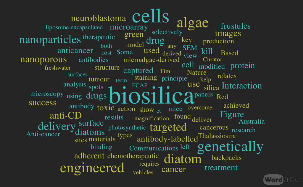
Genetically engineered algae kill cancer cells

By Tim Sandle     Nov 11, 2015 in Science

http://www.digitaljournal.com/science/are-algae-the-solution-to-beating-cancer/article/449045#ixzz3rZQZ7pRj

A research group in Australia has developed algae nanoparticles. The algae have been found to kill 90 percent of cancer cells in cultured human cells. Based on this success, the modified algae have been shown to destroy cancerous tumors in mice.

 Algae is a general term for a range of single-celled microscopic photosynthetic organisms such as diatoms, and larger, multi-cellular plants, like kelp. The use of algae in cancer treatment relates to drug delivery. Some success has been achieved with the use of nanoparticles (such as nanoporous silica) to deliver anti-cancer medicines. One downside with this relates to the cost of production and the fact that some nanoparticles can be toxic. To overcome this, researchers based at the University of South Australia have used algae to model an alternative drug delivery system.
Success has been achieved with microalgae-derived nanoporous biosilica. The bio-material was derived from Thalassiosira pseudonana, which is a species of marine diatom. Diatoms are are among the most common types of phytoplankton, and they are found in the oceans, in freshwater, in soils and on damp surfaces. In studies, modified diatoms have been used as types of backpacks for the targeted delivery of anticancer drugs to tumor sites.

Lead researcher Nico Voelcker, speaking with International Business Times, explained why genetic modification was key in adapting the diatoms: “By genetically engineering diatom algae – tiny, unicellular, photosynthesising algae with a skeleton made of nanoporous silica, we are able to produce an antibody-binding protein on the surface of their shells. Anti-cancer chemotherapeutic drugs are often toxic to normal tissues.”

 Although the results are interesting, experiments in cell culture and even on mice do not necessarily translate into success in humans. A series of clinical trials will be required to verify the results and to streamline any potential treatment options.

The research is published in the journal Nature Communications, in a paper headed “Targeted drug delivery using genetically engineered diatom biosilica.”

Targeted drug delivery using genetically engineered diatom biosilica


The ability to selectively kill cancerous cell populations while leaving healthy cells unaffected is a key goal in anticancer therapeutics. The use of nanoporous silica-based materials as drug-delivery vehicles has recently proven successful, yet production of these materials requires costly and toxic chemicals. Here we use diatom microalgae-derived nanoporous biosilica to deliver chemotherapeutic drugs to cancer cells. The diatom Thalassiosira pseudonana is genetically engineered to display an IgG-binding domain of protein G on the biosilica surface, enabling attachment of cell-targeting antibodies. Neuroblastoma and B-lymphoma cells are selectively targeted and killed by biosilica displaying specific antibodies sorbed with drug-loaded nanoparticles. Treatment with the same biosilica leads to tumour growth regression in a subcutaneous mouse xenograft model of neuroblastoma. These data indicate that genetically engineered biosilica frustules may be used as versatile ‘backpacks’ for the targeted delivery of poorly water-soluble anticancer drugs to tumour sites.


Figure 1: The principle of action of the genetically engineered biosilica therapeutic nanoparticles

The principle of action of the genetically engineered biosilica therapeutic nanoparticles.

http://www.nature.com/ncomms/2015/151110/ncomms9791/carousel/ncomms9791-f1.jpg

Genetically engineered diatom biosilica (green) containing liposome-encapsulated drug molecules (yellow) can be targeted to both adherent neuroblastoma cells (red) and lymphocyte cells in suspension (purple) by functionalizing the biosilica


Figure 2: SEM images of T. pseudonana biosilica and analysis of IgG–HRP binding to T. pseudonana biosilica

SEM images of T. pseudonana biosilica and analysis of IgG-HRP binding to T. pseudonana biosilica.

http://www.nature.com/ncomms/2015/151110/ncomms9791/carousel/ncomms9791-f2.jpg

(a) Side view and (b) top view of cylinder-shaped biosilica from a single diatom cell. (c,d) Details of the biosilica structure showing the highly porous surface. (e) Schematic structure of the S–T8–GFP–GB1 fusion protein. S, N-terminal…


Figure 4: Interaction of anti-CD20 antibody-labelled GB1–biosilica with B and T cells captured on an anti-CD45 antibody microarray.

Interaction of anti-CD20 antibody-labelled GB1-biosilica with B and T cells captured on an anti-CD45 antibody microarray.

http://www.nature.com/ncomms/2015/151110/ncomms9791/carousel/ncomms9791-f4.jpg

The left panels show epifluorescence microscopy images of cells captured on microarray spots after incubation with anti-CD20 antibody-labelled biosilica (n=3). The right panels show higher magnification images within the spots. Green: b…


Figure 6: Interaction of anti-p75NTR–GB1–biosilica frustules with adherent cells

Interaction of anti-p75NTR-GB1-biosilica frustules with adherent cells.

http://www.nature.com/ncomms/2015/151110/ncomms9791/carousel/ncomms9791-f6.jpg

(a) Confocal fluorescence microscopy image of SH-SY5Y neuroblastoma cells and (b) BSR fibroblast cells (n=3). Red: actin (phalloidin staining, green: biosilica (GFP-labelled), blue: nucleus (Hoechst 33342 staining). (c) The left panel s…

Read Full Post »

Plant to Animal Viral Transmission

Larry H. Bernstein, MD, FCAP, Curator

LPBI


Algae Virus May Be Linked to Human Cognitive Decline

http://www.genengnews.com/gen-news-highlights/algae-virus-may-be-linked-to-human-cognitive-decline/81251885/


A research team led by scientists at the University of Nebraska-Lincoln (UNL) has provided the first direct evidence that an algae-infecting virus can invade and potentially replicate within some mammalian cells. Known as Acanthocystis turfacea chlorella virus 1, or ATCV-1, the pathogen is among a class of chloroviruses long believed to take up residence only in green algae. That thinking changed with a 2014 study from Johns Hopkins University and UNL that found gene sequences resembling those of ATCV-1 in throat swabs of human participants.

The new study (“Response of mammalian macrophages to challenge with the Chlorovirus ATCV-1), published in the Journal of Virology, introduced ATCV-1 to macrophage cells that serve critical functions in the immune responses of mice, humans, and other mammals. By tagging the virus with fluorescent dye and assembling three-dimensional images of mouse cells, the authors determined that ATCV-1 successfully infiltrated them.

The authors also measured a threefold increase in ATCV-1 within 24 hours of introducing the virus. The relatively modest spike nevertheless suggests that ATCV-1 can replicate within the macrophage cells, according to co-author David Dunigan, Ph.D.

Though a few studies have documented viruses jumping from one biological kingdom to another, chloroviruses were previously thought to have a limited “host range” that stopped well short of the animal kingdom, said Dr. Dunigan.

“A few years ago, no one I know would have made a prediction like this,” noted Dr. Dunigan, research professor of plant pathology and member of the Nebraska Center for Virology. “You probably would’ve been laughed out of the room. But we are now in the middle of something that is so very interesting.”

The macrophage cells underwent multiple changes characteristic of those breached by a virus, he continued. These changes eventually included a form of programmed death that virologists consider an innate “scorched earth” defense against the spread of viruses, which require living cells to survive and replicate.

Before dying, the cells exhibited multiple signs of stress that tentatively support links to mild cognitive impairments first reported in the 2014 paper. The new study measured a post-viral rise in interleukin 6, which previous research has linked with diminished spatial learning and certain neurological diseases. The authors also reported an increase in nitric oxide, an important signaling molecule that has been associated with memory impairments when produced in excess.

The 2014 investigation, which was initially designed to test the cognitive functioning of human participants, found that those with the ATCV-1 DNA performed slightly worse on measures of visual processing and visual motor speed. Mice inoculated with the virus showed similar deficits in memory and attention while navigating mazes. The 2014 paper further suggested that ATCV-1 altered the expression of more than 1,000 genes in the rodent hippocampus, an area of the brain tied to memory and spatial navigation.

The new study’s authors are continuing their collaboration with Johns Hopkins in the hope of ultimately confirming whether and how the virus contributes to any cognitive deficits suggested by the initial studies.

“It is still unclear whether the factors induced by the cell-based virus challenge could also be induced in the whole animal, and whether the induced factors cause cognitive impairments in the animal or the human,” said co-author Tom Petro, Ph.D., professor of microbiology and immunology at the University of Nebraska Medical Center.

Dr. Dunigan said he and his colleagues are also searching for other cellular responses to ATCV-1 while investigating how these responses might drive systemic changes in mice.


Read Full Post »

%d bloggers like this: