Feeds:
Posts
Comments

Posts Tagged ‘hedgehog’

Targeting Epithelial To Mesenchymal Transition (EMT) As A Therapy Strategy For Pancreatic Cancer

Curator: David Orchard-Webb, PhD


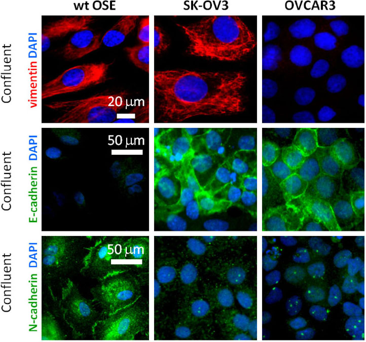
Epithelial to mesenchymal transition (EMT) is a mechanism by which cells of an epithelial phenotype dedifferentiate to a plastic mesenchymal phenotype. The epithelial cell rearranges its actin cytoskeleton from a cortical tight junction associated ring to form elongated stress fibres, redistributes and down regulates its cell-cell contacts, loses its polarity, and upregulates mesenchymal markers including α-smooth muscle actin (α-SMA) and vimentin [1]. The cell changes the composition of its extracellular matrix (ECM) contacts and secretes matrix metalloproteinases [2]. EMT has a role during development [3], chronic fibrotic disorders [4], and a postulated role in epithelial cancer metastasis [5].


 c6-tgf
Mouse mammary cell line induced to EMT with TGFβ1. Image Source: David Orchard-Webb.


Inflammatory signalling associated with pancreatitis is a driver of both pancreatic cancer and EMT [4,8]. Pancreatic cancer has a large stromal component that has therapeutic implications such as reduced drug tumour penetrance [9]. EMT is a mechanism of pancreatic stroma generation and may generate cancer stem-like cells [10]. This suggests a strategy for success in pancreatic cancer therapy. Cancer stem cells and stroma are major impediments to current therapeutics therefore targeting EMT is strategically viable to enhance their effectiveness.


A number of drug candidates have entered clinical trial which target EMT pathways. Curcumin which can reverse the EMT phenotype in vitro, has been shown to enhance the effectiveness of gemcitabine, the first FDA approved chemotherapeutic for pancreatic cancer [11]. Prism Pharma Co., Ltd. has developed a Wnt pathway inhibitor that may be effective in pancreatic cancer, however the associated phase I trial had to be terminated in 2015 due to low enrolment [14]. There are ongoing clinical trials targeting the hedgehog pathway which plays a role in EMT, in combination with gemcitabine and nab-paclitaxel (Abraxane) [12, 13].


STATs are transcription factors which are normally present in the cytoplasm and activated by inflammatory signalling associated with EMT which leads to their nuclear import [15]. STAT3 expression is maintained and constitutive activation has been reported in at least 30% of pancreatic cancers [6]. STAT3 is not active in normal pancreatic tissue but its activation is required in the early stages of pancreatic cancer progression. A means to eliminate STAT3 has been developed by Astrazeneca – stable systemically delivered siRNA which has completed phase I clinical trials [7]. This may prove beneficial in combination with standard chemotherapeutics.


In summary a number of EMT pathway targeting therapeutics are in development which have the potential to target pancreatic cancer stem cells, which could reduce cancer recurrence, and deplete the cancer associated stroma which should improve the penetrance of existing therapeutics and may help relieve suppression of the immune system by pancreatic tumours.


REFERENCES

  1. Savagner, P. 2001. Leaving the neighborhood: molecular mechanisms involved during Epithelial-Mesenchymal Transition. BioEssays. 23: 912-923.
  2. LaGamba, D. Nawshad, A. and Hay, E.D. 2005. Microarray analysis of gene expression during Epithelial-Mesenchymal Transformation. Dev Dyn. 234: 132-42
  3. Hay, E.D. 1995. An overview of Epithelio-Mesenchymal Transformation. Acta Anat (Basel). 154: 8-20.
  4. Kalluri, R. and Neilson, E.G. 2003. Epithelial-Mesenchymal Transition and its implications for fibrosis. J Clin Invest. 112: 1776-84.
  5. Thiery, J.P. 2002. Epithelial-Mesenchymal Transitions in tumour progression. Nat Rev Cancer. 2: 442–454.
  6. Corcoran, R. B., G. Contino, V. Deshpande, A. Tzatsos, C. Conrad, C. H. Benes, D. E. Levy, J. Settleman, J. A. Engelman, and N. Bardeesy. ‘STAT3 Plays a Critical Role in KRAS-Induced Pancreatic Tumorigenesis’. Cancer Research 71, no. 14 (15 July 2011): 5020–29. doi:10.1158/0008-5472.CAN-11-0908.
  7. Hong, David, Razelle Kurzrock, Youngsoo Kim, Richard Woessner, Anas Younes, John Nemunaitis, Nathan Fowler, et al. ‘AZD9150, a next-Generation Antisense Oligonucleotide Inhibitor of STAT3 with Early Evidence of Clinical Activity in Lymphoma and Lung Cancer’. Science Translational Medicine 7, no. 314 (18 November 2015): 314ra185. doi:10.1126/scitranslmed.aac5272.
  8. Guerra, Carmen, Alberto J. Schuhmacher, Marta Cañamero, Paul J. Grippo, Lena Verdaguer, Lucía Pérez-Gallego, Pierre Dubus, Eric P. Sandgren, and Mariano Barbacid. ‘Chronic Pancreatitis Is Essential for Induction of Pancreatic Ductal Adenocarcinoma by K-Ras Oncogenes in Adult Mice’. Cancer Cell 11, no. 3 (March 2007): 291–302. doi:10.1016/j.ccr.2007.01.012.
  9. Xie, Dacheng, and Keping Xie. ‘Pancreatic Cancer Stromal Biology and Therapy’. Genes & Diseases 2, no. 2 (June 2015): 133–43. doi:10.1016/j.gendis.2015.01.002.
  10. Dangi-Garimella, Surabhi, Seth B. Krantz, Mario A. Shields, Paul J. Grippo, and Hidayatullah G. Munshi. ‘Epithelial-Mesenchymal Transition and Pancreatic Cancer Progression’. In Pancreatic Cancer and Tumor Microenvironment, edited by Paul J. Grippo and Hidayatullah G. Munshi. Trivandrum (India): Transworld Research Network, 2012. http://www.ncbi.nlm.nih.gov/books/NBK98932/.
  11. Osterman, Carlos J. Díaz, and Nathan R. Wall. ‘Curcumin and Pancreatic Cancer: A Research and Clinical Update’. Journal of Nature and Science 1, no. 6 (2015): 124. http://www.jnsci.org/files/html/e124.htm.
  12. ‘Hedgehog Inhibitors for Metastatic Adenocarcinoma of the Pancreas – Full Text View – ClinicalTrials.gov’. Accessed 18 April 2016. https://clinicaltrials.gov/ct2/show/NCT01088815.
  13. Singh, Brahma N., Junsheng Fu, Rakesh K. Srivastava, and Sharmila Shankar. ‘Hedgehog Signaling Antagonist GDC-0449 (Vismodegib) Inhibits Pancreatic Cancer Stem Cell Characteristics: Molecular Mechanisms’. PLOS ONE 6, no. 11 (8 November 2011): e27306. doi:10.1371/journal.pone.0027306.
  14. ‘Safety and Efficacy Study of PRI-724 in Subjects With Advanced Solid Tumors – Full Text View – ClinicalTrials.gov’. Accessed 18 April 2016. https://clinicaltrials.gov/ct2/show/NCT01302405.
  15. Kaplan, Mark H. ‘STAT Signaling in Inflammation’. JAK-STAT 2, no. 1 (January 2013): e24198. doi:10.4161/jkst.24198.


Other Related Articles Published In This Open Access Online Journal Include The Following:


https://pharmaceuticalintelligence.com/2012/11/30/histone-deacetylase-inhibitors-induce-epithelial-to-mesenchymal-transition-in-prostate-cancer-cells/

https://pharmaceuticalintelligence.com/2015/02/24/inhibiting-the-gene-protein-kinase-d1-pkd1-and-its-protein-could-stop-spread-of-this-form-of-pancreatic-cancer/

https://pharmaceuticalintelligence.com/2014/06/01/locally-advanced-pancreatic-cancer-efficacy-of-folfirinox/

https://pharmaceuticalintelligence.com/2014/04/10/consortium-of-european-research-institutions-and-private-partners-will-develop-a-microfluidics-based-lab-on-a-chip-device-to-identify-pancreatic-cancer-circulating-tumor-cells-ctc-in-blood/

https://pharmaceuticalintelligence.com/2013/10/21/whats-new-in-pancreatic-cancer-research-and-treatment/

https://pharmaceuticalintelligence.com/2013/04/11/update-on-pancreatic-cancer/

https://pharmaceuticalintelligence.com/2015/04/10/targeting-the-wnt-pathway-7-11/

https://pharmaceuticalintelligence.com/2015/10/29/gene-amplification-and-activation-of-the-hedgehog-pathway/


Read Full Post »

Screen Shot 2021-07-19 at 6.30.31 PM

Role of Primary Cilia in Ovarian Cancer


Curator: Aashir Awan, PhD


Word Cloud By Danielle Smolyar

Each year, over 20 000 women in the US are diagnosed with ovarian cancer while there are nearly 15 000 deaths (http://www.cdc.gov/uscs). The 5-year (post-diagnosis) mortality rate is less than 46% partly due to the fact that early detection still remains difficult (Jemal et al., 2010). So, it is not surprising to see biomedical research putting a much needed focus on this deadly killer.

As such, a joint collaboration between Dr. Søren T Christensen’s lab (University of Copenhagen, Denmark) and the Laboratory of Reproductive Biology (LRB, RigsHospital, Copenhagen) decided to investigate into the specific molecular mechanisms behind ovarian cancer. This resulted in the recent publication linking aberrant hedgehog/PDGF signaling and ovarian cancer (Egeberg et al., 2012). Specifically, the paper shows that there are specific defects associated with the primary cilium of these cancer cell lines which then lead to a perturbation of two critical signaling systems that were examined in the paper. The starting materials included ovarian cancer lines OVCAR3 and SK-OV3. And, the control WT cells are ovarian surface epithelium (OSE which are thought to be the cells of origin for ovarian cancer) cells that were obtained at LRB where young girls that are diagnosed with cancer can deposit and cryopreserve their ovaries for future implantation after being deemed cancer-free.

Biomarkers evaluated in the study included

1. Gli proteins – glioma transcription factors specific to hedgehog signaling (regulating cell proliferation/differentiation). In the off stage, they are thought to act as repressors while in the on state, the proteins are modified at the primary cilium to become activators (Satir el al., 2010).

2. CHFR (checkpoint with forkhead-associated and ring finger domains) – a tumor suppressor gene with multiple functions in checkpoints during mitosis (Kang et al., 2002)

3. AURA (Aurora Kinase A) mark centrosome maturation, mitotic entry an spindle assembly (Fu et al., 2007).

4. PDGF (platelet derived growth factor) is a signaling pathway involved in cell growth/survival, differentiation, migration and wound healing (Schneider et al., 2010).

Fig5aThe paper begins with characterizing the epithelial nature of OSE to confirm the validity of the primary OSE cell cultures. Of note is the fact that OSE lack E-cadherins present in other epithelial cells while expressing N-cadherin and vimentin which together suggest OSE have a more mesenchymal characteristic in comparison to other epithelial tissues. Meawhile, OVCAR3 was negative for vimentin while both cancer cell lines expressed N-cadherin and E-cadherin (more so) indicating these cancer cell lines retained more of a classic epithelial characteristic in comparison to WT OSE which might imply lack of plasticity (Wong et al., 1999).

Fig1aNext, ciliogenesis (ability to form a primary cilium) was evaluated in the cancer cell lines vs WT OSE. Under serum starvation (used to induce cilia formation), far fewer cancer cells are able to form a primary cilium. Since primary cilium are formed in mitotically inactive cells (usually at G0), it was speculated that the lack of cilia in the cancer lines implied greater proliferation rates. However, Ki67, phosphorylated retinoblastoma protein (p-RB) and PCNA (cell proliferation markers) stainings showed that under serum starvation conditions, the cancer cells still have the ability to enter growth arrest.

Fig3a

So, there had be another explanation accounting for the difference between WT OSE and the cancer cell lines such as a interruption of signal between the primary cilium and the growth arrest apparatus. Thus, the hedgehog signaling was then examined. Full-length Gli2 acts as a repressor while cleavage at the primary cilia results in the activator form. WT OSE had very little full-length Gli2 whereas the cleaved repressor form is present. This indicates that the hedgehog pathway is mostly inactive in WT OSE. On the other hand, the two cancer lines had higher levels of the full length activator form and little of the cleaved repressor form. Serum starvation decreased this activation of the hedgehog signal as expected (due to the presence of the primary cilium cleaving the Gli2) but not down to the same levels as in WT OSE. This was confirmed by RT-PCR results which in summary stated that there is a higher basal level of hedgehog signaling in the cancer lines vs WT OSE.

Fig3aPDGFa had previously been reported to be upregulated during serum starvation and consequently is seen accumulating at the primary cilium (Schneider et al., 2005). While this phenomenon is observed In WT OSE, not only are there lower levels of PDGF overall, but there is no accumulation of this protein upon serum starvation. Therefore, PDGF signaling is perturbed in the cancer cell lines.

Fig4aNext, the expression of AURA was examined. This kinase is responsible for ciliary disassembly which is a necessary step before entering mitosis. The cancer OSE cells were shown to have higher AURA levels as compared to WT OSE. Given that these cancer cell lines have fewer primary cilia, one can deduce that the higher AURA protein levels suppress ciliogenesis while in WT OSE, they have a role in ciliary disassembly. This was further confirmed by the fact that knockdown of AURA in the cancer cell lines resulted in increased ciliogenesis.

Fig7aThen, the paper looks upstream of AURA at CHFR which is responsible for the ubiquitination/ proteosomal degradation of AURA. During serum starvation (i.e. primary cilia is formed), CHFR is found at the base of the cilium in WT OSE and this localization disappears during mitosis. In the transformed hTERT-RPE1 cells, CHFR is found both in growth-arrested as well as cells in mitosis. Therefore, this result hints at a AURA/CHFR interaction at the centrosome regulating ciliary dynamics.

Thus, an axis linking the primary cilium to different signal transduction systems and eventually leading to cancer is shown below:

Cilia defectarrow5 Hedgehog/PDGFarrow5 uncontrolled proliferationarrow5 cancer

The cilia defect is likely due to a mutation in the AURA/CHFR axis or even further upstream. While this mechanistic explanation summarizes the findings of this paper,its logic can be applied to other cancer types also (Veland et al., 2009).

REFERENCES

Egeberg DL, Lethan M, Manguso R, Schneider L, Awan A, Jørgensen TS, Byskov AG, Pedersen LB, Christensen ST (2012) Primary cilia and aberrant cell signaling in epithelial ovarian cancer. Cilia 1:1-15.

Fu J, Bian M, Jiang Q, Zhang C (2007) Roles of Aurora kinases in mitosis and tumorigenesis. Mol Cancer Res 5:1–10.

Jemal A, Siegel R, Xu J, Ward E (2010) Cancer statistics, 2010. CA Cancer J Clin 60:277–300.

Kang D, Chen J, Wong J, Fang G (2002) The checkpoint protein Chfr is a ligase that ubiquitinates Plk1 and inhibits Cdc2 at the G2 to M transition. J Cell Biol 156:249–259.

Satir P, Pedersen LB, Christensen ST (2010) The primary cilium at a glance. J Cell Sci. 123:499-503.

Schneider L, Clement CA, Teilmann SC, Pazour GJ, Hoffmann EK, Satir P, Christensen ST (2005) PDGFRalphaalpha signaling is regulated through the primary cilium in fibroblasts. Curr Biol 15:1861-1866.

Schneider L, Cammer M, Lehman J, Nielsen SK, Guerra CF, Veland IR, Stock C, Hoffmann EK, Yoder BK, Schwab A, Satir P, Christensen ST (2010) Directional cell migration and chemotaxis in wound healing response to PDGF-AA are coordinated by the primary cilium in fibroblasts. Cell Physiol Biochem 25:279-292.

Veland IR, Awan A, Pedersen LB, Yoder BK, Christensen ST (2009) Primary cilia and signaling pathways in mammalian development, health and disease. Nephron Physiol 111: 39-53.

Wong AS, Maines-Bandiera SL, Rosen B, Wheelock MJ, Johnson KR, Leung PC, Roskelley CD, Auersperg N (1999) Constitutive and conditional cadherin expression in cultured human ovarian surface epithelium: influence of family history of ovariancancer. Int J Cancer 81:180–188.

Read Full Post »

Author: Aashir Awan, PhD

The primary cilium is organelle that has garnered much attention in the field of cell biology during the last 15 years. It is a slender, solitary hair-like organelle that extends 5-10 uM from each mammalian cell (in the G0 cell cycle state) that is microtubule-based (9 outer doublets arranged in a circular fashion) and dependent on a process called Intraflagellar Transport (IFT). IFT is the bidirectional movement of motors (kinesin-2 in the anterograde and dynein-2 in the retrograde direction) responsible for the assembly and maintenance of the cilium (Pedersen et al., 2006).

Until this time, it had been labeled a ‘vestigial’ organelle not worthy of research. Yet, a breakthrough into the sensory role of the primary cilium came in 2000 based on Dr. Rosenbaum’s research on Chlamydomonas and the motile cilium or flagella. Along with Dr. George Whitman’s group, they were able to show the importance of Tg737 (IFT88) protein to the pathology of polycystic kidney disease in mouse (Pazour et al., 2000). Since then, research into the primary cilium has exploded and has been linked to diverse pathologies (collectively known as ciliopathies) such as

  • retinitis pigmentosa,
  • hydrocephaly,
  • situs inversus,
  • ovarian and pancreatic cancers among others (Nielsen et al., 2008; Edberg et al., 2012). Also, various
  • signal transduction pathways have been found to be coordinated by the primary cilia such as hedgehog, wnt, PDGF among others (Veland et al., 2008).

Thus, in 2006, the Christensen lab at the University of Copenhagen (Denmark) with the collaboration of Dr. Peter Satir’s group at Albert Einstein College of Medicine (Bronx, NY) began to investigate whether the human embryonic stem cells (hESCs) possess primary cilium and then to begin preliminary molecular dissections of the role that this organelle could play in the proliferation and differentiation profiles of these pluripotent cells. The Albert Einstein group, due to NIH restrictions, had to work with two federally-sanctioned cell lines. Working with the Laboratory of Reproductive Biology at RigsHospital, the Danish side had access to in-house derived stem cell lines from discarded blastocysts. The advantage for the Danish side was obvious since these newer cell lines hadn’t undergone as many passages as the NIH cell lines and were thus more robust. To begin preliminary characterizations of these lines, some basic hallmarks of hESCs (Bernhardt et al., 2012) had to be localized to the nucleus such as the transcription factor (TF) Oct4 (Fig. 1).

In addition, a single primary cilium can be seen denoted by the acetylated tubulin staining emanating from each cell in the micrographs. Also, the base of the cilium is marked by the presence of pericentrin and centrin which demarcate the centriole.

Fig1 Fig. 1 Primary cilia stained with anti-acetylated tubulin (tb, red) are indicated by arrows and undifferentiated stem cells are identified by nuclear colocalization of OCT-4 (green) and DAPI (dark blue) in the merged image (light blue). A primary cilium (tb, red, arrow) in undifferentiated hESCs emerges from one of the centrioles (asterisks) marked with anti-centrin (centrin, green). Inset shows anti-pericentrin localization to base of cilia (Pctn, green).

Together, the three labs were the first to discover primary cilia in stem cells while other groups have since then confirmed these findings (Kiprilov et  al. 2008; Han et al. 2008). Attention was then to characterize different signal transduction pathways in the stem cell cilium. Since the hedgehog pathway has been shown to be important for differentiation and proliferation (Cerdan and Bhatia, 2012), the groups characterized this signal pathway in these cells using immunofluorescence, electron microscopy and qPCR techniques. One particularly interesting experiment to show that the hedgehog pathway was functional in these cells was to add the hedgehog agonist, SAG (Smoothened agonist), and then to isolate the cells for immunofluorescence at different times.

Gradually, one can see the appearance of the smoothened protein into the cilium as indicated by increasing intensity of the immunofluorescence staining. Conversely, patched levels in the cilium, decreased. This is a hallmark of hedgehog activation (Fig. 2).
Fig. 2 copiaFig. 2 Immunofluorescence micrographs of hESC showing smoothened (green), acetylated tubulin (red) and DAPI (blue). The micrographs from left to right represents SAG treatments at t = 0, 1 and 4 hours.

However, an additional interesting observation was made concerning these stem cells. An important characteristic for stem cells is the presence of certain transcription factors which render these cells in the pluripotent or undifferentiated state. These include Oct4, Sox2, and Nanog whose localization had been observed in the nucleus as expected for other TFs.

However, the Danish groups curiously found a subpopulation of stem cells where these TFs were additionally localized to the primary cilium (Fig. 3). This had never been observed or investigated before.  Additionally, proper negative controls were  carried out to exclude this phenomenon from being an artifact (e.g. bleed through).
Fig. 3 copia Fig. 3 Stem cell markers (Sox2, Nanog, and Oct4) localizing to the nucleus and the primary cilia (arrows) of hESC line LRB003. This and the previous figure show shifted overlay images whereby the green and red channels have been slightly shifted so that the red channel doesn’t swamp out the intensity of the green channels.

Thus, it raises an intriguing possibility that perhaps the primary cilia plays a previously uncharacterized role in the differentiation/proliferation state of the hESCs via possible modifications of these TFs perhaps analogous to the processing of the Gli transcription factors (Hui and Angers, 2011). Another curious observation is that the subpopulation of cells whose primary cilia are positive for these TFs always occur in clusters which might hint at its mechanistic explanation.  In conclusion, since stem cells are now being more routinely used for regenerative medicine such as repair of severed spinal cord (Lu et al. 2012), it behooves us to better learn the molecular mechanisms that keeps these invaluable cells in an undifferentiated state so that we can harness their full therapeutic potential.

REFERENCES

Awan A, Oliveri RS, Jensen PL, Christensen ST, Andersen CY. 2010 Immunoflourescence and mRNA analysis of human embryonic stem cells (hESCs) grown under feeder-free conditions. Methods Mol Biol. 584:195-210.

Bernhardt M, Galach M, Novak D, Utikal J. 2012 Mediators of induced pluripotency and their role in cancer cells – current scientific knowledge and future perspectives. Biotechnol J. 7:810-821.

Cerdan C, Bhatia M. 2010 Novel roles for Notch, Wnt and Hedgehog in hematopoesis derived from human pluripotent stem cells. Int J Dev Biol. 54:955-963.

Han YG, Spassky N, Romaguera-Ros M, Garcia-Verdugo JM, Aguilar A, Schneider-Maunoury S, Alvarez-Buylla A. 2008 Hedgehog signaling and primary cilia are required for the formation of adult neural stem cells.Nat Neurosci. 11:277-284.

Hui CC, Angers S. 2011 Gli proteins in development and disease. Annu Rev Cell Dev Biol. 27:513-537.

Kiprilov EN, Awan A, Desprat R, Velho M, Clement CA, Byskov AG, Andersen CY, Satir P, Bouhassira EE, Christensen ST, Hirsch RE 2008 Human embryonic stem cells in culture possess primary cilia with hedgehog signaling machinery. J Cell Biol. 2008 180:897-904.

Lu P, Wang Y, Graham L, McHale K, Gao M, Wu D, Brock J, Blesch A, Rosenzweig ES, Havton LA, Zheng B, Conner JM, Marsala M, Tuszynski MH. 2012 Long-distance growth and connectivity of neural stem cells after severe spinal cord injury. Cell 150:1264-73.

Nielsen SK, Møllgård K, Clement CA, Veland IR, Awan A, Yoder BK, Novak I, Christensen ST. 2008 Characterization of primary cilia and Hedgehog signaling during development of the human pancreas and in human pancreatic duct cancer cell lines. Dev Dyn. 237:2039-52.

Pazour GJ, Dickert BL, Vucica Y, Seeley ES, Rosenbaum JL, Witman GB, Cole DG. 2000 Chlamydomonas IFT88 and its mouse homologue, polycystic kidney disease gene tg737, are required for assembly of cilia and flagella. J Cell Biol 151: 709-18.

Pedersen LB, Veland IR, Schrøder JM, Christensen ST. 2008 Assembly of primary cilia. Dev Dyn. 237:1993-2006.

Veland IR, Awan A, Pedersen LB, Yoder BK, Christensen ST. 2009 Primary cilia and signaling pathways in mammalian development, health and disease. Nephron Physiol. 111: 39-53.

Read Full Post »