Feeds:
Posts
Comments

Posts Tagged ‘optical images’

A step forward in diagnostics

Larry H. Bernstein, MD, FCAP, Curator

LPBI


3D Imaging of Cancer Cells Could Lead to Improved Ability of Pathologists and Radiologists to Plan Cancer Treatments and Monitor Cell Interactions

DARK DAILY   4/8/2016   info@DarkDaily.com  http://www.darkdaily.com/#axzz45En6Xbfr


Imaging research is one step closer to giving clinicians a way to do high-resolution scans of malignant cells in order to diagnose cancer and help identify useful therapies. If this technology were to prove successful in clinical studies, it might change how anatomic pathologists and radiologists diagnose and treat cancer.

Researchers at the University of Texas Southwestern Medical Center developed a way to create near-isotropic, high-resolution scans of cells within their microenvironments. The process involves utilizing a combination of two-photonBessel beams and specialized filtering.

New Imaging Approach Could be Useful to Both Pathologists and Radiologists

In a recent press release, senior author Reto Fiolka, PhD, said “there is clear evidence that the environment strongly affects cellular behavior—thus, the value of cell culture experiments on glass must at least be questioned. Our microscope is one tool that may bring us a deeper understanding of the molecular mechanisms that drive cancer cell behavior, since it enables high-resolution imaging in more realistic tumor.”

In a study in Developmental Cell, Erik S. Welf, PhD, et al, described the new microenvironmental selective plane illumination microscopy (meSPIM). When developing the technology, the team outlined three goals:

1. The microscope design must not prohibitively constrain microenvironmental properties.

2. Spatial and temporal resolution must match the cellular features of interest.

3. Spatial resolution must be isotropic to avoid spatial bias in quantitative measurements.

This new technology offers pathologists and medical laboratory scientists a new look at cancer cells and other diseases. The study notes that meSPIM eliminates the influence of stiff barriers, such as glass slide covers, while also allowing a level of control over both mechanical and chemical influences that was previously impossible.

Early meSPIM Research Reveals New Cell Behaviors

Early use of meSPIM in observing melanoma cells is already offering new insights into the relationship between the cell behavior of cellular- and subcellular-scale mechanisms and the microenvironment in which these cells exist. The study notes, “The ability to image fine cellular details in controllable microenvironments revealedmorphodynamic features not commonly observed in the narrow range of mechanical environments usually studied in vitro.”

One such difference is the appearance of blebbing. Created by melanoma cells and lines, these small protrusions are thought to aid in cell mobility and survival. Using meSPIM, observers could follow the blebbing process in real-time. Formation of blebs on slides and within an extracellular matrix (ECM) showed significant differences in both formation and manipulation of the surrounding microenvironment.

The team is also using meSPIM to take a look at membrane-associated biosensorand cytosolic biosensor signals in 3D. They hope that investigation of proteins such as phosphatidylinositol 3-kinase (PI3K) and protein kinase C will help to further clarify the roles these signals play in reorientation of fibroblasts.


meSPIM-500ppi

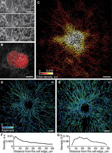
meSPIM combined with computer vision enables imaging, visualization, and quantification of how cells alter collagen fibers over large distances within an image volume measuring 100 mm on each side. (Photo Copyright: Welf and Driscoll et al.)   http://www.darkdaily.com/wp-content/uploads/meSPIM-500ppi-220×300.jpg


Seeing cancer cells in 3-D (w/ Video)

February 22, 2016

Cancer in 3-D

Extracted surfaces of two cancer cells. (Left) A lung cancer cell colored by actin intensity near the cell surface. Actin is a structural molecule that is integral to cell movement. (Right) A melanoma cell colored by PI3-kinase activity near the cell surface. PI3K is a signaling molecule that is key to many cell processes. Credit: Welf and Driscoll et al.  http://cdn.phys.org/newman/csz/news/800/2016/cancerin3d.png

Cancer cells don’t live on glass slides, yet the vast majority of images related to cancer biology come from the cells being photographed on flat, two-dimensional surfaces—images that are sometimes used to make conclusions about the behaviour of cells that normally reside in a more complex environment. But a new high-resolution microscope, presented February 22 in Developmental Cell, now makes it possible to visualize cancer cells in 3D and record how they are signaling to other parts of their environment, revealing previously unappreciated biology of how cancer cells survive and disperse within living things.

“There is clear evidence that the environment strongly affects cellular behavior—thus, the value of cell culture experiments on glass must at least be questioned,” says senior author Reto Fiolka, an optical scientist at the University of Texas Southwestern Medical Center. “Our is one tool that may bring us a deeper understanding of the molecular mechanisms that drive cancer cell behavior, since it enables high-resolution imaging in more realistic tumor environments.”

In their study, Fiolka and colleagues, including co-senior author Gaudenz Danuser, and co-first authors Meghan Driscoll and Erik Welf, also of UT Southwestern, used their microscope to image different kinds of skin cancer cells from patients. They found that in a 3D environment (where cells normally reside), unlike a glass slide, multiple melanoma cell lines and primary melanoma cells (from patients with varied genetic mutations) form many small protrusions called blebs. One hypothesis is that this blebbing may help the survive or move around and could thus play a role in skin cancer cell invasiveness or drug resistance in patients.

The researchers say that this is a first step toward understanding 3D biology in tumor microenvironments. And since these kinds of images may be too complicated to interpret by the naked eye alone, the next step will be to develop powerful computer platforms to extract and process the information.

“When we conceived of this project, we first asked what we wanted to measure and then designed a microscope and analytical platform to achieve this goal,” says co-first author Erik Welf, a cell biologist. “We hope that now instead of asking what we can measure, scientists will ask what we must measure in order to make meaningful contributions to cancer cell biology.”

The microscope control software and image analytical code are freely available to the scientific community.

More information: Developmental Cell, Welf and Driscoll et al.: “Quantitative Multiscale Cell Imaging in Controlled 3D Microenvironments” dx.doi.org/10.1016/j.devcel.2016.01.022

Read more at: http://phys.org/news/2016-02-cancer-cells-d-video.html#jCp

Quantitative Multiscale Cell Imaging in Controlled 3D Microenvironments

Erik S. Welf4, Meghan K. Driscoll4, Kevin M. Dean, Claudia Schäfer, Jun Chu, Michael W. Davidson, Michael Z. Lin, Gaudenz Danusercorrespondence , Reto Fiolkacorrespondence
Dev Cell  22 Feb 2016;  Volume 36, Issue 4:462–475     DOI: http://dx.doi.org/10.1016/j.devcel.2016.01.022
Highlights
  • meSPIM allows microenvironmentally conscious 3D imaging/analysis of subcellular biology
  • Precisely controlled microenvironments reveal diverse morphological phenotypes
  • Isotropic resolution and high speed enable the quantification of 3D cell signaling and morphodynamics
  • Multiscale quantification of microenvironmental reorganization by cells

Summary

The microenvironment determines cell behavior, but the underlying molecular mechanisms are poorly understood because quantitative studies of cell signaling and behavior have been challenging due to insufficient spatial and/or temporal resolution and limitations on microenvironmental control. Here we introduce microenvironmental selective plane illumination microscopy (meSPIM) for imaging and quantification of intracellular signaling and submicrometer cellular structures as well as large-scale cell morphological and environmental features. We demonstrate the utility of this approach by showing that the mechanical properties of the microenvironment regulate the transition of melanoma cells from actin-driven protrusion to blebbing, and we present tools to quantify how cells manipulate individual collagen fibers. We leverage the nearly isotropic resolution of meSPIM to quantify the local concentration of actin and phosphatidylinositol 3-kinase signaling on the surfaces of cells deep within 3D collagen matrices and track the many small membrane protrusions that appear in these more physiologically relevant environments.

Read more: 3D Imaging of Cancer Cells Could Lead to Improved Ability of Pathologists and Radiologists to Plan Cancer Treatments and Monitor Cell Interactions | Dark Daily http://www.darkdaily.com/3d-imaging-of-cancer-cells-could-lead-to-improved-ability-of-pathologists-and-radiologists-to-plan-cancer-treatments-and-monitor-cell-interactions-301#ixzz45Enp2yT0

The research team believes this opens new possibilities for studying diseases at a subcellular level, saying, “Cell biology is necessarily restricted to studying what we can measure. Accordingly, while the last hundred years have yielded incredible insight into cellular processes, unfortunately most of these studies have involved cells plated onto flat, stiff surfaces that are drastically different from the in vivo microenvironment …

“Here, we introduce an imaging platform that enables detailed subcellular observations without compromising microenvironmental control and thus should open a window for addressing these fundamental questions of cell biology.”

Limitations of meSPIM

One significant issue associated with the use of meSPIM is the need to process the large quantity of data into useful information. Algorithms currently allow for automatic bleb detection. However, manual marking, while time consuming, still provides increased accuracy. Researchers believe the next step in improving the quality of meSPIM scans lie in computer platforms designed to extract and process the scan data.

Until this process is automated, user bias, sample mounting, and data handling will remain risks for introducing errors into the collected data. Yet, even in its early stages, meSPIM offers new options for assessing the state of cancer cells and may eventually provide pathologists and radiologists with additional information when creating treatment plans or assessments.


Read Full Post »

New method of visual communication

Larry H. Bernstein, MD, FCAP, Curator

LPBI


New optical material discovered in secret language of mantis shrimp

http://www.rdmag.com/news/2016/02/new-optical-material-discovered-secret-language-mantis-shrimp    University of Bristol

Using a combination of careful anatomy, light measurements, and theoretical modelling, it was found that mantis shrimp polarizers work by manipulating light across the structure rather than through its depth, which is how typical polarizers work. Courtesy of Roy Caldwell, University of California, Berkeley

http://www.rdmag.com/sites/rdmag.com/files/New_optical_material_discovered_in_secret_language_of_mantis_shrimp_440.jpg

Using a combination of careful anatomy, light measurements, and theoretical modelling, it was found that mantis shrimp polarizers work by manipulating light across the structure rather than through its depth, which is how typical polarizers work. Courtesy of Roy Caldwell, University of California, Berkeley


A study into how animals secretly communicate has led to the discovery of a new way to create a polarizer — an optical device widely used in cameras, DVD players and sunglasses.

Mantis shrimp like to keep their conversations private, which is why they communicate using the polarization of light. These animals have evolved bright reflectors that control the polarization of their visual signals, a property of light not commonly used for animal communication. Most eavesdroppers can’t see this type of light information and so animals that use it are less likely to attract the attention of predators or unwelcomed competition.

In a quest to understand how these uncommon light signals are produced in mantis shrimp, researchers from the Ecology of Vision Group based in the University of Bristol’s School of Biological Sciences discovered that they use a polarizing structure unlike anything ever seen or developed by humans.  The research is published in the journal Scientific Reports.

Using a combination of careful anatomy, light measurements, and theoretical modeling, it was found that the mantis shrimp polarizers work by manipulating light across the structure rather than through its depth, which is how typical polarizers work. Such a photonic mechanism affords the animal with small, microscopically thin and dynamic optical structures that still produce big, bright and colorful polarized signals.

Dr. Nicholas Roberts in the School of Biological Sciences said: “When it comes to developing a new way to make polarizers, nature has come up with optical solutions we haven’t yet thought of.

“Industries working on optical technologies will be interested in this new solution mantis shrimp have found to create a polarizer as new ways for humans to use and control light are developed.”

This research was funded by the US Air Force Office of Scientific Research.

Citation: A shape-anisotropic reflective polarizer in a stomatopod crustacean’ by Thomas M. Jordan,  David Wilby, Tsyr-Huei Chiou, Kathryn D. Feller, Roy L. Caldwell, Thomas W. Cronin and Nicholas W. Roberts in Scientific Reports


A shape-anisotropic reflective polarizer in a stomatopod crustacean

Jordan, T, Wilby, D, Chiou, T-H, Feller, K, Caldwell, R, Cronin, T & Roberts, N, 2016, ‘A shape-anisotropic reflective polarizer in a stomatopod crustacean’. Scientific Reports.

    Many biophotonic structures have their spectral properties of reflection ‘tuned’ using the (zeroth-order) Bragg criteria for phase constructive interference. This is associated with a periodicity, or distribution of periodicities, parallel to the direction of illumination. The polarization properties of these reflections are, however, typically constrained by the dimensional symmetry and intrinsic dielectric properties of the biological materials. Here we report a linearly polarizing reflector in a stomatopod crustacean that consists of 6-8 layers of hollow, ovoid vesicles with principal axes of ~550nm, ~250nm and ~150nm. The reflection of unpolarized normally incident light is blue/green in colour with maximum reflectance wavelength of 520 nm and a degree of polarization greater than 0.6 over most of the visible spectrum. We demonstrate that the polarizing reflection can be explained by a resonant coupling with the first-order, in-plane, Bragg harmonics. These harmonics are associated with a distribution of periodicities perpendicular to the direction of illumination, and, due to the shape-anisotropy of the vesicles, are different for each linear polarization mode. This control and tuning of the polarization of the reflection using shape-anisotropic hollow scatterers is unlike any optical structure previously described and could provide a new design pathway for polarization-tunability in man-made photonic devices.

There is a great diversity of reflective photonic structures throughout the animal kingdom, including biological analogues of periodic photonic crystals1,2,3,4, quasi-ordered amorphous solids5,6,7,8, and one-dimensional multilayer reflectors9,10,11,12. Biophotonic structures frequently serve as optical adaptations that enable animals to communicate through reflective visual signals. It is therefore highly advantageous to be able to control the optical properties of these reflectors and thus the visual information content of the reflection. For example, multilayer reflectors found in fish and cephalopods are ‘spectrally tuned’ via the distribution of layer thicknesses around the quarter-wave criteria to have colours that range from blue to silver9,12,13. However, whilst the principles that control and optimize either the reflected colour or the level of reflectivity are understood, the same cannot be said for how 3-dimensional animal photonic structures could be structured to control the polarization of light.

At normal incidence, the polarization of the light reflected from the majority of photonic structures, whether biological or man-made, remains unchanged. In order to polarize the reflection, the symmetry of the photonic structure with respect to orthogonal polarization modes must be broken. For example, the circularly polarized reflections seen from the chitin structures in the elytra of beetles14 and cholesteric or chiral smectic liquid crystals15 arise due to chirality. In two-dimensional photonic crystals, it is a general result that for light propagation along correctly chosen coordinate axes it is possible to separate orthogonal polarization modes16,17 and geometric shape-anisotropy can be introduced to provide polarization-selective reflection and transmission17. In true 3-dimensional photonic crystals, however, it is in general not possible to obtain the strict separation of orthogonal polarization modes16. Subsequently, engineering a three-dimensional photonic structure to produce a reflection with a high degree of polarization mode separation is a non-trivial task.

One 3-dimensional biophotonic structural reflector that potentially acts as a linear polarizer is found in the maxilliped appendages of certain species of stomatopod crustaceans (known commonly as mantis shrimps). Maxillipeds are frontal sets of modified limbs, generally used to manipulate food or for cleaning, and are involved in sexual and agonistic communication18,19,20. In the genus Haptosquilla, the first maxillipeds possess a striking and conspicuous blue/green colouration, which in some species is also strongly linearly polarized21,22.Figure 1a,b illustrates two species that display both blue/green and polarized reflections of the first maxillipeds, H. trispinosa (a) and H. banggai (b).

Figure 1

Figure 1

http://www.nature.com/article-assets/npg/srep/2016/160217/srep21744/images_hires/w926/srep21744-f1.jpg

The striking polarized blue/green structural colour of the first maxillipeds in species of the stomatopod genus Haptosquilla (a) Haptosquilla trispinosa. Scale bar approx. 15 mm. (b) H. banggai. Scale bar approx. 10 mm.


J Exp Biol. 2012 Feb 15;215(Pt 4):584-9.  http://dx.doi.org:/10.1242/jeb.066019.
A novel function for a carotenoid: astaxanthin used as a polarizer for visual signalling in a mantis shrimp.
Biological signals based on color patterns are well known, but some animals communicate by producing patterns of polarized light. Known biological polarizers are all based on physical interactions with light such as birefringence, differential reflection or scattering. We describe a novel biological polarizer in a marine crustacean based on linear dichroism of a carotenoid molecule. The red-colored, dichroic ketocarotenoid pigment astaxanthin is deposited in the antennal scale of a stomatopod crustacean, Odontodactylus scyllarus. Positive correlation between partial polarization and the presence of astaxanthin indicates that the antennal scale polarizes light with astaxanthin. Both the optical properties and the fine structure of the polarizationally active cuticle suggest that the dipole axes of the astaxanthin molecules are oriented nearly normal to the surface of the antennal scale. While dichroic retinoids are used as visual pigment chromophores to absorb and detect polarized light, this is the first demonstration of the use of a carotenoid to produce a polarizing signal. By using the intrinsic dichroism of the carotenoid molecule and orienting the molecule in tissue, nature has engineered a previously undescribed form of biological polarizer.
Previous work21,22 hypothesised that the blue/green polarizing reflections arise from a quasi-ordered structure found under the cuticular surface of the maxillipeds. The three-dimensional architecture comprises a morphology of ovoid vesicles that exhibit degrees of both positional and orientational order21,22. In this paper, we use a combination of transmission electron microscopy, optical measurements, and theoretical modelling to validate that the reflection arises from this structure, which we categorise as a ‘shape-anisotropic amorphous solid’. Our theoretical model, which is based upon decomposing the optical response of the structure into contributions from different Bragg harmonics, demonstrates that the polarizing reflection can be explained by a resonant coupling between incident light and the in-plane (first-order) Bragg harmonics23,24. The first-order Bragg harmonics arise due to the in-plane periodicity from the spacing between the interior walls of the hollow ovoid vesicles, and, due to the in-plane shape-anisotropy, are different for each linear polarization mode. This in-plane coupling to a first-order Bragg harmonic has not been reported as a mechanism of reflection before in a biophotonic structure. To the best of our knowledge, the apparent ‘tunability’ of the polarization properties of reflection via the in-plane dimensions of the hollow vesicles provides a novel design pathway to control the polarization properties of reflection in a 3-dimensional photonic structure.
Figure 2: Experimental setup to measure the polarized reflectivity of the first maxillipeds of Haptosquilla trispinosa.
Figure 2
(a) Schematic diagram of the experimental setup used to measure the spectral reflections from the maxillipeds as a function of input and output polarization. (b) Optical micrograph of an individual H. trispinosa and the maxillipeds. Scale bar 10 mm. (c) Close up of the maxillipeds with an analyser placed horizontally (left) and vertically (right). Scale bar 1 mm.
Spatially modulated structural colour in bird feathers

Andrew J. Parnell, Adam L. Washington, Oleksandr O. Mykhaylyk,…, , Richard A. L. Jones, J. Patrick. A. Fairclough & Andrew R. Parker

Scientific Reports5, Article number: 18317 (2015)    http://dx.doi.org:/10.1038/srep18317

Eurasian Jay (Garrulus glandarius) feathers display periodic variations in the reflected colour from white through light blue, dark blue and black. We find the structures responsible for the colour are continuous in their size and spatially controlled by the degree of spinodal phase separation in the corresponding region of the feather barb. Blue structures have a well-defined broadband ultra-violet (UV) to blue wavelength distribution; the corresponding nanostructure has characteristic spinodal morphology with a lengthscale of order 150 nm. White regions have a larger 200 nm nanostructure, consistent with a spinodal process that has coarsened further, yielding broader wavelength white reflectance. Our analysis shows that nanostructure in single bird feather barbs can be varied continuously by controlling the time the keratin network is allowed to phase separate before mobility in the system is arrested. Dynamic scaling analysis of the single barb scattering data implies that the phase separation arrest mechanism is rapid and also distinct from the spinodal phase separation mechanism i.e. it is not gelation or intermolecular re-association. Any growing lengthscale using this spinodal phase separation approach must first traverse the UV and blue wavelength regions, growing the structure by coarsening, resulting in a broad distribution of domain sizes.

The vibrancy and variety of structural colours found in nature has long been well-known; what has only recently been discovered is the sophistication of the physics that underlies these effects1,2,3,4,5. Bird feathers have proved particularly important in our understanding of structural colour. Structurally coloured feathers are a stable nanostructured material made from β-keratin that can be studied in detail even after collection, although pigmented sections or layers will degrade over time, importantly any nanostructure will remain. Iridescent feathers such as the tail feather of the male Peacock are a vibrant example of the wealth of possible colours. Robert Hooke, a founding father of optical microscopy was one of the first to examine the Peacock6,7 and Duck8 feathers in his revolutionary text Micrographia. He saw that by exposing them to water he could alter the intensity of the colour9. Clyde Mason performed a comprehensive study of this effect for the case of the Blue Jay (Cyanocitta Cristata). He was able to optically contrast match the permeating solvent to that of the blue feather barbs using Canada balsam (n 1.54)10. Solvents above and below the refractive index of β-keratin (n 1.54)11 showed distinct colour, pale blue in the case of the solvent Carbon disulphide (n 1.627) and a sea green colour for water (n1.33). The conclusion of this repeated solvent exposure is that the blue colour is not due to a pigment, as this would not explain this switching off and on of the colour.

In this paper we primarily focus on a comprehensive study of the Eurasian Jay and the origins of its structural colour. We also detail spatially modulated structures in a number of geographically diverse birds spanning the globe, from the two dominant types of isotropic structural colours found in nature. These structure formation routes are categorized as sphere forming (nucleation and growth) and channel type (spinodal decomposition). Initially we examine the Eurasian Jay, shown in Fig. 1b, with its distinctive flash coloration on the wing feathers. This pattern is the same for both male and female. It is periodic on the macroscopic scale (Fig. 1a) and along an individual feather barb (see Fig. 6a). The purpose of these markings is still unclear but possible explanations include species recognition at a distance or as a sexual selection signal12 where the ultra violet component of the signal could also play a role13. When the feather is seen in cross section (Fig. 1c) it is evident that only a thin layer (~10 μm) is needed to provide the effect. The microstructure of individual barbs shows a network of polygonal cells (Fig. 1d,e), responsible for the structural colour, having an appearance of a thick layer of blue enamel, termed “émail” by Fatio14,15,16.

Figure 1: Optical images of a Eurasian Jay (Garrulus glandarius) feather at different lengthscales.

From: Spatially modulated structural colour in bird feathers

Figure 1

http://www.nature.com/article-assets/npg/srep/2015/151216/srep18317/images_hires/w926/srep18317-f1.jpg

(a) The periodically patterned Jay feather covert, with a period of around 4.5 mm. (b) Photograph of the Jay Garrulus glandarius (Credit: Luc Viatour/www.Lucnix.be) this image is licensed under the Attribution-ShareAlike 3.0 Unported license. The license terms can be found on the following link:https://creativecommons.org/licenses/by-sa/3.0/” (c) A transverse cross section of the light blue part of a Jay feather barb, showing the dorsal portion of the barb with a thin protective outer sheath on top of the vividly blue region, which resembles an arc of colour. d and e, optical microscopy images of the light blue region, the boundaries of these cells are ordinarily invisible under reflected light. In e using a polarizer and an analyser in the optical path, it is possible to distinguish the polygonal cells boundaries, which look distinctly like Voronoi tessellation structures. These range in size from 10 μm–20 μm in diameter and similarly in depth. The blue colour in the barbs is located in the polygonal cells22 due to a porous network with dimensions smaller than the wavelength of light23.
Figure 2: Scanning probe imaging of the dark blue region, showing the “sponge” morphology responsible for the optical properties
Figure 2
(b) The measured reflectance for the different regions of a Jay feather covert.

Read Full Post »