See on Scoop.it – Cardiovascular and vascular imaging
Canadian researchers are calling into question PET/CT’s ability to improve (more)
See on www.auntminnie.com
See on Scoop.it – Cardiovascular and vascular imaging
Canadian researchers are calling into question PET/CT’s ability to improve (more)
See on www.auntminnie.com
Posted in Uncategorized | Leave a Comment »
See on Scoop.it – Cardiovascular and vascular imaging
See on www.ejradiology.com
Posted in Uncategorized | Leave a Comment »
Reporter: Aviva Lev-Ari, PhD, RN
See on Scoop.it – Cardiovascular Disease: PHARMACO-THERAPY
Researchers at the Cockrell School of Engineering at The University of Texas at Austin have built the smallest, fastest and longest-running tiny synthetic motor to date. The team’s nanomotor is an important step toward developing miniature machines that could one day move through the body to administer insulin for diabetics when needed, or target and treat cancer cells without harming good cells.
With the goal of powering these yet-to-be invented devices, UT Austin engineers focused on building a reliable, ultra-high-speed nanomotor that can convert electrical energy into mechanical motion on a scale 500 times smaller than a grain of salt.
Mechanical engineering assistant professor Donglei “Emma” Fan led a team of researchers in the successful design, assembly and testing of a high-performing nanomotor in a nonbiological setting. The team’s three-part nanomotor can rapidly mix and pump biochemicals and move through liquids, which is important for future applications. The team’s study was published in the April issue of Nature Communications.
Fan and her team are the first to achieve the extremely difficult goal of designing a nanomotor with large driving power.
With all its dimensions under 1 micrometer in size, the nanomotor could fit inside a human cell and is capable of rotating for 15 continuous hours at a speed of 18,000 RPMs, the speed of a motor in a jet airplane engine. Comparable nanomotors run significantly more slowly, from 14 RPMs to 500 RPMs, and have only rotated for a few seconds up to a few minutes.
Looking forward, nanomotors could advance the field of nanoelectromechanical systems (NEMS), an area focused on developing miniature machines that are more energy efficient and less expensive to produce. In the near future, the Cockrell School researchers believe their nanomotors could provide a new approach to controlled biochemical drug delivery to live cells.
See on www.engr.utexas.edu
Posted in MicroEngineering Cell-Tissue & Systems, Nanotechnology for Drug Delivery | Leave a Comment »
Reporter: Aviva Lev-Ari, PhD, RN
See on Scoop.it – Cardiovascular Disease: PHARMACO-THERAPY
Researchers at the University of North Carolina (UNC) School of Medicine have found a new target for treating chronic pain: an enzyme called PIP5K1C. In a paper published May 21 in the journal Neuron, a team of researchers led by Mark Zylka, PhD, Associate Professor of Cell Biology and Physiology, shows that PIP5K1C controls the activity of cellular receptors that signal pain.
By reducing the level of the enzyme, researchers showed that the levels of a crucial lipid called PIP2in pain-sensing neurons is also lessened, thus decreasing pain.
They also found a compound that could dampen the activity of PIP5K1C. This compound, currently named UNC3230, could lead to a new kind of pain reliever for the more than 100 million people who suffer from chronic pain in the United States alone.
In particular, the researchers showed that the compound might be able to significantly reduce inflammatory pain, such as arthritis, as well as neuropathic pain – damage to nerve fibers. The latter is common in conditions such as shingles, back pain, or when bodily extremities become numb due to side effects of chemotherapy or diseases such as diabetes.
Brittany Wright, a graduate student in Zylka’s lab, found that the PIP5K1C kinase was expressed at the highest level in sensory neurons compared to other related kinases. Then the researchers used a mouse model to show that PIP5K1C was responsible for generating at least half of all PIP2 in these neurons.
“That told us that a 50 percent reduction in the levels of PIP5K1C was sufficient to reduce PIP2levels in the tissue we were interested in – where pain-sensing neurons are located” Zylka said. “That’s what we wanted to do – block signaling at this first relay in the pain pathway.”
Once Zylka and colleagues realized that they could reduce PIP2 in sensory neurons by targeting PIP5K1C, they teamed up with Stephen Frye, PhD, the Director of the Center for Integrative Chemical Biology and Drug Discovery at the UNC Eshelman School of Pharmacy.
They screened about 5,000 small molecules to identify compounds that might block PIP5K1C. There were a number of hits, but UNC3230 was the strongest. It turned out that Zylka, Frye, and their team members had come upon a drug candidate. They realized that the chemical structure of UNC3230 could be manipulated to potentially turn it into an even better inhibitor of PIP5K1C. Experiments to do so are now underway at UNC.
See on www.neuroscientistnews.com
Posted in Pain: Etiology, Genetics & Innovations in Treatment | Leave a Comment »
See on Scoop.it – Cardiovascular Disease: PHARMACO-THERAPY
Andrew Thompson is CEO and co-founder of Proteus Digital Health, a California-based company building tiny ingestible sensors that can be incorporated into pills to let doctors know when patients take them. This is one of several connected products the company has in the pipeline that should help improve current diagnosis and treatment methods. Andrew was speaking at Wired Health on 29 April, 2014.
What we’ve created is a new category of therapeutic products. Today you have generic products, branded products and soon we’ll have digital products”. We’re in an early phase of commercial release of these products and that will start to expand fairly dramatically over the next couple of years. We created an FDA De Novo device category for an ingestible sensor and we have permission to make use of that technology either as a co-ingested, co-packaged or encapsulated dose form. We’ve also created a new pathway for what I’m going to call a digital NDA [new drug approval], which could lead to a new class of therapeutic product. The first of these digital NDAs will start to appear in 2015. Andrew Thomson states, “What we’ve created is a new category of therapeutic products. Today you have generic products, branded products and soon we’ll have digital products. And digital products are going to by far the most valuable and biggest category — over time.”
Video talk: https://www.youtube.com/watch?v=3aId6jSDSg0
Proteus Digital Health
See on www.wired.co.uk
Posted in Uncategorized | Leave a Comment »
See on Scoop.it – Cardiovascular Disease: PHARMACO-THERAPY
The capacity of designed nucleases, like ZFNs and TALENs, to generate DNA double-stranded breaks (DSBs) at desired positions in the genome has created optimism for therapeutic translation of locus-directed genome engineering. ZFNs and TALENs are chimeric nucleases composed of a custom-designed DNA binding domain fused to the DNA-cleavage domain from the FokI endonuclease that upon dimer formation cleaves the DNA. ZFN- and TALEN-induced DSBs trigger genome editing through cellular repair mechanisms involving either error-prone non-homologous end joining (NHEJ) or homologous recombination (HR) with an available DNA donor template. Designer nucleases have broad applications in biological experimentation (Urnov et al., 2010;Bogdanove and Voytas, 2011) and have been successfully utilized for the production of gene knockout model animals (Doyon et al., 2008; Geurts et al., 2009; Tesson et al., 2011) and in emerging gene therapies (Perez et al., 2008; Li et al., 2011, 2013; Sun et al., 2012).
The safety of designer nucleases is of major concern in relation to their use in treatment of human diseases. Thus far, ZFNs and TALENs have been administered to cells by transfection or electroporation of nucleic acids, DNA or RNA, encoding a pair of nuclease proteins (Urnov et al., 2005; Miller et al., 2011; Carlson et al., 2012) or by exploiting viral gene vehicles such as integrase-deficient lentiviral vectors (IDLVs) (Lombardo et al., 2007), adeno-associated virus-derived vectors (AAV vectors) (Ellis et al., 2013), or adenoviral vectors (Holkers et al., 2013). Successful administration of ZFN- or TALEN-encoding genes leads to high intracellular levels of nucleases and furthermore imposes a risk of random insertion in the genome, resulting potentially in prolonged nuclease expression and accumulating events of off-target cleavage. Ideally, ZFNs and TALENs are provided in a ‘hit-and-run’ fashion allowing short-term and dose-controllable nuclease activity without losing the effectiveness of creating locus-directed DSBs. Towards this goal, ZFNs have been fused to destabilizing domains regulated by small molecules to attenuate ZFN toxicity (Pruett-Miller et al., 2009).
Moreover, by exploiting the cell-penetrating capability of ZFNs, targeted gene disruption has recently been achieved by direct cellular delivery of purified ZFN proteins (Gaj et al., 2012). Although such approach may require multiple treatments due to the reduced cellular uptake of proteins (Mellert et al., 2012), recent findings suggest that ZFN uptake may be further improved by ligand-mediated endocytosis (Chen et al., 2013). However, for gene correction by homology-directed repair such strategies would need to be combined with other means of delivering the donor template.
It has been known for decades that retroviruses can tolerate the incorporation of heterologous proteins (Jones et al., 1990; Weldon et al., 1990). Lentiviral particles (LPs) have been engineered to carry foreign proteins for the purpose of visualizing the intracellular behavior of the virus during infection (McDonald et al., 2002; Jouvenet et al., 2008) and altering the viral integration profile (Bushman, 1994; Goulaouic and Chow, 1996; Bushman and Miller, 1997), as well as for ferrying antiviral (Okui et al., 2000; Ao et al., 2008) and antitumor (Link et al., 2006; Miyauchi et al., 2012) protein therapeutics. As the delicate structural composition of HIV-1-derived lentiviral particles is easily disturbed by an inappropriate load of nonviral proteins, leading to suboptimal vector yields and/or reduced transduction capability, various strategies for transducing heterologous protein cargo have been scrutinized. In early strategies, the accessory HIV-1 protein Vpr was adapted as a carrier of fused proteins (Wu et al., 1995). Recently, Vpr fusions have been shown also to ferry Cre recombinase (Michel et al., 2010) and I-SceI meganuclease (Izmiryan et al., 2011) into transduced cells. However, HIV-1 virions incorporate relatively few copies of Vpr (estimated 700 copies Vpr per virion [Swanson and Malim, 2008]), and the therapeutic potential of such approach may be hampered further by the known toxicity of the Vpr protein (Tachiwana et al., 2006).
Alternatively, nonviral proteins may be packaged in LPs as part of the Gag polypeptide, as was previously shown for reporter proteins like GFP (Aoki et al., 2011) and the apoptosis-inducing caspase 3 protein (Miyauchi et al., 2012). During virion maturation, Gag is processed by the viral proteins into shorter proteins constituting the structural—and most abundant—proteins of the virus particle. It is estimated that each virion contains 5000 copies of Gag and 250 copies of GagPol (Swanson and Malim, 2008). A research team recently adapted LPs for the delivery of the piggybac DNA transposase (Cai et al., 2014). The transposase was released from Gag in the virus particles in a protease-dependent manner and found to be able to facilitate efficient DNA transposition in transduced cells. In yet another strategy, heterologous proteins fused to the integrase in the Pol region of the GagPol polypeptide were successfully delivered by protein transduction (Schenkwein et al., 2010).
This present study describes the use of lentivirus-derived particles as carriers of designer nucleases for safe administration of ZFN and TALEN proteins fused to lentiviral Gag precursors. The researchers produce ZFN-loaded lentiviral particles that induce high-efficiency gene disruption with a favorable on-target/off-target ratio in safe genomic harbors like the CCR5 locus. Also, gene disruption and repair is evident in cells treated with particles carrying TALEN proteins. Successful incorporation of nuclease proteins within lentiviral particles allows co-delivery of nucleases and the donor template for homology-directed repair. The obtained findings demonstrate targeted and programmable gene repair in the human genome by delivery of both ‘scissors’ and ‘patch’ in a single combined protein and gene vehicle.
See on elifesciences.org
Posted in Uncategorized | Leave a Comment »
Reporter: Aviva Lev-Ari, PhD, RN
See on Scoop.it – Cardiovascular Disease: PHARMACO-THERAPY
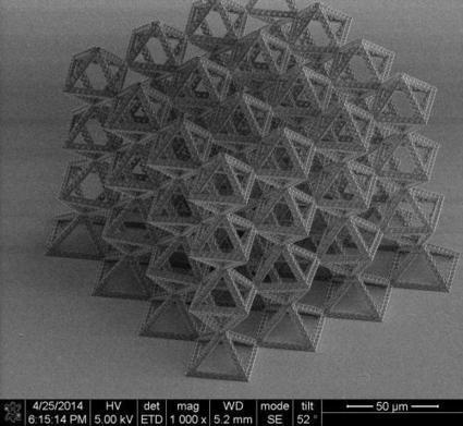
Julia Greer together with her group has developed a three-step process for building such complex structures very precisely. They first use a direct laser writing method called two-photon lithography to “write” a three-dimensional pattern in a polymer, allowing a laser beam to crosslink and harden the polymer wherever it is focused. At the end of the patterning step, the parts of the polymer that were exposed to the laser remain intact while the rest is dissolved away, revealing a three-dimensional scaffold. Next, the scientists coat the polymer scaffold with a continuous, very thin layer of a material—it can be a ceramic, metal, metallic glass, semiconductor, “just about anything,” Greer says. In this case, they used alumina, or aluminum oxide, which is a brittle ceramic, to coat the scaffold. In the final step they etch out the polymer from within the structure, leaving a hollow architecture.
Taking advantage of some of the size effects that many materials display at the nanoscale, these nanotrusses can have unusual, desirable qualities. For example, intrinsically brittle materials, like ceramics, including the alumina shown, can be made deformable so that they can be crushed and still rebound to their original state without global failure.
“Having full control over the architecture gives us the ability to tune material properties to what was previously unattainable with conventional monolithic materials or with foams,” says Greer. “For example, we can decouple strength from density and make materials that are both strong (and tough) as well as extremely lightweight. These structures can contain nearly 99 percent air yet can also be as strong as steel. Designing them into fractals allows us to incorporate hierarchical design into material architecture, which promises to have further beneficial properties.”
See on phys.org
Posted in Nanotechnology for Drug Delivery | Leave a Comment »
Reporter: Aviva Lev-Ari, PhD, RN
See on Scoop.it – Cardiovascular Disease: PHARMACO-THERAPY
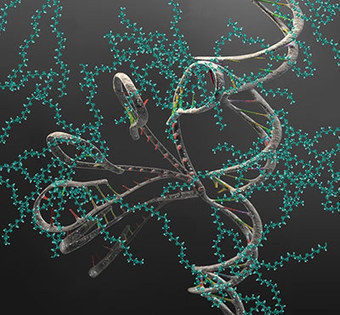
Crowding has notoriously negative effects at large size scales, blamed for everything from human disease and depression to community resource shortages. But relatively little is known about the influence of crowding at the cellular level. A new JILA study shows that a crowded environment has dramatic effects on individual biomolecules.
In the first data on the underlying dynamics (or kinetics)of crowded single biomolecules , reported in Proceedings of the National Academy of Sciences,* JILA researchers found that crowding leads to a 35-fold increase in the folding rate of RNA (ribonucleic acid), while the unfolding rate remains relatively stable.
RNA is a long chain-like molecule that contains genetic information, makes proteins and catalyzes biological reactions. It must fold into the correct 3D shape to function properly. The new results show that while RNA usually spends most of its time unfolded, in a crowded situation it folds much more often, although it remains folded for the usual period of time during each round.
“Cells are 25 to 35 percent filled with ‘stuff’—proteins, nucleic acids, lipids, etc.—and the effect of crowding on simple reactions like folding of nucleic acids and proteins is not well understood,” JILA/NIST Fellow David Nesbitt says. “Almost all detailed kinetic data comes from in vitro studies, that is, not in a living cell.
“But our work at the single-molecule level suggests that the rates and equilibrium constants (where folding and unfolding rates are equal) for simple nucleic acid folding processes may be shifted by up to 400,000 percent or more from what one might expect from such uncrowded solution studies.”
See on www.nist.gov
Posted in Amino acids, Proteins, Proteomics | Leave a Comment »
See on Scoop.it – Cardiovascular Disease: PHARMACO-THERAPY
A new method for using immunotherapy to specifically attack tumor cells that have mutations unique to a patient’s cancer has been developed by scientists at the National Cancer Institute (NCI), part of the National Institutes of Health. The researchers demonstrated that the human immune system can mount a response against mutant proteins expressed by cancers that arise in epithelial cells which can line the internal and external surfaces (such as the skin) of the body. These cells give rise to many types of common cancers, such as those that develop in the digestive tract, lung, pancreas, bladder and other areas of the body. The research provides evidence that this immune response can be harnessed for therapeutic benefit in patients, according to the scientists. The study appeared in the journal Science. “Our study deals with the central problem in human cancer immunotherapy, which is how to effectively attack common epithelial cancers,” said Steven Rosenberg, chief of the Surgery Branch in NCI’s Center for Cancer Research. “The method we have developed provides a blueprint for using immunotherapy to specifically attack sporadic or driver mutations, unique to a patient’s individual cancer.” All malignant tumors harbor genetic alterations, some of which may lead to the production of mutant proteins that are capable of triggering an antitumor immune response. Research led by Rosenberg and his colleagues had shown that human melanoma tumors often contain mutation-reactive immune cells called tumor-infiltrating lymphocytes, or TILs. The presence of these cells may help explain the effectiveness of adoptive cell therapy (ACT) and other forms of immunotherapy in the treatment of melanoma. In ACT, a patient’s own TILs are collected, and those with the best antitumor activity are grown in the laboratory to produce large populations that are infused into the patient. However, prior to this work it had not been clear whether the human immune system could mount an effective response against mutant proteins produced by epithelial cell cancers. These cells comprise more than 80% of all cancers. It was also not known whether such a response could be used to develop personalized immunotherapies for these cancers. In this study, Rosenberg and his team set out to determine whether TILs from patients with metastatic gastrointestinal cancers could recognize patient-specific mutations. They analyzed TILs from a patient with bile duct cancer that had metastasized to the lung and liver and had not been responsive to standard chemotherapy. The patient, a 43-year-old woman, was enrolled in an NIH trial of ACT for patients with gastrointestinal cancers.
See on www.dddmag.com
Posted in Uncategorized | Leave a Comment »
See on Scoop.it – Cardiovascular and vascular imaging
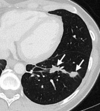
Study shows that incidental chest CT findings can help identify individuals at risk for future heart attacks
See on www.radiologyinfo.org
Posted in Uncategorized | Leave a Comment »