Maladaptive Vascular Remodeling found by four-dimensional (4D) flow MRI: Outflow Patterns, Wall Shear Stress, and Expression of Aortopathy are caused by Congenital bicuspid aortic valve (BAV) Cusp Fusion
Reporter: Aviva Lev-Ari, PhD, RN
- Original Article
- Valvular Heart Disease
Bicuspid Aortic Cusp Fusion Morphology Alters Aortic Three-Dimensional Outflow Patterns, Wall Shear Stress, and Expression of Aortopathy
- Riti Mahadevia, MD;
- Alex J. Barker, PhD;
- Susanne Schnell, PhD;
- Pegah Entezari, MD;
- Preeti Kansal, MD;
- Paul W.M. Fedak, MD;
- S. Chris Malaisrie, MD;
- Patrick McCarthy, MD;
- Jeremy Collins, MD;
- James Carr, MD;
- Michael Markl, PhD
+Author Affiliations
From the Department of Radiology, Feinberg School of Medicine, Northwestern University, Chicago, IL (R.M., A.J.B., SS., P.E., J. Collins, J. Carr, M.M.); Division of Cardiology, Northwestern University, Chicago, IL (P.K.); Department of Cardiac Sciences, Libin Cardiovascular Institute of Alberta, University of Calgary, Canada (P.W.M.F.); Division of Cardiothoracic Surgery, Northwestern University, Chicago, IL (P.W.M.F., S.C.M., P.M.)); and Department Biomedical Engineering, McCormick School of Engineering, Northwestern University, Chicago, IL (M.M.).
- Correspondence to Michael Markl, PhD, Department of Radiology, Northwestern University 737 N Michigan Ave, Suite 1600, Chicago, IL 60611. E-mailmichael.markl@northwestern.edu
Abstract
Background—Aortic 3-dimensional blood flow was analyzed to investigate altered ascending aorta (AAo) hemodynamics in bicuspid aortic valve (BAV) patients and its association with differences in cusp fusion patterns (right-left, RL versus right-noncoronary, RN) and expression of aortopathy.
Methods and Results—Four-dimensional flow MRI measured in vivo 3-dimensional blood flow in the aorta of 75 subjects: BAV patients with aortic dilatation stratified by leaflet fusion pattern (n=15 RL-BAV, mid AAo diameter=39.9±4.4 mm; n=15 RN-BAV, 39.6±7.2 mm); aorta size controls with tricuspid aortic valves (n=30, 41.0±4.4 mm); healthy volunteers (n=15, 24.9±3.0 mm). Aortopathy type (0–3), systolic flow angle, flow displacement, and regional wall shear stress were determined for all subjects. Eccentric outflow jet patterns in BAV patients resulted in elevated regional wall shear stress (P<0.0125) at the right-anterior walls for RL-BAV and right-posterior walls for RN-BAV in comparison with aorta size controls. Dilatation of the aortic root only (type 1) or involving the entire AAo and arch (type 3) was found in the majority of RN-BAV patients (87%) but was mostly absent for RL-BAV patients (87% type 2). Differences in aortopathy type between RL-BAV and RN-BAV patients were associated with altered flow displacement in the proximal and mid AAo for type 1 (42%–81% decrease versus type 2) and distal AAo for type 3 (33%–39% increase versus type 2).
Conclusions—The presence and type of BAV fusion was associated with changes in regional wall shear stress distribution, systolic flow eccentricity, and expression of BAV aortopathy. Hemodynamic markers suggest a physiological mechanism by which the valve morphology phenotype can influence phenotypes of BAV aortopathy.
Key Words:
Introduction
Congenital bicuspid aortic valve (BAV) is the most common congenital cardiovascular abnormality and occurs with an incidence of 1% to 2% of the population.1 This entity is associated with significant morbidity and mortality including valvular stenosis, valvular regurgitation, aortic dilatation, aneurysm, and dissection.2–5 The most common distinct morphologies have been identified based on fusion patterns between the right and left coronary leaflet (RL, frequency ≈80%), the right and noncoronary leaflet (RN, ≈17%), and the less commonly left and noncoronary leaflet (≈2%) fusion patterns.6,7 Additionally, BAV fusion patterns have been shown to be significant as a potential predictive factor in the location and rate of development of aortic complications.6,8 Although studies have linked genetics to the development of aortopathy in patients with BAV, the role of valve-related alterations in aortic hemodynamics and their impact on the underlying aortopathy is a reoccurring topic of debate.9 Studies based on flow-sensitive MRI and, more recently, four-dimensional (4D) flow MRI, provide evidence that the modified hemodynamic environments associated with BAV can cause altered wall shear stress (WSS) in the ascending aorta, which may trigger maladaptive vascular remodeling.10–14
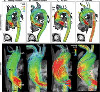
Figure 2.
Top, 3D streamline visualization of peak systolic blood flow in patients with BAV (C and D) in comparison with an aorta size–matched control subject (B) and a healthy volunteer (A). Note the presence of distinctly different 3D outflow flow jet patterns (black dashed arrows) in the ascending aorta (AAo) for patients B and C. Bottom, 3D flow patterns in the left ventricular outflow tract (LVOT) and AAo distal to the aortic valve. Note the different systolic aortic valve outflow flow jet patterns (red indicating high velocities > 1 m/s) and wall impingement zones that correspond to variable exertion of high wall shear forces between different valve groups (C and D) and aorta size–matched controls (B) and healthy volunteers (A). BAV indicates bicuspid aortic valve; RL, right and left coronary leaflet; and RN, right and noncoronary leaflet.
SOURCE for Image
http://circ.ahajournals.org/content/129/6/673.full#sec-28
CLINICAL PERSPECTIVE
The findings of this study show that the presence of bicuspid aortic valve and type of cusp fusion pattern were accompanied by changes in systolic outflow as quantified by flow displacement, flow angles, and regional wall shear stress. Altered aortic hemodynamics were clearly associated with the predominant expression of the aortopathy phenotype (aortic region affected by dilatation) which was different for the right-left in comparison with the less common right-noncoronary cusp fusion morphology. These findings represent new insights regarding the current guidelines using maximal aortic diameter to influence timing and extent of surgical resection. The decision to resect aortic tissues in bicuspid aortic valve aortopathy is difficult, because the degree of aortic dilatation can be highly variable with respect to location on the aorta and the degree of enlargement. The findings of this study indicate that 4-dimensional flow MRI may be used to determine which regional areas of the aorta are most prone to developing aortopathy, and different aortic resection may be indicated in patients with right-left versus right-noncoronary fusion patterns. However, our data also indicate that hemodynamic alterations and the aortopathy phenotype can have variable patterns that could be important for clinical surgical management decisions for the individual patient. Novel metrics of aortic hemodynamics such as flow displacement may be capable of shaping these decisions with respect to the timing and extent of aortic replacement in this diverse group of patients with bicuspid aortic valve.
- Received April 4, 2013.
- Accepted October 30, 2013.
- © 2013 American Heart Association, Inc.
References
- 1.↵
- Fedak PW,
- Verma S,
- David TE,
- Leask RL,
- Weisel RD,
- Butany J
. Clinical and pathophysiological implications of a bicuspid aortic valve. Circulation. 2002;106:900–904.
- 2.↵
- 3.↵
- Mordi I,
- Tzemos N
. Bicuspid aortic valve disease: a comprehensive review. Cardiol Res Pract. 2012;2012:196037.
- 4.↵
- Rudolph AM
. Bicuspid aortic valve and aortic stenosis. Congenital diseases of the heart. Hoboken, NJ: Wiley-Blackwell;2009:225–256.
- 5.↵
- Ward C
. Clinical significance of the bicuspid aortic valve. Heart. 2000;83:81–85.
- 6.↵
- Schaefer BM,
- Lewin MB,
- Stout KK,
- Gill E,
- Prueitt A,
- Byers PH,
- Otto CM
. The bicuspid aortic valve: an integrated phenotypic classification of leaflet morphology and aortic root shape. Heart. 2008;94:1634–1638.
- 7.↵
- 8.↵
- 9.↵
- Girdauskas E,
- Borger MA,
- Secknus MA,
- Girdauskas G,
- Kuntze T
. Is aortopathy in bicuspid aortic valve disease a congenital defect or a result of abnormal hemodynamics? A critical reappraisal of a one-sided argument. Eur J Cardiothorac Surg.2011;39:809–814.
- 10.↵
- 11.↵
- 12.↵
- 13.↵
- Barker AJ,
- Markl M
. The role of hemodynamics in bicuspid aortic valve disease.Eur J Cardiothorac Surg. 2011;39:805–806.
- 14.↵
- 15.↵
- Meierhofer C,
- Schneider EP,
- Lyko C,
- Hutter A,
- Martinoff S,
- Markl M,
- Hager A,
- Hess J,
- Stern H,
- Fratz S
. Wall shear stress and flow patterns in the ascending aorta in patients with bicuspid aortic valves differ significantly from tricuspid aortic valves: a prospective study. Eur Heart J Cardiovasc Imaging. 2013;14:797–804.
- 16.↵
- Barker AJ,
- Markl M,
- Bürk J,
- Lorenz R,
- Bock J,
- Bauer S,
- Schulz-Menger J,
- von Knobelsdorff-Brenkenhoff F
. Bicuspid aortic valve is associated with altered wall shear stress in the ascending aortaclinical perspective. Circ Cardiovasc Imaging.2012;5:457–466.
- 17.↵
- Bissell MM,
- Hess AT,
- Biasiolli L,
- Glaze SJ,
- Loudon M,
- Pitcher A,
- Davis A,
- Prendergast B,
- Markl M,
- Barker AJ,
- Neubauer S,
- Myerson SG
. Aortic dilation in bicuspid aortic valve disease: flow pattern is a major contributor and differs with valve fusion type.Circ Cardiovasc Imaging. 2013;6:499–507.
- 18.↵
- Kang JW,
- Song HG,
- Yang DH,
- Baek S,
- Kim DH,
- Song JM,
- Kang DH,
- Lim TH,
- Song JK
. Association between bicuspid aortic valve phenotype and patterns of valvular dysfunction and bicuspid aortopathy: comprehensive evaluation using MDCT and echocardiography. JACC Cardiovasc Imaging. 2013;6:150–161.
- 19.↵
- 20.↵
- Hiratzka LF,
- Bakris GL,
- Beckman JA,
- Bersin RM,
- Carr VF,
- Casey JDE,
- Eagle KA,
- Hermann LK,
- Isselbacher EM,
- Kazerooni EA,
- Kouchoukos NT,
- Lytle BW,
- Milewicz DM,
- Reich DL,
- Sen S,
- Shinn JA,
- Svensson LG,
- Williams DM
American College of Cardiology Foundation/American Heart Association Task Force on Practice Guidelines, American Association for Thoracic Surgery, American College of Radiology, American Stroke Association, Society of Cardiovascular Anesthesiologists, Society for Cardiovascular Angiography and Interventions, Society of Interventional Radiology, Society of Thoracic Surgeons, and Society for Vascular Medicine. 2010 ACCF/AHA/AATS/ACR/ASA/SCA/SCAI/SIR/STS/SVM guidelines for the diagnosis and management of patients with thoracic aortic disease. A Report of the American College of Cardiology Foundation/American Heart Association Task Force on Practice Guidelines, American Association for Thoracic Surgery, American College of Radiology,American Stroke Association, Society of Cardiovascular Anesthesiologists, Society for Cardiovascular Angiography and Interventions, Society of Interventional Radiology, Society of Thoracic Surgeons, and Society for Vascular Medicine. J Am Coll Cardiol. 2010;55:e27–e129.
- 21.↵
- Bonow RO,
- Carabello BA,
- Chatterjee K,
- de Leon AC,
- Faxon DP,
- Freed MD,
- Gaasch WH,
- Lytle BW,
- Nishimura RA,
- O’Gara PT,
- O’Rourke RA,
- Otto CM,
- Shah PM,
- Shanewise JS
; American College of Cardiology/American Heart Association Task Force on Practice Guidelines. 2008 focused update incorporated into the ACC/AHA 2006 guidelines for the management of patients with valvular heart disease: a report of the American College of Cardiology/American Heart Association Task Force on Practice Guidelines (Writing Committee to Revise the 1998 Guidelines for the Management of Patients With Valvular Heart Disease) Endorsed by the Society of Cardiovascular Anesthesiologists, Society for Cardiovascular Angiography and Interventions, and Society of Thoracic Surgeons. J Am Coll Cardiol. 2008;52:e1–e142.
- 22.↵
- 23.↵
- Bock J,
- Kreher B,
- Hennig J,
- Markl M
. Optimized pre-processing of time-resolved 2d and 3d phase contrast mri data. Proceedings of the 15th Annual Meeting of ISMRM,Berlin, Germany. 2007:3138.
- 24.↵
- 25.↵
- 26.↵
- 27.↵
- 28.↵
- Verma S,
- Yanagawa B,
- Kalra S,
- Ruel M,
- Peterson MD,
- Yamashita MH,
- Fagan A,
- Currie ME,
- White CW,
- Wai Sang SL,
- Rosu C,
- Singh S,
- Mewhort H,
- Gupta N,
- Fedak PW
.Knowledge, attitudes, and practice patterns in surgical management of bicuspid aortopathy: a survey of 100 cardiac surgeons. J Thorac Cardiovasc Surg.2013;146:1033–1040.e4.
- 29.↵
- 30.↵
- 31.↵
- 32.↵
- den Reijer PM,
- Sallee D 3rd.,
- van der Velden P,
- Zaaijer ER,
- Parks WJ,
- Ramamurthy S,
- Robbie TQ,
- Donati G,
- Lamphier C,
- Beekman RP,
- Brummer ME
.Hemodynamic predictors of aortic dilatation in bicuspid aortic valve by velocity-encoded cardiovascular magnetic resonance. J Cardiovasc Magn Reson. 2010;12:4.
Leave a Reply