BioMEMS based Optical Sensors
Author: Danut Dragoi, PhD
Optical sensors are so well developed that many applications can benefit from them. Important applications in medical field that utilizes embedded optical sensors are using the BioMEMS. In this presentation we focus on BioMEMS based optical sensors in ophthalmology, eyes artificial retina, LASIK, micro endoscope, plasmonic devices with single molecules detection utilizing SERS-Surface Enhance Raman Scattering in which photon interaction (scattering) with bio-cells is a major effect of the detection. It will be shown how cancer detection works (utilizing kapa/lambda ratio).
The presentation will focus also on eye vision correction, vision for the blind, and virtual reality for entertainment.
Slide 4 shows the results of the interaction of photons with living cells. Examples are given to illustrate the physical effects of the interaction. The abbreviations used in the text are: abs for absorption, H for Hydrogen, e for electron, m* for excited mass of the living cell, ElemPart for elementary particles.

Photon as an elementary particle, can be found in lasers, the best source of artificial light today. As we can remark on Slide 5 , the intensity of the lasers of very high power in peta watts range, one peta watts is 10 raised to the power of 15 watts, is expected to play a major role in the future of medicine.

Slide 6 shows the world’s most powerful laser fired at Japanese Lab of Osaka University.

On Slide 6 the laser beam is so intense, 2PW, so that the scattering from the air molecules can be seen on very large distances.
Slide 7 shows schematically our natural sensor, the eye, that works on visualizing objects like a tree exposed to natural light of Sun, in which light reflects / scatters in all directions, the lens of the eye focuses some rays on retina that give a signal to the brain through the optical nerve.

Slide 8 gives the location of visual spectrum in the general electromagnetic spectrum, in which color green is in the middle of visible spectrum. The nature selected the maximum of sensitivity of our eyes to be green color that coincides with the color of chlorophyll, the green pigment, present in all green plants.

Slide 9 shows the branch of eye care called Ophthalmology, in which correction of vision is done utilizing eye glasses or LASIK, an eye special surgery on cornea of the eye.

Slide 10 shows three options of eye correction of vision, normal vision gasses, contact lenses, and LASIK..
Slide 11 explains LASIK procedure, which is laser assisted in situ keratomileusis. In the three picture is shown the process of precise cut of the top of cornea, tissue removal using an eximer laser, whose wavelength is so short that does not penetrate the ocular lens, and the last step of flipping back the cut from cornea in the first stage. In many countries milions of people opted for this procedure.

Slide 12 is an attempt to explain the etymology of keratomileusis in the word LASIK.
Slide 13 suggests what beyond LASIK procedure, in which the concept for blindness is given as a solution utilizing implantable photo-detector arrays.

Slide 14 compares natural optical sensors with artificial optical sensor based on Si microelectronic technology.

Slide 15 shows a bio-optical sensor made by Anitoa, a company in Palo Alto CA. What is special about this photo-detector is its high sensitivity pushed toward one photon detection.

Slide 16 shows an endoscope with one fiber optic and two electrical lines, which is recognized as an Optical Coherent Topography device.
 The optical fiber guides a laser beam towards the end of the fiber where a GRIN lens, which is a gradient index lens, focuses the beam on a mirror that rock around an axis in order to scan the beam on the object, then the reflected beam goes back on mirror through the GRIN lens and the fiber again where an image is produced.
Slide 17 shows a MEMS endoscope made by Santec, where we recognise all elements described in previous slide.

Slide 18 compares sensors sensitive to visible spectrum made in nature, fruit flies eye, and those sensors made in the lab utilizing the model of fly eye. Because the resolution of the recreated eye fly is poor we expect that technology to not be used. The actual Si microelectronics is much better in producing high performance photo detectors.

Slide 19 shows a ‘smart’ contact lens to monitors the pressure inside the eye that can produce glaucoma and possibly lose the sight.

Slide 21 shows schematically a prosthetic retina for people who have the photoreceptors retina destroyed, either by disease or by an accidental exposure to a laser beam.

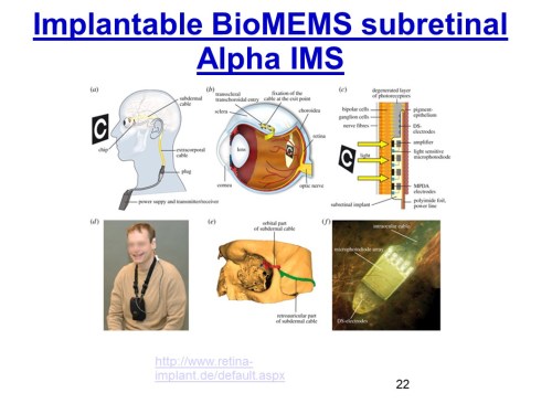
Slide 22 shows an implantable BioMEMS subretinal Alpha IMS for blind people.
Slide 23 shows the number of pixels in natural vision for different types of eyes, starting with low pixels for insect and ending with very high pixels for predatory birds. The horizontal axis describes the number of images . The red lines represent the memory storage of pixels for different vision systems.

Slide 24 shows the implantable retina micro-array from Sandia National Lab.

Slide 25 an artificial retina from Lawrence Livermore Lab.

Slide 26 describe other advanced optical devices based on SERS (Surface Enhanced Raman Scattering) for single molecule detection such as cancer cells, toxic molecules, poison molecule and other.

Slide 27 gives the definition of plasmon and Raman spectroscopy which is s the measurement of the wavelength and intensity of in-elastically scattered light from molecules. The Raman scattered light occurs at wavelengths that are shifted from the incident light by the energies of molecular vibrations.

Slide 28 is for how SERS works.

Slide 29 explains the principle of SERS for detection of single molecules.

Slide 30 shows the principle of SERS enhancement of the spectrum using Ag nano particles.

Slide 31 examples of molecules detected by SERS,

Slide 32 shows a mini-device plasmonic biosensor for leukemia detection.

Slide 33 shows how the optical plasmonic device is tuned to detect cancer cells by measuring IgG-kappa and IgG-lambda ratio.

Slide 34 shows how the ratio IgG-kappa and IgG-lambda is determined in clinical diagnostic utilizing SERS wave guides.

Slide 35 shows a MEMS device as a mini-spectrometers in visible range of the electromagnetic spectrum.

Slide 36 shows how the mini-spectrometers works.

Slide 37 shows a mini-spectrometer at work utilizing a laptop, an absorption cuvette and optical fibers for input into spectrometer and electrical connections between a laptop and spectrometer.

Slide 38 shows a mini MEMS USB spectrometer based WiFi.

Slide 39 shows MEMS USB spectrometer connected to an iPhone.

Slide 40 shows an integrated color sensors for blood glucose meters.

Slide 41 shows an optical device for measuring Oxygen saturation of blood.

Slide 42 shows how the oxymeter works.

Slide 43 shows the glaucoma can destroy the optical nerve producing total blindness.

Slide 44 gives the definition of glaucoma.

Slide 45 shows the micro-systemic approach for glaucoma.

Slide 46 shows bio MEMS coil for glaucoma. The graph on the slide show a calibration curve, resonant frequency of the coil versus pressure in a water testing device, where the pressure of water is well known and the frequency measured precisely with a pressure instrument.

Slide 48 shows the definition of cataract which is a leading eye problem for the older.

Slide 49 shows a BioMEMS artificial lens.

Slide 49 shows how artificial lens is working.

Slide 51 shows a sub-retinal BioMEMS principle of working.

Slide 52 shows a higher complex BioMEMS artificial retina system.

Slide 53 shows a BioMEMS artificial retina system by Professor Wilfried Mokwa of RWTH Aachen University.

Slide 54 shows a BioMEMS and epiretinal stimulation from Retina Implant AG.

Slide 55 shows a Bionic Microchip at the back of the eye with 1500 pixels.

Slide 56 shows a bionic microchip installed on the back of the eye.
Slide 57 shows a schematic of retinal bionic implant, 3×3 mm with a light processing cells, which is the latest generation of a light sensitive chip.

Slide 58 shows a contact lens for Virtual Reality applications. Notice in this application the eye is healthy and normal functioning.

Slide 59 shows a description of contact lenses for Virtual Reality applications.

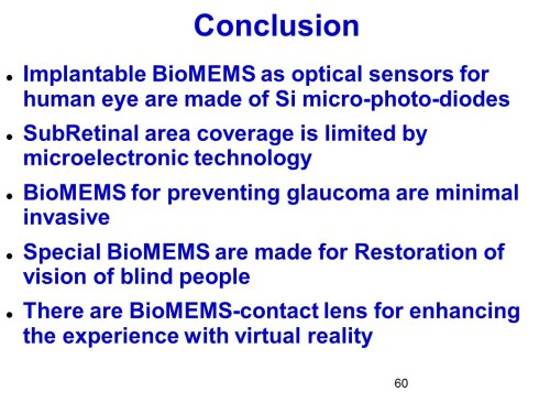
Slide 60 and 61 show the conclusions.

This is the end of the presentation. Thank you!