Feeds:
Posts
Comments

Archive for March, 2013

Amyloidosis with Cardiomyopathy

Author: Larry H Bernstein, MD, FACP
Introduction
Amyloidosis describes the various clinical syndromes that occur as a result of damage by amyloid deposits in tissues and organs throughout the body.  Systemic amyloidosis is a relatively rare multisystem disease caused by the deposition of misfolded protein in various tissues and organs. The term amyloid describes the deposition in the extracellular space of certain proteins in a highly characteristic, insoluble fibrillar form.  The disease entity is a disorder of misfolded or misassembled proteins.  There is extracellular amyloid fiber laid down as cross β-sheets disrupting organ function, which may affect the pancreas, kidney, autonomic nervous system, the heart, and in one form causes carpal tunnel syndrome.
It may present to almost any specialty, and diagnosis is frequently delayed. Cardiac involvement is a leading cause of morbidity and mortality, especially in primary light chain (AL) amyloidosis and in both wild-type and hereditary transthyretin amyloidosis. The heart is also occasionally involved in acquired serum amyloid A type (AA) amyloidosis and other rare hereditary types. Clinical phenotype varies greatly between different types of amyloidosis, and even the cardiac presentation has a great spectrum. The incidence of amyloidosis is uncertain, but it is thought that the most frequently diagnosed AL amyloidosis has an annual incidence of 6 to 10 cases per million population in the United Kingdom and United States.
The molecular basis for this particular phenomenon came with the extensive work done on multiple myeloma, antibody structure, and light chains.  In 1950, the discovery of a familial amyloid polyneuropathy was described in Portugal, and there were similar diseases in Sweden and Japan.  There were 72 known variants of transthyretin (TTR) in 1995, and now there are 100.  In addition, the occurance of different TTR associated variants with and without (amyloid) is found is Brazil, UK, US, Israel, Spain, France, Germany, Denmark, and Africa.  The table of variants, organ damage, and geographic location is too large to place on this document. If we refer to amyloid cardiomyopathy, it is exclusively a primary amyloidopathy, not secondary to light chain disorders or an inflammatory disease.  If we consider amyloidosis, we also have to consider family history, organ dysfunction, and we have to make a distinction between primary cardiac involvement, autonomic nervous system instability, and the two coexisting.  Familial amyloid polyneuropathy (FAP) is an extremely debilitating and progressive disease that is only treatable by liver transplantation.  Primary amyloid cardiomyopathy has been treated by heart transplant.  The qualifying statement here is, it depends.

Primary and Secondary Amyloidoses

Amyloid was originally described by pathologists based on microscopy. Amyloidoses are a systemic primary or secondary disease. There are distinctions to be made based on location and type.  The clinical significance of amyloid disease varies enormously, ranging from incidental asymptomatic deposits to localized disease through to rapidly fatal systemic forms that can affect multiple vital organs.
Common causes of secondary amyloidosis are – light chain production (AL) as in plasma cell dyscrasia, amyloid A (AA), senile systemic amyloidosis (diagnosed rarely in life).  The systemic amyloidoses are designated by a capital A (for amyloid) followed by the abbreviation for the chemical identity of the fibril protein. Thus, TTR amyloidosis is abbreviated ATTR, and immunoglobulin light chain type amyloidosis is abbreviated AL. Both normal-sequence TTR and variant-sequence TTR form amyloidosis. Normal-sequence TTR forms cardiac amyloidosis in elderly people, termed senile cardiac amyloidosis (SCA). When it was recognized that SCA is often accompanied by microscopic deposits in many other organs, the alternative name senile systemic amyloidosis (SSA) was proposed. Both terms are now used.
Currently available therapy is focused on reducing the supply of the respective amyloid fibril precursor protein and supportive medical care, which together have greatly improved survival. Chemotherapy and anti-inflammatory treatment for the disorders that underlie AL and AA amyloidosis are guided by serial measurements of the respective circulating amyloid precursor proteins, i.e. serial serum free light chains in AL and serum amyloid A protein in AA type.
Quality of life and prognosis of some forms of hereditary systemic amyloidosis can be improved by liver and other organ transplants. Various new therapies, ranging from silencing RNA, protein stabilizers to monoclonal antibodies, aimed at inhibiting fibril precursor supply, fibril formation or the persistence of amyloid deposits, are in development; some are already in clinical phase.
Ann Clin Biochem May 2012; 49(3 ): 229-241   http://acb.2011.011225v1 49/3/229

What is transthyretin (TTR)?

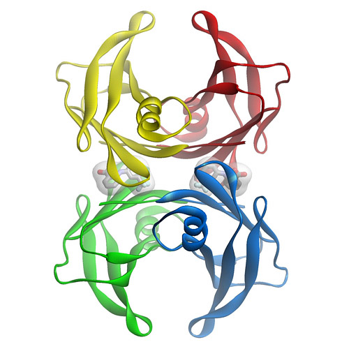
TTR is a  tetramer of 4 127 amino acid subunits synthesized by the liver that circulates as a transporter of thyroxin, and with retinol-binding protein, transports vitamin A.  It was originally defined by the migration in electrophoresis more anodal to albumin, hence, prealbumin.  It is present in cerebrospinal fluid, secreted by the choroid plexus.  The TTR monomer contains 8 antiparallel beta pleated sheet domains. TTR can be found in plasma and in cerebrospinal fluid and is synthesized by the choroid plexus of the brain and, to a lesser degree, by the retina. Its gene is located on the long arm of chromosome 18 and contains 4 exons and 3 introns.
The concentration in serum can be expected to be above 20 mg/dL in a health adult, but the protein decreases by 1 mg/dL/day postoperatively, and it decreases with acute or chronic renal failure, pneumonia or sepsis, rising again with the onset of anabolism.  Patients in the pulmonary intensive care unit have TTR levels that remain low for 7-10 days, but followup data for the remainder of the hospital stay or in relationship to readmission in the six months after release from hospital care was not part of the study.
A decrease in TTR is associated with the systemic inflammatory response, whereby, the liver reprioritizes the synthesis of proteins with an increase in acute phase reactants (APRs), namely, C-reactive protein (CRP) and a-1 acid glycoprotein, and decreased albumin and TTR.  The inflammatory condition maintains a euthyroid status with decreased TTR because of the availability of free thyroxine in equilibrium with the lower binding protein.  This has been referred to sick euthyroid status. The role in thyroxine transport is not insignificant, as chronic protein malnutrition is associated with hypothyroidism, as originally described by Prof. Yves Ingenbleek, Univ. Louis Pateur, Starsbourg, Fr. in Senegalese children with Kwashiorkor.  However, the importance of TTR as a unique biomarker is not to be downgraded because of what is often refered to as “an inverted APR”.
Transthyretin was discovered to be a good reflection of the “lean body mass”, by Vernon Young, MIT, and Ingenbleek, as a result of 3 decades of study. The ratio of S:N being 1:20 in plant proteins and 1:12.5 in animal sources, is closely related to methylation reactions and sustained deficiency of S intake results in elevated homocysteine level.

What is FAP?

Familial amyloid polyneuropathy (FAP), also called transthyretin-related hereditary amyloidosis, transthyretin amyloidosis or Corino de Andrade’s disease, is an autosomal dominant neurodegenerative disease. It is a form of amyloidosis, and was first identified and described by Portuguese neurologist Mário Corino da Costa Andrade, in the 1950s.FAP is distinct from senile systemic amyloidosis (SAS), which is not inherited, and which was determined to be the primary cause of death for 70% of supercentenarians who have been autopsied.
Familial amyloid polyneuropathy (FAP) is an extremely debilitating and progressive disease that is only treatable by liver transplantation.  Primary amyloid cardiomyopathy has been treated by heart transplant.  The qualifying statement here is, it depends.  Those patients with TTR-amyloidopathy have a specific gene substitution in the TTR gene. Consequently, there is circulation TTR, but it is not effectively involved in thyroxine transport.

Characteristics.

Usually manifesting itself between 20 and 40 years of age, it is characterized by pain, paresthesia, muscular weakness and autonomic dysfunction. In its terminal state, the kidneys and the heart are affected. FAP is characterized by the systemic deposition of amyloidogenic variants of the transthyretin protein, especially in the peripheral nervous system, causing a progressive sensory and motor polyneuropathy. The age at symptom onset, pattern of organ involvement, and disease course vary, but most mutations are associated with cardiac and/or nerve involvement. The gastrointestinal tract, vitreous, lungs, and carpal ligament are also frequently affected. When the peripheral nerves are prominently affected, the disease is termed familial amyloidotic polyneuropathy (FAP). When the heart is involved heavily but the nerves are not, the disease is called familial amyloid cardiomyopathy (FAC). Regardless of which organ is primarily targeted, the general term is simply amyloidosis-transthyretin type, abbreviated ATTR.

Genetics.

  1. TTR mutations accelerate the process of TTR amyloid formation and are the most important risk factor for the development of clinically significant ATTR. More than 85 amyloidogenic TTR variants cause systemic familial amyloidosis. The variant TTR is mostly produced by the liver. Amyloidogenic TTR mutations destabilize TTR monomers or tetramers, allowing the molecule to more easily attain an amyloidogenic intermediate conformation. The tetramer has to dissociate into misfolded monomers to aggregate into a variety of structures including amyloid fibrils. Because most patients are heterozygotes, they deposit both mutant and wild type TTR subnits.
  2. Familial amyloid polyneuropathy has an autosomal dominant pattern of inheritance. FAP is caused by a mutation of the TTR gene, located on human chromosome 18q12.1-11.2. A replacement of valine by methionine at position 30 (TTR V30M) is the mutation most commonly found in FAP.
  3. The disease in the TTR V30M kindreds was termed FAP because early symptoms arose from peripheral neuropathy, but these patients actually have systemic amyloidosis, with widespread deposits often involving the heart, gastrointestinal tract, eye, and other organs.
  4. TTR V122I: This variant, carried by 3.9% of African Americans and over 5% of the population in some areas of West Africa, increases the risk of late-onset (after age 60 years) cardiac amyloidosis. It appears to be the most common amyloid-associated TTR variant worldwide. Affected patients usually do not have peripheral neuropathy.
  5. TTR T60A: This variant causes late-onset systemic amyloidosis with cardiac, and sometimes neuropathic, involvement. This variant originated in northwest Ireland and is found in Irish and Irish American patients.
  6. TTR L58H: Typically affecting the carpal ligament and nerves of the upper extremities, this variant originated in Germany. It has spread throughout the United States but is most common in the mid-Atlantic region.
  7. TTR G6S: This is the most common TTR variant, but it appears to be a neutral polymorphism not associated with amyloidosis. It is carried by about 10% of people of white European descent.

Cardiac transthyretin (TTR) amyloidosis

Cardiac amyloidosis of transthyretin fibril protein (ATTR) type is an infiltrative cardiomyopathy characterised by ventricular wall thickening and diastolic heart failure. More than 27 different precursor proteins have the propensity to form amyloid fibrils. The particular precursor protein that misfolds to form amyloid fibrils defines the amyloid type and predicts the patient’s clinical course. Several types of amyloid can infiltrate the heart, resulting in progressive diastolic and systolic dysfunction, congestive heart failure, and death.  Increased access to cardiovascular magnetic resonance imaging has led to a marked increase in referrals to St George’s University of London, London (Dr. Jason Dungu) of Caucasian patients with wild-type ATTR (senile systemic) amyloidosis and Afro-Caribbean patients with the hereditary ATTR V122I type. Both subtypes present predominantly as isolated cardiomyopathy. The differential diagnosis includes cardiac amyloid light-chain (AL) amyloidosis, which has a poorer prognosis and can be amenable to chemotherapy.

Clinical Presentation

Cardiac amyloidosis, irrespective of type, presents as a restrictive cardiomyopathy characterized by progressive diastolic and subsequently systolic biventricular dysfunction and arrhythmia.1 Key “red flags” to possible systemic amyloidosis include nephrotic syndrome, autonomic neuropathy (eg, postural hypotension, diarrhea), soft-tissue infiltrations (eg, macroglossia, carpal tunnel syndrome, respiratory disease), bleeding (eg, cutaneous, such as periorbital, gastrointestinal), malnutrition/cachexia and genetic predisposition (eg, family history, ethnicity). Initial presentations may be cardiac, with progressive exercise intolerance and heart failure. Other organ involvement, particularly in AL amyloidosis, may cloud the cardiac presentation (eg, nephrotic syndrome, autonomic neuropathy, pulmonary or bronchial involvement). Pulmonary edema is not common early in the disease process, but pleural and pericardial effusions and atrial arrhythmias are often seen. Syncope is common and a poor prognostic sign. It is typically exertional or postprandial as part of restrictive cardiomyopathy, sensitivity to intravascular fluid depletion from loop diuretics combined with autonomic neuropathy, or conduction tissue involvement (atrioventricular or sinoatrial nodes) or ventricular arrhythmia. The latter may rarely cause recurrent syncope. Disproportionate septal amyloid accumulation mimicking hypertrophic cardiomyopathy with dynamic left ventricular (LV) outflow tract obstruction is rare but well documented. Myocardial ischemia can result from amyloid deposits within the microvasculature. Atrial thrombus is common, particularly in AL amyloidosis

Diagnosis and Treatment

imaging – Cardiovascular Magnetic Resonance in Cardiac Amyloidosis*.

Cardiac amyloidosis can be diagnostically challenging. Cardiovascular magnetic resonance (CMR) can assess abnormal myocardial interstitium. In cardiac amyloidosis, CMR shows a characteristic pattern of global subendocardial late enhancement coupled with abnormal myocardial and blood-pool gadolinium kinetics. The findings agree with the transmural histological distribution of amyloid protein and the cardiac amyloid load.
 *AM Maceira; J Joshi; SK Prasad; J Charles Moon, et al. Royal Brompton Hospital, London;
The diagnosis of amyloidosis requires histological identification of amyloid deposits. Congo Red staining renders amyloid deposits salmon pink by light microscopy, with a characteristic apple green birefringence under polarized light conditions. Additional immunohistochemical staining for precursor proteins identifies the type of amyloidosis.  Ultimately, immunogold electron microscopy and mass spectrometry confer the greatest sensitivity and specificity for amyloid typing.
Treatment of cardiac amyloidosis is dictated by the amyloid type and degree of involvement. Consequently, early recognition and accurate classification are essential.
Novel diagnostic and surveillance approaches using imaging (echocardiography, cardiovascular magnetic resonance), biomarkers (brain natriuretic peptide [BNP], high-sensitivity troponin), new histological typing techniques, and current and future treatments, including approaches directly targeting the amyloid deposits.

Etiology

Amyloidosis is caused by the extracellular deposition of autologous protein in an abnormal insoluble β-pleated sheet fibrillary conformation—that is, as amyloid fibrils. More than 30 proteins are known to be able to form amyloid fibrils in vivo, which cause disease by progressively damaging the structure and function of affected tissues. Amyloid deposits also contain minor nonfibrillary constituents, including serum amyloid P component (SAP), apolipoprotein E, connective tissue components (glycosaminoglycans, collagen), and basement membrane components (fibronectin, laminin). Amyloid deposits can be massive, and cardiac or other tissues may become substantially replaced. Amyloid fibrils bind Congo red stain, yielding the pathognomonic apple-green birefringence under cross-polarized light microscopy that remains the gold standard for identifying amyloid deposits.

AL Amyloidosis

AL amyloidosis is caused by deposition of fibrils composed of monoclonal immunoglobulin light chains and is associated with clonal plasma cell or other B-cell dyscrasias. The spectrum and pattern of organ involvement is very wide, but cardiac involvement occurs in half of cases and is sometimes the only presenting feature. Cardiac AL amyloidosis may be rapidly progressive. Low QRS voltages, particularly in the limb leads, are common. Thickening of the LV wall is typically mild to moderate and is rarely >18 mm even in advanced disease. Cardiac AL amyloid deposition is accompanied by marked elevation of the biomarkers BNP and cardiac troponin, even at an early stage. Involvement of the heart is the commonest cause of death in AL amyloidosis and is a major determinant of prognosis; without cardiac involvement, patients with AL amyloidosis have a median survival of around 4 years, but the prognosis among affected patients with markedly elevated BNP and cardiac troponin (Mayo stage III disease) is on the order of 8 months.

Hereditary Amyloidoses

Mutations in several genes, such as transthyretin, fibrinogen, apolipoprotein A1, and apolipoprotein A2 can be responsible for hereditary amyloidosis, but by far the most common cause is variant ATTR amyloidosis (variant ATTR) caused by mutations in the transthyretin gene causing neuropathy and, often, cardiac involvement.

TTR gene mutation

 The most common is the Val122Ile mutation. In a large autopsy study that included individuals with cardiac amyloidosis, the TTR Val122Ile allele was present in 3.9% of all African Americans and 23% of African Americans with cardiac amyloidosis. Penetrance of the mutation is not truly known and is associated with a late-onset cardiomyopathy that is indistinguishable from senile cardiac amyloidosis.

Pathology, Presentation, and Management of Amyloidoses

More than 100 genetic variants of TTR are associated with amyloidosis. Most present as the clinical syndrome of progressive peripheral and autonomic neuropathy. Unlike wild-type ATTR or variant ATTR Val122Ile, the features of other variant ATTR include vitreous amyloid deposits or, rarely, deposits in other organs. Cardiac involvement in variant ATTR varies by mutations and can be the presenting or indeed the only clinical feature. For example, cardiac involvement is rare in variant ATTR associated with Val30Met (a common variant in Portugal or Sweden), but it is almost universal and develops early in individuals with variant ATTR due to Thr60Ala mutation (a mutation common in Ireland).

Senile Systemic Amyloidosis (Wild-Type ATTR)

Wild-type TTR amyloid deposits are found at autopsy in about 25% of individuals >80 years of age.  The prevalence of wild-type TTR deposits leading to the clinical syndrome of wild-type ATTR cardiac amyloidosis is unknown. Wild-type ATTR is a predominantly cardiac disease, and the only other significant extracardiac feature is a history of carpal tunnel syndrome, often preceding heart failure by 3 to 5 years. Extracardiac involvement is most unusual.
Both wild-type ATTR and ATTR due to Val122Ile are diseases of the >60-year age group and are often misdiagnosed as hypertensive heart disease. Wild-type ATTR has a strong male predominance, and the natural history remains poorly understood, but studies suggest a median survival of about 7 years from presentation. Recent developments in cardiac magnetic resonance (CMR), which have greatly improved detection of cardiac amyloid during life, suggest that wild-type ATTR is more common than previously thought: It accounted for 0.5% of all patients seen at the UK amyloidosis center until 2001 but now accounts for 7% of 1100 cases with amyloidosis seen since the end of 2009. There appears to be an association between wild-type ATTR and history of myocardial infarctions, G/G (Val/Val) exon 24 polymorphism in the alpha2-macroglobulin (alpha2M), and the H2 haplotype of the tau gene36; the association of tau with Alzheimer’s disease raises interesting questions as both are amyloid-associated diseases of aging.
ECG of a patient with cardiac AL amyloidosis showing small QRS voltages (defined as ≤6 mm height), predominantly in the limb leads and pseudoinfarction pattern in the anterior leads.
Echocardiography is characteristic. Typical findings include concentric ventricular thickening with right ventricular involvement, poor biventricular long-axis function with normal/near-normal ejection fraction and valvular thickening (particularly in wild-type or variant ATTR). Diastolic dysfunction is the earliest echocardiographic abnormality and may occur before cardiac symptoms develop. Biatrial dilatation in presence of biventricular, valvular, and interatrial septal thickening 53 is a useful clue to the diagnosis.
Transthoracic echocardiogram with speckle tracking. The red and yellow lines represent longitudinal motion in the basal segments, whereas the purple and green lines represent apical motion. This shows loss of longitudinal ventricular contraction at the base compared to apex.

Biomarkers.

High-sensitivity troponin is abnormal in >90% of cardiac AL patients, and the combination of BNP/NT-proBNP plus troponin measurements is used to stage and risk-stratify patients with AL amyloidosis at diagnosis. Very interestingly, the concentration of BNP/NT-proBNP in AL amyloidosis may fall dramatically within weeks after chemotherapy that substantially reduces the production of amyloidogenic light chains. The basis for this very rapid phenomenon, which is not mirrored by changes on echocardiography or CMR, remains uncertain, but a substantial fall is associated with improved outcomes.

Cardiac Magnetic Resonance.

CMR provides functional and morphological information on cardiac amyloid in a similar way to echocardiography, though the latter is superior for evaluating and quantifying diastolic abnormalities. An advantage of CMR is in myocardial tissue characterization. Amyloidotic myocardium reveals subtle precontrast abnormalities (T1, T2), but extravascular contrast agents based on chelated gadolinium provide the key information.

CMR with the classic amyloid global, subendocardial late gadolinium enhancement pattern in the left ventricle with blood and mid-/epimyocardium nulling together.
Recently, the technique of equilibrium contrast CMR has demonstrated much higher extracellular myocardial volume in cardiac amyloid than any other measured disease. It is anticipated that accurate measurements of the expanded interstitium in amyloidosis will prove useful in serial quantification of cardiac amyloid burden.
Sequential static images from a CMR TI scout sequence. As the inversion time (TI) increases, myocardium nulls first (arrow in image 3), followed by blood afterwards (arrow in image 6), implying that there is more gadolinium contrast in the myocardium than blood—a degree of interstitial expansion such that the “myocrit” is smaller than the hematocrit.

Tissue biopsy.

To confirm amyloidosis, including familial TTR amyloidosis, the demonstration of amyloid deposition on biopsied tissues is essential. With Congo red staining, amyloid deposits show a characteristic yellow-green birefringence under polarized light. Tissues suitable for biopsy include: subcutaneous fatty tissue of the abdominal wall, skin, gastric or rectal mucosa, sural nerve, and peritendinous fat from specimens obtained at carpal tunnel surgery. Sensitivity of endoscopic biopsy of gastrointestinal mucosa is around 85%; biopsy of the sural nerve is less sensitive. It is ideal to show that these amyloid deposits are specifically immunolabeled by anti-TTR antibodies.

Serum variant TTR protein.

TTR protein normally circulates in serum or plasma as a soluble protein having a tetrameric structure [Kelly 1998, Rochet & Lansbury 2000]. Normal plasma TTR concentration is 20-40 mg/dL (0.20-0.40 mg/mL).  Pathogenic mutations in TTR cause conformational change in the TTR protein molecule, disrupting the stability of the TTR tetramer, which is then more easily dissociated into pro-amyloidogenic monomers.

After immunoprecipitation with anti-TTR antibody, serum variant TTR protein can be detected by mass spectrometry. Approximately 90% of TTR variants so far identified are confirmed by this method. Mass shift associated with each variant TTR protein is indicated.

Molecular genetic testing.

  • TTR is the only gene in which mutations are known to cause familial TTR amyloidosis.
  • Identified in many individuals of different ethnic backgrounds; found in large clusters in Portugal, Sweden, and Japan.
  • The gene has four exons; and all the hitherto-identified mutations are in exons 2, 3, or 4.
GeneReviews designates a molecular genetic test as clinically available only if the test is listed in the GeneTests Laboratory Directory by either a US CLIA-licensed laboratory or a non-US clinical laboratory.
  • Molecular genetic testing of TTR by sequence analysis (may be preceded by targeted mutation analysis)
  • Although deletion/duplication testing is available clinically, no exonic or whole-gene deletions or duplications involving TTR have been reported to cause familial transthyretin amyloidosis.
  • However, with newly available deletion/duplication testing methods, it is theoretically possible that such mutations may be identified in affected individuals in whom prior testing by sequence analysis of the entire coding region was negative.
  • Predictive testing for at-risk asymptomatic adult family members requires prior identification of the disease-causing mutation in the family.
  • Prenatal diagnosis and preimplantation genetic diagnosis (PGD) for at-risk pregnancies require prior identification of the disease-causing mutation in the family.

Genetically Related (Allelic) Disorders

Familial euthyroid hyperthyroxinemia is caused by normal allelic variants in TTR, including Gly6Ser, Ala109Thr, Ala109Val, and Thr119Met (see Table 5) [Nakazato 1998, Benson 2001, Saraiva 2001]. The TTR protein binds approximately 15% of serum thyroxine. These mutations increase total serum thyroxine concentration because of their increased affinity for thyroxine, however, they increase neither free thyroxine nor free triiodothyronine. Therefore, individuals with these sequence variants develop no clinical symptoms (i.e., they are euthyroid).
Senile systemic amyloidosis (SSA; previously called senile cardiac amyloidosis) results from the pathologic deposition of wild-type TTR, predominantly in the heart. Pathologic deposits are also seen in the lungs, blood vessels, and the renal medulla of the kidneys [Westermark et al 2003]. SSA affects mainly the elderly but is rarely diagnosed during life.
Sensorimotor neuropathy and autonomic neuropathy progress over ten to 20 years. Various types of cardiac conduction block frequently appear. Cachexia is a common feature at the late stage of the disease. Affected individuals usually die of cardiac failure, renal failure, or infection.

Cardiac amyloidosis.

Cardiac amyloidosis, mainly characterized by progressive cardiomyopathy, has been reported with more than two thirds of TTR mutations. In some families with specific TTR mutations, such as Asp18Asn, Val20Ile, Pro24Ser, Ala45Thr, Ala45Ser, His56Arg, Gly57Arg, Ile68Leu, Ala81Thr, Ala81Val, His88Arg, Glu92Lys, Arg103Ser, Leu111Met, or Val122Ile, cardiomyopathy without peripheral neuropathy is a main feature of the disease.

Cardiac amyloidosis is usually late onset. Most individuals develop cardiac symptoms after age 50 years; cardiac amyloidosis generally presents with restrictive cardiomyopathy. The typical electrocardiogram shows a pseudoinfarction pattern with prominent Q wave in leads II, III, aVF, and V1-V3, presumably resulting from dense amyloid deposition in the anterobasal or anteroseptal wall of the left ventricle. The echocardiogram reveals left ventricular hypertrophy with preserved systolic function. The thickened walls present “a granular sparkling appearance.”
Among the mutations responsible for cardiac amyloidosis, Val122Ile is notable for its prevalence in African Americans. Approximately 3.0%-3.9% of African Americans are heterozygous for Val122Ile . The high frequency of Val122Ile partly explains the observation that in individuals in the US older than age 60 years, cardiac amyloidosis is four times more common among blacks than whites.

Leptomeningeal (oculoleptomeningeal) amyloidosis.

Amyloid deposition is seen in the pial and arachnoid membrane, as well as in the walls of vessels in the subarachnoid space associated with TTR mutations including Leu12Pro, Asp18Gly, Ala25Thr, Val30Gly, Ala36Pro, Gly53Glu, Gly53Ala, Phe64Ser, Tyr69His, or Tyr114Cys.  Individuals with leptomeningeal amyloidosis show CNS signs and symptoms including: dementia, psychosis, visual impairment, headache, seizures, motor paresis, ataxia, myelopathy, hydrocephalus, or intracranial hemorrhage. When associated with vitreous amyloid deposits, leptomeningeal amyloidosis is known as familial oculolepto-meningeal amyloidosis (FOLMA). In leptomeningeal amyloidosis protein concentration in the cerebrospinal fluid is usually high, and gadolinium-enhanced MRI typically shows extensive enhancement of the surface of the brain, ventricles, and spinal cord.

Genotype-Phenotype Correlations.

In subsets of families with the Val30Met mutation, considerable variation in phenotypic manifestations and age of onset is observed. It is hypothesized that genetic modifiers and non-genetic factors contribute to the pathogenesis and progression of familial TTR amyloidosis. The vast majority of individuals with familial TTR amyloidosis are heterozygous for a TTR mutation. It has been clinically and experimentally demonstrated that the normal allelic variant c.416C>T (Thr119Met) has a protective effect on amyloidogenesis in individuals who have the Val30Met mutation.

Cardiac amyloidosis is caused by Asp18Asn, Val20Ile, Pro24Ser, Ala45Thr, Ala45Ser, His56Arg, Gly57Arg, Ile68Leu, Ala81Thr, Ala81Val, His88Arg, Glu92Lys, Arg103Ser, Leu111Met, or Val122Ile. Peripheral and autonomic neuropathy are absent or less evident in persons with these mutations.
Leptomeningeal amyloidosis is associated with Leu12Pro, Asp18Gly, Ala25Thr, Val30Gly, Ala36Pro, Gly53Glu, Gly53Ala, Phe64Ser, Tyr69His, or Tyr114Cys.

Penetrance.

It is generally accepted that the penetrance is much higher in individuals in endemic foci than outside of endemic foci. In Portugal, cumulative disease risk in individuals with the Val30Met mutation is estimated at 80% by age 50 and 91% by age 70 years, whereas the risk in French heterozygotes is 14% by age 50 and 50% by age 70 years. In Sweden, the penetrance is much lower: 1.7% by age 30, 5% by age 40, 11% by age 50, 22% by age 60, 36% by age 70, 52% by age 80, and 69% by age 90, respectively.

Nomenclature

The neuropathy associated with TTR mutations, now called familial TTR amyloidosis, was formerly referred to as one of the following:
  • Familial amyloid polyneuropathy type I (or the Portuguese-Swedish-Japanese type)
  • Familial amyloid polyneuropathy type II (or the Indiana/Swiss or Maryland/German type)

Prevalence

The Val30Met mutation, found worldwide, is the most widely studied TTR variant and is responsible for the well-known large foci of individuals with TTR amyloid polyneuropathy in Portugal, Sweden, and Japan. Numerous families with various non-Val30Met mutations have also been identified worldwide.

 Small transthyretin (TTR) ligands as possible therapeutic agents in TTR amyloidoses.

Almeida MR, Gales L, Damas AM, Cardoso I, Saraiva MJ. Porto, Portugal.
Curr Drug Targets CNS Neurol Disord. 2005 Oct;4(5):587-96.
In transthyretin (TTR) amyloidosis TTR variants deposit as amyloid fibrils giving origin, in most cases, to peripheral polyneuropathy, cardiomyopathy, carpal tunnel syndrome and/or amyloid deposition in the eye. The amino acid substitutions in the TTR variants destabilize the tetramer, which may dissociate into non native monomeric intermediates that aggregate and polymerize in amyloid fibrils that further elongate. Since this is a multi-step process there is the possibility to impair TTR amyloid fibril formation at different stages of the process namely by tetramer stabilization, inhibition of fibril formation or fibril disruption. Based on the proposed mechanism for TTR amyloid fibril formation we discuss the action of some of the proposed TTR stabilizers such as derivatives of some NSAIDs (diflunisal, diclofenac, flufenamic acid, and derivatives) and the action of amyloid disrupters such as 4′-iodo-4′-deoxydoxorubicin (I-DOX) and tetracyclines. Among all these compounds, TTR stabilizers seem to be the most interesting since they would impair very early the process of amyloid formation and could also have a prophylactic effect.

Clusterin regulates transthyretin amyloidosis.

Lee KW, Lee DH, Son H, Kim YS, Park JY, et al.  Gyeongnam National University, South Korea
Biochem Biophys Res Commun 2009;388(2):256-60.   http://dx.doi.org/10.1016/j.bbrc.2009.07.166.
Clusterin has recently been proposed to play a role as an extracellular molecular chaperone, affecting the fibril formation of amyloidogenic proteins. The ability of clusterin to influence amyloid fibril formation prompted us to investigate whether clusterin is capable of inhibiting TTR amyloidosis. Here, we report that clusterin strongly interacts with wild-type TTR and TTR variants V30M and L55P under acidic conditions, and blocks the amyloid fibril formation of TTR variants. In particular, the amyloid fibril formation of V30M TTR in the presence of clusterin is reduced to level similar to wild-type TTR. We also demonstrated that clusterin is an effective inhibitor of L55P TTR amyloidosis, the most aggressive form of TTR diseases. The mechanism by which clusterin inhibits TTR amyloidosis appears to be through stabilization of TTR tetrameric structure.

Prognosis.

Cardiac amyloidosis in general has a poor prognosis, but this differs according to amyloid type and availability and response to therapy. Treatment may be classified as follows: supportive therapy (ie, modified heart-failure treatment including device therapy); therapies that suppress production of the respective amyloid fibril precursor protein (eg, chemotherapy in AL amyloidosis); and novel strategies to inhibit amyloid fibril formation or to directly target the amyloid deposits or stabilize the precursor protein (especially in ATTR with drugs such as tafamidis or diflunisal). Cardiac transplantation, although rarely feasible, can be very successful in carefully selected patients.

Reducing Amyloid Fibril Precursor Protein Production

Treatment of amyloidosis is currently based on the concept of reducing the supply of the respective amyloid fibril precursor protein. In AL amyloidosis, therapy is directed toward the clonal plasma cells using either cyclical combination chemotherapy or high-dose therapy with autologous stem cell transplantation.
The newer treatment options include bortezomib (a proteosome inhibitor)105 and the newer immunomodulatory drugs lenalidomide and pomalidomide. Bortezomib combinations appear to be especially efficient in amyloidosis with high rates of near-complete clonal responses, which appear to translate into early cardiac responses.106–108 Phase II (bortezomib in combination with cyclophosphamide or doxorubicin) and phase III (bortezomib, melphalan, and dexamethasone compared to melphalan and dexamethasone as front-line treatment) trials are underway.
AA amyloidosis is the only other type of amyloidosis in which production of the fibril precursor protein can be effectively suppressed by currently available therapies. Anti-inflammatory therapies, such as anti-tumor necrosis factor agents in rheumatoid arthritis, can substantially suppress serum amyloid A protein production, but very little experience has been obtained regarding cardiac involvement, which is very rare in this particular type of amyloidosis.
TTR is produced almost exclusively in the liver, and TTR amyloidosis has lately become a focus for novel drug developments aimed at reducing production of TTR through silencing RNA and antisense oligonucleotide therapies. ALN-TTR01, a systemically delivered silencing RNA therapeutic, is already in phase I clinical trial. Liver transplantation has been used as a treatment for variant ATTR for 20 years, to remove genetically variant TTR from the plasma. Although this is a successful approach in ATTR Val30Met, it has had disappointing results in patients with other ATTR variants, which often involve the heart. The procedure commonly results in progressive cardiac amyloidosis through ongoing accumulation of wild-type TTR on the existing template of variant TTR amyloid. The role of liver transplantation in non-Val30Met–associated hereditary TTR amyloidosis thus remains very uncertain.

Inhibition of Amyloid Formation

Amyloid fibril formation involves massive conformational transformation of the respective precursor protein into a completely different form with predominant β-sheet structure. The hypothesis that this conversion might be inhibited by stabilizing the fibril precursor protein through specific binding to a pharmaceutical has lately been explored in TTR amyloidosis. A key step in TTR amyloid fibril formation is the dissociation of the normal TTR tetramer into monomeric species that can autoaggregate in a misfolded form. In vitro studies identified that diflunisal, a now little used nonsteroidal anti-inflammatory analgesic, is bound by TTR in plasma, and that this enhances the stability of the normal soluble structure of the protein. Studies of diflunisal in ATTR are in progress. Tafamidis is a new compound without anti-inflammatory analgesic properties that has a similar mechanism of action. Tafamidis has just been licensed for neuropathic ATTR, but its role in cardiac amyloidosis remains uncertain, and clinical trial results are eagerly awaited. Higher-affinity “superstabilizers” are also in development.

Conclusion

Cardiac amyloidosis remains challenging to diagnose and to treat. Key “red flags” that should raise suspicion include clinical features indicating multisystem disease and concentric LV thickening on echocardiography in the absence of increased voltage on ECG; the pattern of gadolinium enhancement on CMR appears to be very characteristic. Confirmation of amyloid type is now possible in most cases through a combination of immunohistochemistry, DNA analysis, and proteomics. A variety of novel specific therapies are on the near horizon, with potential to both inhibit new amyloid formation and enhance clearance of existing deposits.

Future Prospects

Jeffery W. Kelly, the former Dean of Graduate Studies (2000-2008) and Vice President of Academic Affairs (2000-2006), currently is the Chairman of Molecular and Experimental Medicine and the Lita Annenberg Hazen Professor of Chemistry within the Skaggs Institute of Chemical Biology at The Scripps Research Institute in La Jolla, California.
The work on folding proteins by the Kelly Group focuses on
[1] understanding protein misfolding and aggregation and on developing both chemical
[2] and biological strategies
[3] to ameliorate diseases caused by protein misfolding and/or aggregation.
Besides studying the structural and energetic basis behind protein folding, his laboratory also studies the etiology of neurodegenerative diseases linked to protein aggregation, including Alzheimer’s disease, Parkinson’s Disease, and the familial gelsolin and transthyretin-based amyloidoses–publishing over 250 peer-reviewed papers in this area to date. He has also provided insight into genetic diseases associated with loss of protein function, such as lysosomal storage diseases.
Kelly has cofounded three biotechnology companies, FoldRx Pharmaceuticals (with Susan Lindquist), now owned by Pfizer, Proteostasis Therapeutics, Inc. (with Andrew Dillin and Richard Morimoto) (a private corporation) and Misfolding Diagnostics (with Xin Jiang and Justin Chapman; a private corporation). The Kelly laboratory discovered the first regulatory agency-approved drug that slows the progression of a human amyloid disease using a structure-based design approach. This drug, now called Tafamidis or Vyndaqel, slowed the progression of familial amyloid polyneuropathy in an 18 month placebo controlled trial and in an 18 month extension study sponsored by FoldRx Pharmaceuticals (acquired by Pfizer in 2010). Vyndaqel or Tafamidis  was approved for the treatment of Familial amyloid Polyneuropathy by the European Medicines Agency in late 2011. Kelly also discovered that diflunisal kinetically stabilizes transthyretin, enabling a placebo controlled clinical trial with it to ameliorate familial amyloid polyneuropathy–the results of which will be announced in 2013. Proteostasis Therapeutics, Inc. is developing first-in-class drugs that adapt the proteostasis network to ameliorate both loss-of-function misfolding diseases and gain-of-toxic function diseases linked to protein aggregation.
In addition to discovering the first drug that slows the progression of a human amyloid disease, the Kelly Laboratory is credited with demonstrating that transthyretin conformational changes alone are sufficient for amyloidogenesis, discovering the first example of functional amyloid in mammals, making major contributions toward understanding β-sheet folding, discovering the “enhanced aromatic sequon”–sequences that are more efficiently glycosylated by cells and sequences which stabilize the proteins that they are incorporated into as a consequence of N-glycosylation and was corresponding author on and contributed some of the key experimental data demonstrating that altering cellular proteostasis capacity has the potential to alleviate protein misfolding and aggregation diseases.
Native state kinetic stabilization as a strategy to ameliorate protein misfolding diseases: a focus on the transthyretin amyloidoses. Johnson SM, Wiseman RL, Sekijima Y, Green NS, Adamski-Werner SL, Kelly JW.  http://www.ncbi.nlm.nih.gov/pubmed/16359163
Small molecule-mediated protein stabilization inside or outside of the cell is a promising strategy to treat protein misfolding/misassembly diseases. Herein we focus on the transthyretin (TTR) amyloidoses and demonstrate that preferential ligand binding to and stabilization of the native state over the dissociative transition state raises the kinetic barrier of dissociation (rate-limiting for amyloidogenesis), slowing and in many cases preventing TTR amyloid fibril formation. Since T119M-TTR subunit incorporation into tetramers otherwise composed of disease-associated subunits also imparts kinetic stability on the tetramer and ameliorates amyloidosis in humans, it is likely that small molecule-mediated native state kinetic stabilization will also alleviate TTR amyloidoses.
Energetic characteristics of the new transthyretin variant A25T may explain its atypical central nervous system pathology.
Sekijima Y, Hammarström P, Matsumura M, Shimizu Y, Iwata M, Tokuda T, Ikeda S, Kelly JW.
Lab Invest. 2003 Mar;83(3):409-17.   http://www.ncbi.nlm.nih.gov/pubmed/12649341
Transthyretin (TTR) is a tetrameric protein that must misfold to form amyloid fibrils. Misfolding includes rate-limiting tetramer dissociation, followed by fast tertiary structural changes that enable aggregation. Amyloidogenesis of wild-type (WT) TTR causes a late-onset cardiac disease called senile systemic amyloidosis. The aggregation of one of > 80 TTR variants leads to familial amyloidosis encompassing a collection of disorders characterized by peripheral neuropathy and/or cardiomyopathy. Prominent central nervous system (CNS) impairment is rare in TTR amyloidosis. Herein, we identify a new A25T TTR variant in a Japanese patient who presented with CNS amyloidosis at age 42 and peripheral neuropathy at age 44. The A25T variant is the most destabilized and fastest dissociating TTR tetramer published to date, yet, surprising, disease onset is in the fifth decade. Quantification of A25T TTR in the serum of this heterozygote reveals low levels relative to WT, suggesting that protein concentration influences disease phenotype. Another recently characterized TTR CNS variant (D18G TTR) exhibits strictly analogous characteristics, suggesting that instability coupled with low serum concentrations is the signature of CNS pathology and protects against early-onset systemic amyloidosis. The low A25T serum concentration may be explained either by impaired secretion from the liver or by increased clearance, both scenarios consistent with A25T’s low kinetic and thermodynamic stability. Liver transplantation is the only known treatment for familial amyloid polyneuropathy. This is a form of gene therapy that removes the variant protein from serum preventing systemic amyloidosis. Unfortunately, the choroid plexus would have to be resected to remove A25T from the CSF-the source of the CNS TTR amyloid. Herein we demonstrate that small-molecule tetramer stabilizers represent an attractive therapeutic strategy to inhibit A25T misfolding and CNS amyloidosis. Specifically, 2-[(3,5-dichlorophenyl)amino]benzoic acid is an excellent inhibitor of A25T TTR amyloidosis in vitro.
Prevention of Transthyretin Amyloid Disease by Changing Protein Misfolding Energetics
Per Hammarström*, R. Luke Wiseman*, Evan T. Powers, Jeffery W. Kelly†
Science 31 Jan 2003; 299(5607):713-716    http://dx.doi.org/10.1126/science.1079589
Genetic evidence suggests that inhibition of amyloid fibril formation by small molecules should be effective against amyloid diseases. Known amyloid inhibitors appear to function by shifting the aggregation equilibrium away from the amyloid state. Here, we describe a series of transthyretin amyloidosis inhibitors that functioned by increasing the kinetic barrier associated with misfolding, preventing amyloidogenesis by stabilizing the native state. The trans-suppressor mutation, threonine 119 → methionine 119, which is known to ameliorate familial amyloid disease, also functioned through kinetic stabilization, implying that this small-molecule strategy should be effective in treating amyloid diseases.
R104H may suppress transthyretin amyloidogenesis by thermodynamic stabilization, but not by the kinetic mechanism characterizing T119 interallelic trans-suppression.
Sekijima Y, Dendle MT, Wiseman RL, White JT, D’Haeze W, Kelly JW.
Amyloid. Jun 2006;13(2):57-66.    http://www.ncbi.nlm.nih.gov/pubmed/16911959
The tetrameric protein transthyretin (TTR) forms amyloid fibrils upon dissociation and subsequent monomer misfolding, enabling misassembly. Remarkably, the aggregation of one of over 100 destabilized TTR variants leads to familial amyloid disease. It is known that trans-suppression mediated by the incorporation of T119M subunits into tetramers otherwise composed of the most common familial variant V30M, ameliorates disease by substantially slowing the rate of tetramer dissociation, a mechanism referred to as kinetic stabilization of the native state. R104H TTR has been reported to be non-pathogenic, and recently, this variant has been invoked as a trans-suppressor of amyloid fibril formation. Here, we demonstrate that the trans-suppression mechanism of R104H does not involve kinetic stabilization of the tetrameric structure, instead its modest trans-suppression most likely results from the thermodynamic stabilization of the tetrameric TTR structure. Thermodynamic stabilization increases the fraction of tetramer at the expense of the misfolding competent monomer decreasing the ability of TTR to aggregate into amyloid fibrils. As a consequence of this stabilization mechanism, R104H may be capable of protecting patients with modestly destabilizing mutations against amyloidosis by slightly lowering the overall population of monomeric protein that can misfold and form amyloid.
Amyloidosis, Node, Congo Red. The amyloid depo...

Amyloidosis, Node, Congo Red. The amyloid deposits are strongly congophilic when viewed before white light. (Photo credit: Wikipedia)

Amyloidosis

Amyloidosis (Photo credit: Boonyarit Cheunsuchon)

English: Intermed. mag. (H&E). Image:Cardiac a...

English: Intermed. mag. (H&E). Image:Cardiac amyloidosis high mag he.jpg (Photo credit: Wikipedia)

English: Intermed. mag. (H&E). Image:Cardiac a...

English: Intermed. mag. (H&E). Image:Cardiac amyloidosis high mag he.jpg (Photo credit: Wikipedia)

Read Full Post »

Curator: Aviva Lev-Ari, PhD, RN

Evidence of HDL Modulation of eNOS in Humans

 Whereas the functional link between HDL and eNOS has been appreciated only recently, the relationship between HDL and endothelium-dependent vasodilation has been known for some time. In studies of coronary vasomotor responses to acetylcholine, it was noted in 1994 that patients with elevated HDL have greater vasodilator and attenuated vasoconstrictor responses (Zeiher et al., 1994).

Circulation, 89:2525–2532.

Studies of flow-mediated vasodilation of the brachial artery have also shown that HDL cholesterol is an independent predictor of endothelial function (Li et al., 2000).

Int. J. Cardiol., 73:231–236

Induction of NO Production and Stimulation of eNOS

Mechanism of Action (MOA) for Nitric Oxide (NO) and endothelial Nitric Oxide Syntase (eNOS) are described in George T. and P. Ramwell, (2004). Nitric Oxide, Donors, & Inhibitors. Chapter 19 in Katzung, BG., Basic & Clinical Pharmacology. McGraw-Hill, 9th Edition, pp. 313 – 318

http://books.google.com/books/about/Basic_and_Clinical_Pharmacology.html?id=4O7ghcthkt4C

The direct, short-term impact of HDL on endothelial function also has recently been investigated in humans. One particularly elegant study recently evaluated forearm blood flow responses in individuals who are heterozygous for a loss-of-function mutation in the ATP-binding cassette transporter 1 (ABCA1) gene. Compared with controls, ABCA1 heterozygotes (six men and three women) had HDL levels that were decreased by 60%, their blood flow responses to endothelium-dependent vasodilators were blunted, and endothelium-independent responses were unaltered. After a 4-hour infusion of apoAI/phosphatidylcholine disks, their HDL level increased threefold and endothelium-dependent vasomotor responses were fully restored (Bisoendial et al., 2003). It has also been observed that endothelial function is normalized in hypercholesterolemic men with normal HDL levels shortly following the administration of apoAI/phosphatidylcholine particles (Spieker et al., 2002).

Circulation, 105:1399–1402.

Thus, evidence is now accumulating that HDL is a robust positive modulator of endothelial NO production in humans (Shaul & Mineo, 2004).

J Clin Invest., 15; 113(4): 509–513.

HDL is more than an eNOS Agonist

 In addition to the modulation of NO production by signaling events that rapidly dictate the level of enzymatic activity, important control of eNOS involves changes in the abundance of the enzyme. In a clinical trial by the Karas laboratory of niacin therapy in patients with low HDL levels (nine males and two females), flow-mediated dilation of the brachial artery was improved in association with a rise in HDL of 33% over 3 months (Kuvin et al., 2002).

Am. Heart J., 144:165–172.

They also demonstrated that eNOS expression in cultured human endothelial cells is increased by HDL exposure for 24 hours. They further showed that the increase in eNOS is related to an increase in the half-life of the protein, and that this is mediated by PI3K–Akt kinase and MAPK (Ramet et al., 2003).

J. Am. Coll. Cardiol., 41:2288–2297.

Thus, the same mechanisms that underlie the acute activation of eNOS by HDL appear to be operative in upregulating the expression of the enzyme.

The current understanding of the mechanism by which HDL enhances endothelial NO production is summarized in Shaul & Mineo (2004), Figure 1.

J Clin Invest., 15; 113(4): 509–513.

It describes the mechanism of action for HDL enhancement of NO production by eNOS in vascular endothelium.

(a)   HDL causes membrane-initiated signaling, which stimulates eNOS activity. The eNOS protein is localized in cholesterol-enriched (orange circles) plasma membrane caveolae as a result of the myristoylation and palmitoylation of the protein. Binding of HDL to SR-BI via apoAI causes rapid activation of the nonreceptor tyrosine kinase src, leading to PI3K activation and downstream activation of Akt kinase and MAPK. Akt enhances eNOS activity by phosphorylation, and independent MAPK-mediated processes are additionally required (Duarte, et al., 1997). .Eur J Pharmacol, 338:25–33. HDL also causes an increase in intracellular Ca2+ concentration (intracellular Ca2+ store shown in blue; Ca2+ channel shown in pink), which enhances binding of calmodulin (CM) to eNOS. HDL-induced signaling is mediated at least partially by the HDL-associated lysophospholipids SPC, S1P, and LSF acting through the G protein–coupled lysophospholipid receptor S1P3. HDL-associated estradiol (E2) may also activate signaling by binding to plasma membrane–associated estrogen receptors (ERs), which are also G protein coupled. It remains to be determined if signaling events are also directly mediated by SR-BI (Yuhanna et al., 2001), (Nofer et al., 2004), (Gong et al., 2003), (Mineo et al., 2003).

Nat. Med.7:853–857.

J. Clin. Invest.,113:569–581.

J. Clin. Invest., 111:1579–1587.

J. Biol. Chem., 278:9142–9149.

(b)   HDL regulates eNOS abundance and subcellular distribution. In addition to modulating the acute response, the activation of the PI3K–Akt kinase pathway and MAPK by HDL upregulates eNOS expression (open arrows). HDL also regulates the lipid environment in caveolae (dashed arrows). Oxidized LDL (OxLDL) can serve as a cholesterol acceptor (orange circles), thereby disrupting caveolae and eNOS function. However, in the presence of OxLDL, HDL maintains the total cholesterol content of caveolae by the provision of cholesterol ester (blue circles), resulting in preservation of the eNOS signaling module (Ramet et al., 2003), (Blair et al., 1999), (Uittenbogaard et al., 2000).

J. Am. Coll. Cardiol., 41:2288–2297.

J. Biol. Chem., 274:32512–32519.

J. Biol. Chem., 275:11278–11283.

Source for HDL-eNOS Figure: Shaul & Mineo (2004).


HDL enhances NO production by eNOS in vascular endothelium.

FIGURE SOURCE:

Shaul, PW and Mineo, C, (2004). HDL action on the vascular wall: is the answer NO? J Clin Invest., 15; 113(4): 509–513.

eNOS is not Activated by Nebivolol in Human Failing Myocardium.

Nebivolol is a highly selective beta(1)-adrenoceptor blocker with additional vasodilatory properties, which may be due to an endothelial-dependent beta(3)-adrenergic activation of the endothelial nitric oxide synthase (eNOS). beta(3)-adrenergic eNOS activation has been described in human myocardium and is increased in human heart failure. Therefore, this study investigated whether nebivolol may induce an eNOS activation in cardiac tissue. Immunohistochemical stainings were performed using specific antibodies against eNOS translocation and eNOS serine(1177) phosphorylation in rat isolated cardiomyocytes, human right atrial tissue (coronary bypass-operation), left ventricular non-failing (donor hearts) and failing myocardium after application of the beta-adrenoceptor blockers nebivolol, metoprolol and carvedilol, as well as after application of BRL 37344, a specific beta(3)-adrenoceptor agonist. BRL 37344 (10 muM) significantly increased eNOS activity in all investigated tissues (either via translocation or phosphorylation or both). None of the beta-blockers (each 10 muM), including nebivolol, increased either translocation or phosphorylation in any of the investigated tissues. In human failing myocardium, nebivolol (10 muM) decreased eNOS activity. In conclusion, nebivolol shows a tissue-specific eNOS activation. Nebivolol does not activate the endothelial eNOS in end-stage human heart failure and may thus reduce inhibitory effects of NO on myocardial contractility and on oxidative stress formation. This mode of action may be of advantage when treating heart failure patients.

Brixius K, Song Q, Malick A, Boelck B, Addicks K, Bloch W, Mehlhorn U, Schwinger R, (2006). eNOS is not activated by nebivolol in human failing myocardium.

Life Sci. 2006 Apr 25

REFERENCES

Brixius K, Song Q, Malick A, Boelck B, Addicks K, Bloch W, Mehlhorn U, Schwinger R, (2006). eNOS is not activated by nebivolol in human failing myocardium.

Life Sci. 2006 Apr 25

Mineo C, Yuhanna IS, Quon MJ, Shaul PW., (2003). HDL-induced eNOS activation is mediated by Akt and MAP kinases. J. Biol. Chem., 278:9142–9149.

Shaul, PW and Mineo, C, (2004). HDL action on the vascular wall: is the answer NO? J Clin Invest., 15; 113(4): 509–513.

Read Full Post »

Modulating Stem Cells with Unread Genome: microRNAs

Author, Demet Sag, PhD

Life is simple but complicated. Both simple specific sequences and the big picture approach as a system are necessary in applications for a coherent outcome. Thus, providing an engineered whole cell as a system of correction for “Stem Cell Therapy” may resolve unmet health problems.  Only 1% of the genome is read and the remaining 99% is not a junk but useful. The energy is never getting lost and there is a tight conservation economy in living organisms.  As an example microRNAs that are one of the families of untranslated sequences can be utilized for a stem cell therapy for cancer.  Their power lies at transcription control that may direct the cell expression at exact time, and place for diagnosing, imaging and treatment.  The development of cell biology and understanding of genetic data from model organisms will assist to design a well-working mechanism.

In 1964, after their elegant experiment Till et. al demonstrated that special stimulating factors caused the differentiation and made new colonies. They suggested that “…since stem cells are responsible for continued cell production, it would appear probable that such stem cells are the sites of action for control mechanisms.”  They also pointed out simply that some cells do continue to be stem cells and some do loose the plasticity as they differentiate. Regardless of the two major unescapable events, “the birth” and “the death”, even though can be less predictable than the other, life must go on.  This nature brought an attention to regenerate the cells for our need.

One of the main issues in stem cell biology is figuring out how to re-activate once upon a time fast dividing cells, while the rest of the cells were not even active. The short answer is escaping the control gates with the precise keys without creating any immune responses or toxicity. The easiest and safest method is to re-write instructions of the cells for making a function based on comparative system biology and development. These retrained, resensitized and reprogrammed cells make possible changes to produce right amount of protein(s) on time and its place.

Functional genomics approach to a system within conserved life mechanisms of organisms (C elegans, D. melanogaster, A. nidulans, S. cerevisiae and M. musculus) is necessary for sound principles development.

The first resolution comes from the worm, C. elegans.  The early founding fathers of these special 20-22 bp untranslated specific sequences that control time in development and possible mRNA regulation are called microRNAs. This significant signature sequences and biomarkers control gene regulation for a proper protein expression even though these whistles and bells are not even expressed. Since they are included in 99% of the genome, they must have a voice in the system.  These miRNAs are shown first time in C.elegans were lin4 and let7.  When they were mutated, the cells went onto extra cell proliferation like it would in cancer. Later, in many metazoans it was discovered and shown that these special RNAs negatively regulate specific gene expression during important developmental stages of life such as cell proliferation, apoptosis and stress response.  For example the famous Drosha and Dicer, members of the RNA H III family, is acting sequentially in Drosophila bind to un-translated region of mRNA that either preventing the expression of the protein or causing to be degraded by RISC (He and Hannon 2004).

Dicer is important in biogenesis of miRNA pathway and Drosophila ovary is a great tool to study embryonic stem cells.   Analysis of Dicer-1 (dcr-1) germline mutants showed that these mutants have fewer cysts because at G1/S checkpoint the activity of Decapo, a cyclin kinase inhibitor, depends on Dicer-1.  As a result, cell division mechanisms require functional miRNA. In addition, these miRNAs also make the cells “insensitive” to the environmental influences. The new epigenetic studies  include their function for oncology RD to increase efficacy and survival rate of the treatment along with personalized genomic data.

The new technologies screening of the genome or doing chromosome walk became less labor intense and more informative like miccroarray technology, faster sequencing. Lu’s group designed a microarray analysis on comparative differential expression of miRNAs between healthy and tumor in human.  Their data show that there is a difference between these populations besides having specific loci for miRNAs in the genome (Lu et al. 2005).  The study by O’Dennel’s group reaffirmed their finding. Microarray screening showed several miRNAs are residing at the chromosome 13 region.  These miRNAs are also interacting specifically with MYC to modulate the cell genesis during cancer development (O’Dennel et al. 2005).

Yet, recent evidences show that miRNAs also manipulate regulation of transcription and epigenetics (Wang et. al 2013).  As a result, nanomolecules without affecting the cellular life with specific miRNAs help us to imagine of this complexity and to receive the snapshot of the condition (Conde et al. 2013).

Furthermore, there is a complexity to be included in the design of molecules.  The system mechanism may bring solutions for human health.  Thus, modulated stem cells with engineered special future based on not only one gene-one enzyme theory but also many/one gene, one/many enzyme. For example, Schwartz group showed that polycomb group of genes made up of several hundred genes manipulate a complete function in the system of organism (Schwartz et al. 2007). First polycombs were found in fruit flies (Drosophila), but they are recognized that they function to regulate homeotic genes both in mammals and insects. Now, it is known that these polycomb complexes play a huge global role in organizing epigenetics by enforcing repressed states, but balanced by Trithorax.  Interestingly, even same genes function in both germline and somatic sex determination pathway, there are different cell-cell communications, signal transductions and players in regulation mechanisms of Drosophila (Salz 2013; Ng et al. 2013).

Therefore, the studies modulating cells by engineering oligos may fix a health problem. Immunomodulation of immune cells APC (antigen presenting cells) / DC (dentritic cells) / T (T/B), reprogramming stem cells and restructuring of the membrane receptors for increased sensitivity to protect/locate/activate are few examples of possible platforms to develop products.

Life is simple but complex, also there is a simple solution, since human is the most resilient living who will answer how to cure what is broken to survive.

References:

  1. A Stochastic model of stem cell proliferation,based on th egrowth of spleen xcolony-forming cells. J. E. Till, E. A. McCulloch, L. Siminovitch Proc Natl Acad Sci U S A. 1964 January; 51(1): 29–36.  PMCID: PMC300599. (http://www.ncbi.nlm.nih.gov/pmc/articles/PMC300599/)
  2. MicroRNAs: Small RNAs with a big role in gene regulation L. He, G.J. Hannon Nat. Rev. Genet., 5 (2004), pp. 522–531 (http://www.nature.com/nrg/journal/v5/n7/full/nrg1379.html)
  3. Stem cell division is regulated by the microRNA pathway.  S.D. Hatfield, H.R. Shcherbata, K.A. Fischer, K. Nakahara, R.W. Carthew, H. Ruohola-Baker Nature, 435 (2005), pp. 974–978 (http://www.nature.com/nature/journal/v435/n7044/full/nature03816.html)
  4. MicroRNA expression profiles classify human cancers. J. Lu, G. Getz, E.A. Miska, E. Alvarez-Saavedra, J. Lamb, D. Peck, A. Sweet-Cordero, B.L. Ebert, R.H. Mak, A.A. Ferrando et al. Nature, 435 (2005), pp. 834–838 (http://www.nature.com/nature/journal/v435/n7043/full/nature03702.html)
  5. c-Myc-regulated microRNAs modulate E2F1 expression. K.A. O’Donnell, E.A. Wentzel, K.I. Zeller, C.V. Dang, J.T. Mendell Nature, 435 (2005), pp. 839–843 (http://www.nature.com/nature/journal/v435/n7043/full/nature03677.html)
  6. Gold-nanobeacons for simultaneous gene specific silencing and intracellular tracking of the silencing events. J. Conde, J, Rosa, J. M. la Fuente, P. V. Baptista.  Biomaterials, Vol. 34, issue 10, March 2013, pp. 2516-2523 (http://www.sciencedirect.com/science/article/pii/S0142961212013956)
  7. Transcriptional and epigenetic regulation of human microRNAs.
  8. Zifeng Wang,Hong Yao, Sheng Lin, Xiao Zhu, Zan Shen, Gang Lu, Wai Sang Poon, Dan Xie, Marie Chia-mi Lin, Hsiang-fu KungCancer Letters Volume 331, Issue 1 , Pages 1-10, 30 April 2013. (http://www.cancerletters.info/article/S0304-3835(12)00723-9/abstract)
  9. The MSC: An Injury Drugstore. A. I. Caplan and D. Correa Cell Stem Cell. 2011 July 8; 9(1): 11–15. doi:10.1016/j.stem.2011.06.008.
  10. Polycomb silencing mechanisms and the management of genomic programmes. Schwartz YB, Pirrotta V (January 2007). Nat. Rev. Genet. 8 (1): 9–22. doi:10.1038/nrg1981. PMID 17173055. (http://www.ncbi.nlm.nih.gov/pubmed/17173055)
  11. Sex, stem cells and tumors in the Drosophila ovary. HK Salz, Fly, 2013 (http://www.landesbioscience.com/journals/fly/article/22687/)
  12. In Vivo Epigenomic Profiling of Germ Cells Reveals Germ Cell Molecular Signatures. J. Ng, V. Kumar, M. Muratani, P. Kraus, JC. Yeo, L-P. Yaw, K. XUe, T. Lufkin, S. Prabhakar, H-H, Ng. Developmental Cell, Vol. 24, Issue 3, 11 February 2013, Pages 324–333. (http://www.sciencedirect.com/science/article/pii/S1534580712005850)

Other related article appeared on this Open Access Online Scientific Journal, including:


When Clinical Application of miRNAs?

Larry H Bernstein, MD, FACP, 3/3/2013


Read Full Post »

Genomic Model of Organogenesis: Computer Modeling of the Gene Regulatory Networks

Curator: Larry H Bernstein, MD, FACP


Caltech biologists created the first predictive computational model of gene networks that control the development of sea-urchin embryos. This model outlines the paths cells take in forming different body parts—muscles, bones, heart. In the process the organ development follows a genetic blueprint, which consists of complex webs of interacting genes called gene regulatory networks.

This model, the scientists say, does a remarkably good job of calculating what these networks do to control the fates of different cells in the early stages of sea-urchin development—confirming that the interactions among a few dozen genes suffice to tell an embryo how to start the development of different body parts in their respective spatial locations. The model is also a powerful tool for understanding gene regulatory networks in a way not previously possible, allowing scientists to better study the genetic bases of both development and evolution.

“We have never had the opportunity to explore the significance of these networks before,” says Eric Davidson, the Norman Chandler Professor of Cell Biology at Caltech. “The results are amazing to us.”

The researchers described their computer model in a paper in the Proceedings of the National Academy of Sciences that appeared as an advance online publication on August 27.

The model encompasses the gene regulatory network that controls the first 30 hours of the development of endomesoderm cells, which eventually form the embryo’s gut, skeleton, muscles, and immune system. This network—so far the most extensively analyzed developmental gene regulatory network of any animal organism—consists of about 50 regulatory genes that turn one another on and off.

To create the model, the researchers distilled everything they knew about the network into a series of logical statements that a computer could understand. “We translated all of our biological knowledge into very simple Boolean statements,” explains Isabelle Peter, a senior research fellow and the first author of the paper. In other words, the researchers represented the network as a series of if-then statements that determine whether certain genes in different cells are on or off (i.e., if gene A is on, then genes B and C will turn off).

By computing the results of each sequence hour by hour, the model determines when and where in the embryo each gene is on and off. Comparing the computed results with experiments, the researchers found that the model reproduced the data almost exactly. “It works surprisingly well,” Peter says.

Some details about the network may still be uncovered, the researchers say, but the fact that the model mirrors a real embryo so well shows that biologists have indeed identified almost all of the genes that are necessary to control these particular developmental processes. The model is accurate enough that the researchers can tweak specific parts—for example, suppress a particular gene—and get computed results that match those of previous experiments.

Allowing biologists to do these kinds of virtual experiments is precisely how computer models can be powerful tools, Peter says. Gene regulatory networks are so complex that it is almost impossible for a person to fully understand the role of each gene without the help of a computational model, which can reveal how the networks function in unprecedented detail.

Studying gene regulatory networks with models may also offer new insights into the evolutionary origins of species. By comparing the gene regulatory networks of different species, biologists can probe how they branched off from common ancestors at the genetic level.

So far, the researchers have only modeled one gene regulatory network, but their goal is to model the networks responsible for every part of a sea-urchin embryo, to build a model that covers not just the first 30 hours of a sea urchin’s life but its entire embryonic development. Now that this modeling approach has been proven effective, Davidson says, creating a complete model is just a matter of time, effort, and resources. 

The title of the PNAS paper is “Predictive computation of genomic logic processing functions in embryonic development.”

http://www.pnas.org/content/109/41/16434.abstract

In addition to Peter and Davidson, the other author on the PNAS paper is Emmanuel Faure, a former Caltech postdoctoral scholar who is now at the École Polytechnique in France. This work was supported by the National Institute of Child Health and Human Development and the National Institute of General Medical Sciences.

A small part of the network is shown here. Image: Caltech/Davidson Lab
After a decade detailing how these gene networks control development in sea-urchin embryos, they   constructed a computational model of sea-urchin embryonic development.
VIEW VIDEO Courtesy of genenetwork

Introduction to Gene Network

GeneNetwork is a group of linked data sets and tools used to study complex networks of genes, molecules, and higher order gene function and phenotypes. GeneNetwork combines more than 25 years of legacy data generated by hundreds of scientists together with sequence data (SNPs) and massive transcriptome data sets (expression genetic or eQTL data sets). The quantitative trait locus (QTL) mapping module that is built into GN is optimized for fast on-line analysis of traits that are controlled by combinations of gene variants and environmental factors.

Historic Highlights of Organ Development

Otto Warburg. Improved manometric techniques of Van Slyke and Haldane in 1920’s and used tissue slices of 100-150 layers of cells, allowing measurement of energy reactions using oxygen without damaging cells.  He demonstrated the rate of oxygen utilization and the respiration of sea urchin egg can increase up to sixfold after fertilization.

Otto Warburg. Hans Krebs.  Clarendon Press. 1981.
Thomas Hunt Morgan.  Explored the mechanism of heredity in accounting for the transmission of variations from 1910 -1928, and claimed that while Mendelian theory could predict breeding results, it could not describe the true processes of heredity.
N William Ingalls (1918)
Carnegie Institution No. 23 – Contributions to Embryology
The conditions found here in the cloacal membrane are such as would be expected from the gradual and not entirely regular transformation of the streak into the membrane. All that is required is an arrest of mesoderm formation and the subsequent separation of the upper and middle germ-layers. The entoderm below is a perfectly distinct layer the cells of which have nuclei larger and paler than those of the other layers.
The embryo which we have just described represents an extremely interesting and instructive stage in the ontogenesis of man. In it are found as many important features of early development as could well be expected in one and the same specimen.  Any discussion of the findings in this embryo naturally revolves around the question of gastrulation and the formation of the germ-layers. One should not conclude too much from a single stage, either as to antecedent or later conditions; but every stage must be in harmony with those which precede or follow.
 Hans Spemann (1869 – 1941). Awarded a Nobel Prize in Physiology or Medicine in 1935 for his discovery of the effect now known as embryonic induction. Spemann found that one half of two blastomeres could form a whole embryo, but observed that the plane of division was crucial. This gave support to the concept of a morphogenetic field, a concept of which Spemann learned from Paul Alfred Weiss.  He and colleagues described an area in the embryo, the portions of which, upon transplantation into a second embryo, organized or “induced” secondary embryonic primordia regardless of location.
NOBEL PRIZE FOR GENETICS OF DEVELOPMENT  By Sean Henahan, Access Excellence
Three biologists have been awarded the 1995 Nobel Prize in Medicine for their pioneering work on the genetic control of embryonic development. The researchers work with the Drosophila melanogaster fruit fly provided key information on factors influencing human embryology and birth defects. The recipients of this year’s prize are Drs. Edward Lewis, of the California Institute of Technology; Christiane Nuesslein-Volhard, of Germany’s Max-Planck Institute; and Eric Wieschaus, at Princeton. Each of the three were involved in the early research to find the genes controlling development.
The genes were arranged in the same order on the chromosomes as the body segments they controlled. The first genes in a complex of developmental genes controlled the head region, genes in the middle controlled abdominal segments while the last genes controlled the posterior (“tail”) region.  The fertilized egg is spherical. It divides rapidly to form 2, 4 , 8 cells and so on. Up until the 16-cell stage the early embryo is symmetrical and all cells are equal. Beyond this point, cells begin to specialize and the embryo becomes asymmetrical. Within a week it becomes clear what will form the head and tail regions and what will become the ventral and dorsal sides of the embryo. Somewhat later in development the body of the embryo forms segments and the position of the vertebral column is fixed.
The results of Nuesslein-Volhard and Wieschaus, first published in the English scientific journal Nature during the fall of 1980, established that genes controlling development could be systematically identified. The number of genes involved was limited and they could be classified into specific functional groups.
In 1978 Lewis summarized his results in a review article and formulated theories about how homeotic genes interact, how the gene order corresponded to the segment order along the body axis, and how the individual genes were expressed. This induced other scientists to examine families of analogous genes in higher organisms. In mammalians, the gene clusters first found in Drosophila have been duplicated into four complexes known as the HOX genes. Human genes in these complexes are sufficiently similar to their Drosophila analogues they can restore some of the normal functions of mutant Drosophila genes.
The individual genes within the four HOX gene families in vertebrates occur in the same order as they do in Drosophila, and they exert their influence along the body axis in agreement with the colinearity principle first discovered by Lewis in Drosophila. It is likely that mutations in such important genes are responsible for some of the early, spontaneous abortions that occur in man, and for some of the about 40% of the congenital malformations that develop due to unknown reasons.
Lewis, E.B. (1978) A Gene Complex Controlling Segmentation in Drosophila. Nature 276, 565-570 Nuesslein-Volhard, C., Wieschaus, E. (1980). Mutations Affecting Segment Number and Polarity in Drosophila. Nature 287, 795-801
 Sir John B Gurdon and Shinya Yamanaka. 2012 Nobel Prize for Physiology and Medicine.
the specialisation of cells is reversible,and mature, specialized cells can be reprogrammed.

Work by the Stanford Group on Gene Networks Computational Model

Protein-folding Simulation: Stanford’s Framework for Testing and Predicting Evolutionary Outcomes in Living Organisms – Work by Marcus Feldman

Other Notable and Related Research.
LASAGNA-Search: An integrated web tool for transcription factor binding site search and visualization.  C Lee and Chun-Hsi Huang.
Length-Aware Site Alignment Guided by Nucleotide Association (LASAGNA)
1. unaligned variable-length TF binding sites
2. used for high throughput techniques, such as ChIP-seq
3. a collection of 1726 models
4. automatic promoter sequence retrieval
5. visualization
Biotechniques Rapid Dispatches   http://dx.doi.org/10.2144/000113999

Gene Splicing by Overlap Extension: Tailor-Made Genes Using PCR

RM Horton, Z Cai, SN Ho, LR Pease.  Biotechniques Nov 1990; 8(5):528-535.
Gene splicing by Overlap Extension or “geneSOEing” is a PCR-based recombining DNA sequences without reliance on restriction sites and of directly generated DNA fragments in vitro.  Method relies on modifying the sequences incorporated into the 5′-ends of the primers. Strands from two different fragments can hybridize together forming and overlap.

Democritixing Flow Cytometry.

Reported by M O’Neill. Geneg News Mar 15, 2013; 33(6): p12
Due to advances on many fronts
1. microfluidics
2. software
Flow cytometry is being simplified so that it can be used by a broader range of scientist and clinicians.
Eight Hox genes of D. melanogaster (fruitfly).

Eight Hox genes of D. melanogaster (fruitfly). (Photo credit: Wikipedia)

Read Full Post »

Treatment for Infective Endocarditis

Curator: Larry H Bernstein, MD, FACP

UPDATED on 3/4/2019

WATCH VIDEO

https://consultqd.clevelandclinic.org/tricuspid-valve-reconstruction-for-infective-endocarditis-operative-highlights-video/amp/?__twitter_impression=true

Tricuspid Valve Reconstruction for Infective Endocarditis: Operative Highlights (Video)

There are no easy solutions for acute infective tricuspid valve endocarditis in IV drug users, as the risk of prosthetic endocarditis in this population is high. Complete valve resection without replacement is feasible but leads to progressive right-sided heart failure. Reconstruction of the tricuspid valve with autologous pericardium is an alternative option, as demonstrated in the video case study below.

A 29-year-old female drug abuser with fever, hemoptysis and MRSA bacteremia was started on IV antibiotics. She looked frail and had prominent jugular venous pressure as well as 95 percent saturation on 2 liters of nasal cannula oxygen. She was not on inotropes and had a pulmonary artery pressure of 40/20 mmHg with a good cardiac index. Chest CT showed a large left pleural effusion with associated atelectasis of the left lung. The right lung had manifestations of septic emboli and a smaller pleural effusion.

A Cleveland Clinic surgical team led by cardiothoracic surgeon Faisal Bakaeen, MD, proceeded to excise the patient’s extensive infected and devitalized tissue around the tricuspid valve, leaving only a portion of the anterior leaflet to serve as a reference for reconstruction using autologous pericardium. Dr. Bakaeen walks us through the essential surgical steps — and their underlying rationale — in the narrated operative video below.

SOURCE

https://consultqd.clevelandclinic.org/tricuspid-valve-reconstruction-for-infective-endocarditis-operative-highlights-video/amp/?__twitter_impression=true


An article that appeared in NEJM compares early surgery versus conventional treatment for infective endocarditis.
Early Surgery versus Conventional Treatment for Infective Endocarditis
Duk-Hyun Kang, Yong-Jin Kim, Sung-Han Kim, Byung Joo Sun, et al.

N Engl J Med June 28, 2012; 366:2466-2473. http://doi.org/10.1056/NEJMoa1112843

Background and Purpose: While current guidelines advocate surgical management for complicated left-sided infective endocarditis and early surgery for patients with infective endocarditis and congestive heart failure, the indications for surgical intervention to prevent systemic embolism remain unclear. Surgery is favored by experience with complete excision of infected tissue and valve repair, and low operative mortality, but it does not remove concerns about residual active infection, which results in two sets of guidelines, the 2006 ACC-AHA for class IIa indication only for recurrent emboli and persistent vegetation, and the 2009 ESC guidelines for class IIb indication for very large, isolated vegetations. The Early Surgery versus Conventional Treatment in Infective Endocarditis (EASE) trial was conducted to determine whether early surgical intervention woulddecrease rate of death or embolic events.

Patient Enrollment: The study enrolled 76 consecutive patients, 18 years of age or older, with left-sided, native-valve infective endocarditis and a high risk of embolism. For all patients with suspected infective endocarditis, blood cultures were obtained and transthoracic echocardiography was performed within 24 hours after hospitalization. Patients were only eligible for enrollment if they had received a diagnosis of definite infective endocarditis and had severe mitral valve or aortic valve disease and vegetation with a diameter greater than 10 mm. Patients were excluded if they had moderate-to-severe congestive heart failure, infective endocarditis complicated by heart block, annular or aortic abscess, destructive penetrating lesions requiring urgent surgery, or fungal endocarditis, or were over 80 years age, or coexisting major embolic stroke with a risk of hemorrhagic transformation at the time of diagnosis, and a serious coexisting condition. Patients were also excluded if they had infective endocarditis involving a prosthetic valve, right-sided vegetations, or small vegetations (diameter, ≤10 mm) or had been referred from another hospital more than 7 days after the diagnosis of infective endocarditis.
The protocol specified that patients who were assigned to the early-surgery group should undergo surgery within 48 hours after randomization. Patients assigned to the conventional-treatment group were treated according to the AHA guidelines, and surgery was performed only if complications requiring urgent surgery developed during medical treatment or if symptoms persisted after the completion of antibiotic therapy. Details of the study procedures are provided in the Supplementary Appendix, available at NEJM.org.

Study End Points: The primary end point was a composite of in-hospital death or clinical embolic events that occurred within 6 weeks after randomization. An embolic event was defined as a systemic embolism fulfilling both prespecified criteria: the acute onset of clinical symptoms or signs of embolism and the occurrence of new lesions, as confirmed by follow-up imaging studies. Prespecified secondary end points, at 6 months of follow-up, included death from any cause, embolic events, recurrence of infective endocarditis, and repeat hospitalization due to the development of congestive heart failure.

Clinical and Echocardiographic Characteristics of the Patients at Baseline, According to Treatment Group:

The mean age of the patients was 47 years, and 67% were men. The mitral valve was involved in 45 patients, the aortic valve in 22, and both valves in 9. Severe mitral regurgitation was observed in 45 patients, severe aortic regurgitation in 23, severe aortic stenosis in 3, severe mitral regurgitation and stenosis in 1, and both severe mitral regurgitation and aortic regurgitation in 4. The median diameter of vegetation was 12 mm (interquartile range, 11 to 17). All patients met the Duke criteria for definite endocarditis; the most common pathogens in both groups were viridans streptococci (in 30% of all patients), other streptococci (in 30%), and Staphylococcus aureus (in 11%). Characteristics of Antibiotic Therapy, According to Treatment Group: There were no significant between-group differences in terms of control of the underlying infection, the antibiotic regimen used, or the duration of antibiotic therapy.

Surgical Procedures: All patients in the early-surgery group underwent valve surgery within 48 hours after randomization; the median time between randomization and surgery was 24 hours (interquartile range, 7 to 45). Of the 22 patients with involvement of the mitral valve, 8 patients underwent mitral-valve repair and 14 underwent mitral-valve replacement with a mechanical valve. Of the 15 patients with involvement of the aortic valve or both the mitral and aortic valves, 14 underwent mechanical-valve replacement and 1 underwent valve replacement with a biologic prosthesis. Concomitant coronary-artery bypass grafting at the time of valve surgery was performed in 2 patients (5%).

Conventional Therapy: Of the 39 patients assigned to the conventional-treatment group, 30 (77%) underwent surgery during the initial hospitalization (27 patients) or during follow-up (3). The surgical procedures included 11 mitral-valve repairs, 6 mitral-valve replacements (with 5 patients receiving a mechanical valve and 1 a biologic prosthesis), 11 aortic-valve replacements (with 9 patients receiving a mechanical valve and 2 a biologic prosthesis), and 2 combined aortic-valve replacements (with 1 patient receiving a mechanical valve and 1 a biologic prosthesis) and mitral-valve repairs. In 8 patients (21%), indications for urgent surgery developed during hospitalization (median time to surgery after randomization, 6.5 days [interquartile range, 6 to 10]). Elective surgery was performed in an additional 22 patients owing to symptoms or left ventricular dysfunction more than 2 weeks after randomization. Surgical results are shown in the Supplementary Appendix.

Primary End Point: The primary end point of in-hospital death or embolic events within the first 6 weeks after randomization occurred in one patient (3%) in the early-surgery group, as compared with nine (23%) in the conventional-treatment group (hazard ratio, 0.10; 95% confidence interval [CI], 0.01 to 0.82; P=0.03). In the early-surgery group, one patient died in the hospital and no patients had embolic events; in the conventional-treatment group, one patient died in the hospital and eight patients had embolic events (Table 3TABLE 3).
http://www.nejm.org/na101/home/literatum/publisher/mms/journals/content/nejm/2012/nejm_2012.366.issue-26/nejmoa1112843/production/images/small/nejmoa1112843_t3.gif

At 6 weeks after randomization, the rate of embolism was 0% in the early-surgery group, as compared with 21% in the conventional-treatment group (P=0.005). No patient in either group had an embolic event or was hospitalized for congestive heart failure during follow-up. Recurrence of infective endocarditis within 6 months after discharge was not observed in any patient in the early-surgery group but was reported in 1 patient in the conventional-treatment group. Among the 11 patients (28%) in the conventional-treatment group who were treated medically and discharged without undergoing surgery, 1 (3%) died suddenly, 7 (18%) had symptoms related to severe valve disease or recurrence of infective endocarditis (3 of whom underwent surgery during follow-up), and 3 (8%) had no symptoms or embolic events (Table S3 in the Supplementary Appendix).
There was no significant difference between the early-surgery and conventional-treatment groups in all-cause mortality at 6 months (3% and 5%, respectively; hazard ratio, 0.51; 95% CI, 0.05 to 5.66; P=0.59) (Figure 2AFIGURE 2).
http://www.nejm.org/na101/home/literatum/publisher/mms/journals/content/nejm/2012/nejm_2012.366.issue-26/nejmoa1112843/production/images/small/nejmoa1112843_f2.gif
Kaplan–Meier Curves for the Cumulative Probabilities of Death and of the Composite End Point at 6 Months, According to Treatment Group.

At 6 months, the rate of the composite of death from any cause, embolic events, recurrence of infective endocarditis, or repeat hospitalization due to the development of congestive heart failure was 3% in the early-surgery group, as compared with 28% in the conventional-treatment group (hazard ratio, 0.08; 95% CI, 0.01 to 0.65; P=0.02). The estimated actuarial rate of end points was significantly lower in the early-surgery group than in the conventional-treatment group (P=0.009 by the log-rank test) (Figure 2B).

Conclusion: Early surgery performed within 48 hours after diagnosis reduced the composite primary end point of death from any cause or embolic events by effectively reducing the risk of systemic embolism. Moreover, these improvements in clinical outcomes were achieved without an increase in operative mortality or recurrence of infective endocarditis.

Read Full Post »

Curator: Aviva Lev-Ari, PhD, RN

On 3/13/2013 Forbes Science Writer, Metthew Herper, presented a curated article about the protein Cas9. With a compelling title like 

This Protein Could Change Biotech Forever, we drew over 40 comments. 

A tiny molecular machine used by bacteria to kill attacking viruses could change the way that scientists edit the DNA of plants, animals and fungi, revolutionizing genetic engineering. The protein, called Cas9, is quite simply a way to more accurately cut a piece of DNA.

“This could significantly accelerate the rate of discovery in all areas of biology, including gene therapy in medicine, the generation of improved agricultural goods, and the engineering of energy-producing microbes,” says Luciano Marraffini of Rockefeller University.

The ability to make modular changes in the DNA of bacteria and primitive algae has resulted in drug and biofuel companies such as Amyris and LS9. But figuring out how to make changes in the genomes of more complicated organisms has been tough.

http://www.forbes.com/sites/matthewherper/2013/03/19/the-protein-that-could-change-biotech-forever/?goback=.gde_48920_member_227143277

In this article we bring all the pieces to one place, telling the evolution of a series of discoveries, which together may have the Protein, Cas9,  changing the Biotech Industry forever with its contributions to Diagnosing Diseases and Gene Therapy by Precision Genome Editing and Cost-effective microRNA Profiling. 

MicroRNA detection on the cheap

MIT alumni’s startup provides rapid, cost-effective microRNA profiling, which is beneficial for diagnosing diseases.
Rob Matheson, MIT News Office
March 28, 2013
Current methods of detecting microRNA (miRNA) — gene-regulating molecules implicated in the onset of various diseases — can be time-consuming and costly: The custom equipment used in such tests costs more than $100,000, and the limited throughput of these systems further hinders progress.
Two MIT alumni are helping to rectify these issues through their fast-growing, Cambridge-headquartered startup, Firefly BioWorks Inc., which provides technology that allows for rapid miRNA detection in a large number of samples using standard lab equipment. This technology has helped the company thrive — and also has the potential to increase the body of research on miRNA, which could help lead to better disease diagnosis and screening.The company’s core technology, called Optical Liquid Stamping (OLS) — which was invented at MIT by Firefly co-founder and Chief Technical Officer Daniel C. Pregibon PhD ’08 — works by imprinting (or stamping) microparticle structures onto photosensitive fluids. The resulting three-dimensional hydrogel particles, encoded with unique “barcodes,” can be used for the detection of miRNAs across large numbers of samples. These particles are custom-designed for readout in virtually any flow cytometer, a cost-effective device that’s accessible to most scientists.“Our manufacturing process allows us to make very sophisticated particles that can be read on the most basic instruments,” says co-founder and CEO Davide Marini PhD ’03.The company’s first commercial product, FirePlex miRSelect, an miRNA-detection kit that uses an assay based on OLS-manufactured particles and custom software, began selling about a year ago. Since then, the company has drawn a steady influx of customers (primarily academic and clinical scientists) while seeing rapid revenue growth.

To date, most of the company’s revenue has come from backers who see value in Firefly’s novel technology. In addition to a cumulative $2.5 million awarded through Small Business Innovation Research grants — primarily from the National Cancer Institute — the company has attracted $3 million from roughly 20 independent investors. Its most recent funding came from a $500,000 grant from the Massachusetts Life Sciences Center.

Pregibon developed the technology in the lab of MIT chemical engineering professorPatrick Doyle, a Firefly co-founder who serves on the company’s scientific advisory board. Firefly’s intellectual property is partially licensed through the Technology Licensing Office at MIT, along with several other Firefly patents. Firefly’s technology, from OLS to miRNA detection, has been described in papers published in several leading journals, including ScienceNature MaterialsNature Protocols and Analytical Chemistry.

Shifting complexity from equipment to particle

The success of the technology, Marini says, derives from an early business decision to focus attention on the development of the hydrogel particle instead of the equipment needed. Essentially, this allowed the co-founders to focus on developing a high-quality miRNA assay and hit the market quickly with particles that are universally readable on basic lab instrumentation.

“Imagine sticking a microscopic barcode on a microscopic product,” Marini says. “How do you scan it? At the beginning we thought we would have to build our own scanner. This would have been an expensive proposition. Instead, by using a few clever tricks, we redesigned the barcode to make it readable by existing instruments. You can write these ‘barcodes,’ and all you need is one scanner to read different codes. To quote an investor: ‘It shifts the complexity from the equipment to the particle.’”

Firefly’s particles appear to a standard flow cytometer as a series of closely spaced cells; these data are recorded and the company’s FireCode software then regroups them into particle information, including miRNA target identification and quantity.

But why, specifically, did the company choose a flow cytometer as its primary “scanner”? Pregibon answers: “To start, there are nearly 100,000 cytometers worldwide. In addition, we are now seeing a trend where flow cytometers are getting smaller and closer to the bench — closer to the actual researcher. We’re finding that people are tight for money because of the economy and are trying to conserve capital as much as possible. In order to use our products, they can either buy a very inexpensive bench-top flow cytometer or use one that already exists in their core facility.”

In turn, opting out of equipment development and manufacturing costs has helped the company stay financially sound, says Marini, who worked in London’s financial sector before coming to MIT. As an additional perk, the manufacturers of flow cytometers have begun “courting” Firefly, Marini says, because “our products help expand the capability of their systems, which are now exclusively used to analyze cells.”

The company’s FirePlex kit allows researchers to assay (or analyze) roughly 70 miRNA targets simultaneously across 96 samples of a wide variety — including serum, plasma and crude cell digests — in approximately three hours.

This is actually a “middle-ground” assaying technique, Pregibon says, and saves researchers time and money: Until now, scientists were forced to use separate techniques to look at a few miRNA targets over thousands of samples, or vice versa.

Marini adds that if a scientist suspects a number of miRNAs, perhaps 50 or so, could be involved in a pancreatic-cancer pathway, the only way to know for sure is to test those 50 targets over hundreds of samples. “There’s nowhere to do this today in a cost-effective, timely manner. Our tech now allows that,” he says.

‘Over the bridge of validation’

Because miRNAs are so important in the regulation of genes, and ultimately proteins, they have implications in a broad range of diseases, from cancer to Alzheimer’s disease. Several studies have suggested these relationships, but the field currently lacks the validation required to definitively demonstrate clinical utility.

With that in mind, Pregibon hopes that Firefly’s technology will help push miRNA-based diagnoses “over the bridge of validation,” giving scientists the means to validate miRNA signatures they discover in diagnosing diseases such as cancer. “That’s where we want to fit in,” he says. “With the help of a technology like ours, you’ll start to see more tests hitting the market and ultimately, more people benefitting from early cancer detection.”

Firefly’s aim is to strengthen preventive medicine in the United States. “In the long term, we see these products helping in the shift from reactive to preventative medicine,” Marini says. “We believe we will see a proliferation of tools for detection of diseases. We want to move away from the system we have now, which is curing before it’s too late.”

Pregibon says Firefly’s technology can be used across several molecule classes that are important in development and disease research: proteins, messenger RNA and DNA, among many others. “Essentially, the possibilities are endless,” Pregibon says.

Editing the genome with high precision

New method allows scientists to insert multiple genes in specific locations, delete defective genes.
Anne Trafton, MIT News Office
 
Researchers at MIT, the Broad Institute and Rockefeller University have developed a new technique for precisely altering the genomes of living cells by adding or deleting genes. The researchers say the technology could offer an easy-to-use, less-expensive way to engineer organisms that produce biofuels; to design animal models to study human disease; and  to develop new therapies, among other potential applications.To create their new genome-editing technique, the researchers modified a set of bacterial proteins that normally defend against viral invaders. Using this system, scientists can alter several genome sites simultaneously and can achieve much greater control over where new genes are inserted, says Feng Zhang, an assistant professor of brain and cognitive sciences at MIT and leader of the research team.“Anything that requires engineering of an organism to put in new genes or to modify what’s in the genome will be able to benefit from this,” says Zhang, who is a core member of the Broad Institute and MIT’s McGovern Institute for Brain Research.Zhang and his colleagues describe the new technique in the Jan. 3 online edition ofScience. Lead authors of the paper are graduate students Le Cong and Ann Ran.Early effortsThe first genetically altered mice were created in the 1980s by adding small pieces of DNA to mouse embryonic cells. This method is now widely used to create transgenic mice for the study of human disease, but, because it inserts DNA randomly in the genome, researchers can’t target the newly delivered genes to replace existing ones.

In recent years, scientists have sought more precise ways to edit the genome. One such method, known as homologous recombination, involves delivering a piece of DNA that includes the gene of interest flanked by sequences that match the genome region where the gene is to be inserted. However, this technique’s success rate is very low because the natural recombination process is rare in normal cells.

More recently, biologists discovered that they could improve the efficiency of this process by adding enzymes called nucleases, which can cut DNA. Zinc fingers are commonly used to deliver the nuclease to a specific location, but zinc finger arrays can’t target every possible sequence of DNA, limiting their usefulness. Furthermore, assembling the proteins is a labor-intensive and expensive process.

Complexes known as transcription activator-like effector nucleases (TALENs) can also cut the genome in specific locations, but these complexes can also be expensive and difficult to assemble.

Precise targeting

The new system is much more user-friendly, Zhang says. Making use of naturally occurring bacterial protein-RNA systems that recognize and snip viral DNA, the researchers can create DNA-editing complexes that include a nuclease called Cas9 bound to short RNA sequences. These sequences are designed to target specific locations in the genome; when they encounter a match, Cas9 cuts the DNA.

This approach can be used either to disrupt the function of a gene or to replace it with a new one. To replace the gene, the researchers must also add a DNA template for the new gene, which would be copied into the genome after the DNA is cut.

Each of the RNA segments can target a different sequence. “That’s the beauty of this — you can easily program a nuclease to target one or more positions in the genome,” Zhang says.

The method is also very precise — if there is a single base-pair difference between the RNA targeting sequence and the genome sequence, Cas9 is not activated. This is not the case for zinc fingers or TALEN. The new system also appears to be more efficient than TALEN, and much less expensive.

The new system “is a significant advancement in the field of genome editing and, in its first iteration, already appears comparable in efficiency to what zinc finger nucleases and TALENs have to offer,” says Aron Geurts, an associate professor of physiology at the Medical College of Wisconsin. “Deciphering the ever-increasing data emerging on genetic variation as it relates to human health and disease will require this type of scalable and precise genome editing in model systems.”

The research team has deposited the necessary genetic components with a nonprofit called Addgene, making the components widely available to other researchers who want to use the system. The researchers have also created a website with tips and tools for using this new technique.

Engineering new therapies

Among other possible applications, this system could be used to design new therapies for diseases such as Huntington’s disease, which appears to be caused by a single abnormal gene. Clinical trials that use zinc finger nucleases to disable genes are now under way, and the new technology could offer a more efficient alternative.

The system might also be useful for treating HIV by removing patients’ lymphocytes and mutating the CCR5 receptor, through which the virus enters cells. After being put back in the patient, such cells would resist infection.

This approach could also make it easier to study human disease by inducing specific mutations in human stem cells. “Using this genome editing system, you can very systematically put in individual mutations and differentiate the stem cells into neurons or cardiomyocytes and see how the mutations alter the biology of the cells,” Zhang says.

In the Science study, the researchers tested the system in cells grown in the lab, but they plan to apply the new technology to study brain function and diseases.

The research was funded by the National Institute of Mental Health; the W.M. Keck Foundation; the McKnight Foundation; the Bill & Melinda Gates Foundation; the Damon Runyon Cancer Research Foundation; the Searle Scholars Program; and philanthropic support from MIT alumni Mike Boylan and Bob Metcalfe, as well as the newscaster Jane Pauley.

SOURCE:
Published online 2012 September 4. doi:  10.1073/pnas.1208507109
PMCID: PMC3465414
PNAS Plus

Cas9–crRNA ribonucleoprotein complex mediates specific DNA cleavage for adaptive immunity in bacteria

ABSTRACT

Clustered, regularly interspaced, short palindromic repeats (CRISPR)/CRISPR-associated (Cas) systems provide adaptive immunity against viruses and plasmids in bacteria and archaea. The silencing of invading nucleic acids is executed by ribonucleoprotein complexes preloaded with small, interfering CRISPR RNAs (crRNAs) that act as guides for targeting and degradation of foreign nucleic acid. Here, we demonstrate that the Cas9–crRNA complex of the Streptococcus thermophilus CRISPR3/Cas system introduces in vitro a double-strand break at a specific site in DNA containing a sequence complementary to crRNA. DNA cleavage is executed by Cas9, which uses two distinct active sites, RuvC and HNH, to generate site-specific nicks on opposite DNA strands. Results demonstrate that the Cas9–crRNA complex functions as an RNA-guided endonuclease with RNA-directed target sequence recognition and protein-mediated DNA cleavage. These findings pave the way for engineering of universal programmable RNA-guided DNA endonucleases.

Keywords: nuclease, site-directed mutagenesis, RNA interference, DNA interference

Comparison with Other RNAi Complexes

The mechanism proposed here for the cleavage of dsDNA by the Cas9–crRNA complex differs significantly from that for the type I-E (former “Ecoli”) system (7). In the E. coli type I-E system crRNA and Cas proteins assemble into a large ribonucleoprotein complex, Cascade, that facilitates target recognition by enhancing sequence-specific hybridization between the crRNA and complementary target sequences (7). Target recognition is dependent on the PAM and governed by the seed crRNA sequence located at the 5′ end of the spacer region (24). However, although the Cascade–crRNA complex alone is able to bind dsDNA containing a PAM and a protospacer, it requires an accessory Cas3 protein for DNA cleavage. Cas3 is an ssDNA nuclease and helicase that is able to cleave ssDNA, producing multiple cuts (10). It has been demonstrated recently that Cas3 degrades E. coli plasmid DNA in vitro in the presence of the Cascade–crRNA complex (25). Thus, current data clearly show that the mechanistic details of the interference step for the type I-E system differ from those of type II systems, both in the catalytic machinery involved and the nature of the molecular mechanisms.

In type IIIB CRISPR/Cas systems, present in many archaea and some bacteria, Cmr proteins and cRNA assemble into an effector complex that targets RNA (612). In Pyrococcus furiosus the RNA-silencing complex, comprising six proteins (Cmr1–Cmr6) and crRNA, binds to the target RNA and cleaves it at fixed distance from the 3′ end. The cleavage activity depends on Mg2+ ions; however, individual Cmr proteins responsible for target RNA cleavage have yet to be identified. The effector complex of Sulfolobus solfataricus, comprising seven proteins (Cmr1–Cmr7) and crRNA, cuts invading RNA in an endonucleolytic reaction at UA dinucleotides (13). Importantly, these two archaeal Cmr–crRNA complexes perform RNA cleavage in a PAM-independent manner.

Overall, we have shown that the Cas9–crRNA complex in type II CRISPR/Cas systems is a functional homolog of Cascade in type I systems and represents a minimal DNAi complex. The simple modular organization of the Cas9–crRNA complex, in which specificity for DNA targets is encoded by crRNAs and the cleavage enzymatic machinery is brought by a single, multidomain Cas protein, provides a versatile platform for engineering universal RNA-guided DNA endonucleases. Indeed, by altering the RNA sequence within the Cas9–crRNA complex, programmable endonucleases can be designed both for in vitro and in vivo applications. To provide proof of principle of such a strategy, we engineered de novo into a CRISPR locus a spacer targeted to a specific sequence on a plasmid and demonstrated that such a plasmid is cleaved by the Cas9–crRNA complex at a sequence specified by the designed crRNA. Experimental demonstration that RuvC and HNH active-site mutants of Cas9 are functional as strand-specific nicking enzymes opens the possibility of generating programmed DNA single-strand breaks de novo. Taken together, these findings pave the way for the development of unique molecular tools for RNA-directed DNA surgery.

SOURCE:

Cheap and easy technique to snip DNA could revolutionize gene therapy

By Robert Sanders, Media Relations | January 7, 2013

BERKELEY —A simple, precise and inexpensive method for cutting DNA to insert genes into human cells could transform genetic medicine, making routine what now are expensive, complicated and rare procedures for replacing defective genes in order to fix genetic disease or even cure AIDS.

Cas9 protein on DNA
The bacterial enzyme Cas9 is the engine of RNA-programmed genome engineering in human cells. Graphic by Jennifer Doudna/UC Berkeley.
IMAGE SOURCE:

Discovered last year by Jennifer Doudna and Martin Jinek of the Howard Hughes Medical Institute and University of California, Berkeley, and Emmanuelle Charpentier of the Laboratory for Molecular Infection Medicine-Sweden, the technique was labeled a “tour de force” in a 2012 review in the journal Nature Biotechnology.

That review was based solely on the team’s June 28, 2012, Science paper, in which the researchers described a new method of precisely targeting and cutting DNA in bacteria.

Two new papers published last week in the journal Science Express demonstrate that the technique also works in human cells. A paper by Doudna and her team reporting similarly successful results in human cells has been accepted for publication by the new open-access journal eLife.

“The ability to modify specific elements of an organism’s genes has been essential to advance our understanding of biology, including human health,” said Doudna, a professor of molecular and cell biology and of chemistry and a Howard Hughes Medical Institute Investigator at UC Berkeley. “However, the techniques for making these modifications in animals and humans have been a huge bottleneck in both research and the development of human therapeutics.

“This is going to remove a major bottleneck in the field, because it means that essentially anybody can use this kind of genome editing or reprogramming to introduce genetic changes into mammalian or, quite likely, other eukaryotic systems.”

“I think this is going to be a real hit,” said George Church, professor of genetics at Harvard Medical School and principal author of one of the Science Express papers. “There are going to be a lot of people practicing this method because it is easier and about 100 times more compact than other techniques.”

“Based on the feedback we’ve received, it’s possible that this technique will completely revolutionize genome engineering in animals and plants,” said Doudna, who also holds an appointment at Lawrence Berkeley National Laboratory. “It’s easy to program and could potentially be as powerful as the Polymerase Chain Reaction (PCR).”

The latter technique made it easy to generate millions of copies of small pieces of DNA and permanently altered biological research and medical genetics.

Cruise missiles

Two developments – zinc-finger nucleases and TALEN (Transcription Activator-Like Effector Nucleases) proteins – have gotten a lot of attention recently, including being together named one of the top 10 scientific breakthroughs of 2012 by Science magazine. The magazine labeled them “cruise missiles” because both techniques allow researchers to home in on a particular part of a genome and snip the double-stranded DNA there and there only.

Researchers can use these methods to make two precise cuts to remove a piece of DNA and, if an alternative piece of DNA is supplied, the cell will plug it into the cut instead. In this way, doctors can excise a defective or mutated gene and replace it with a normal copy. Sangamo Biosciences, a clinical stage biospharmaceutical company, has already shown that replacing one specific gene in a person infected with HIV can make him or her resistant to AIDS.

Both the zinc finger and TALEN techniques require synthesizing a large new gene encoding a specific protein for each new site in the DNA that is to be changed. By contrast, the new technique uses a single protein that requires only a short RNA molecule to program it for site-specific DNA recognition, Doudna said.

In the new Science Express paper, Church compared the new technique, which involves an enzyme called Cas9, with the TALEN method for inserting a gene into a mammalian cell and found it five times more efficient.

“It (the Cas9-RNA complex) is easier to make than TALEN proteins, and it’s smaller,” making it easier to slip into cells and even to program hundreds of snips simultaneously, he said. The complex also has lower toxicity in mammalian cells than other techniques, he added.

“It’s too early to declare total victory” over TALENs and zinc-fingers, Church said, “but it looks promising.”

Based on the immune systems of bacteria

Doudna discovered the Cas9 enzyme while working on the immune system of bacteria that have evolved enzymes that cut DNA to defend themselves against viruses. These bacteria cut up viral DNA and stick pieces of it into their own DNA, from which they make RNA that binds and inactivates the viruses.

UC Berkeley professor of earth and planetary science Jill Banfield brought this unusual viral immune system to Doudna’s attention a few years ago, and Doudna became intrigued. Her research focuses on how cells use RNA (ribonucleic acids), which are essentially the working copies that cells make of the DNA in their genes.

Doudna and her team worked out the details of how the enzyme-RNA complex cuts DNA: the Cas9 protein assembles with two short lengths of RNA, and together the complex binds a very specific area of DNA determined by the RNA sequence. The scientists then simplified the system to work with only one piece of RNA and showed in the earlier Science paper that they could target and snip specific areas of bacterial DNA.

“The beauty of this compared to any of the other systems that have come along over the past few decades for doing genome engineering is that it uses a single enzyme,” Doudna said. “The enzyme doesn’t have to change for every site that you want to target – you simply have to reprogram it with a different RNA transcript, which is easy to design and implement.”

The three new papers show this bacterial system works beautifully in human cells as well as in bacteria.

“Out of this somewhat obscure bacterial immune system comes a technology that has the potential to really transform the way that we work on and manipulate mammalian cells and other types of animal and plant cells,” Doudna said. “This is a poster child for the role of basic science in making fundamental discoveries that affect human health.”

Doudna’s coauthors include Jinek and Alexandra East, Aaron Cheng and Enbo Ma of UC Berkeley’s Department of Molecular and Cell Biology.

Doudna’s work was sponsored by the Howard Hughes Medical Institute.

RELATED INFORMATION

SOURCE:
http://newscenter.berkeley.edu/2013/01/07/cheap-and-easy-technique-to-snip-dna-could-revolutionize-gene-therapy/

Matthew Herper, Forbes Staff on 3/24/2013

 A Cancer Patient’s Quest Hits DNA Pay Dirt


Kathy Giusti

Kathy Giusti has faced her cancer with the verve of an entrepreneur. Now her fight with multiple myeloma has moved to a new front: DNA.

Giusti was a 37-year-old marketing executive at Searle (now part of Pfizer) when she was diagnosed in 1996 with myeloma, a deadly blood and bone marrow cancer. She had a 1-year-old daughter. Sixty percent of myeloma patients die within five years, but Giusti beat the odds, living for a decade and a half through multiple rounds of drug therapy and a bone marrow transplant from her twin sister.

She has also changed the way her disease is treated. Giusti founded an advocacy group, the Multiple Myeloma Research Foundation, that works with companies like NovartisCelgene, and Merck to develop new treatments. It played a key role in the development of Velcade and Revlimid, two of the biggest advances in treating the disease, which is diagnosed in 20,000 patients a year.

Now a new research effort, funded with $14 million of MMRF money, has revealed new hints at what causes the disease and potential avenues for treating it. “This is going to be the next wave of how health care gets changed over time,” Giusti says. The results are published in the current issue of Nature.

Working with patient samples collected by the MMRF and using DNA sequencers made by Illumina of San Diego, researchers at the Broad Institute of MIT and Harvard sequenced the genes of 38 myeloma tumors and the DNA of the patients in whom they were growing. Tumors are twisted versions of the people in which they are growing; their DNA is mutated and disfigured, turning them deadly. By comparing DNA from healthy cells with malignant ones, researchers can find genetic differences that might be what led the tumors to go bad in the first place.

This experiment would have been unthinkable just a few years ago, when sequencing a human being was so expensive that all the people whose DNA had been read out could fit in a small room. In 2005, the idea of producing 38 DNA sequences was laughable. Now it’s par for the course, and researchers expect thousands of genomes will be sequenced by the end of the year – and experiments like this are expected to become commonplace.

What’s so exciting is that sometimes the DNA changes scientists find are completely unexpected. “There were genes we found to be recurrently mutated and yet no one had any clue that they had anything to do with multiple myeloma or any other cancer,” says Todd Golub, the Broad researcher who led the study. He splits his time with the Dana-Farber Cancer Institute.

One gene, called FAM46C, was mutated in 13% of the cancers, but has never been studied in humans. “It appears no one had been working on it,” says Golub, but from studies in yeast and bacteria it appears that it has to do with how the recipes in genes are used to make proteins, the building blocks of just about everything in the body.

Another surprise gene, called BRAF, is generating excitement because it is the target of a skin cancer drug developed by Plexxikon, a small biotech firm that is partnered with Roch and is being purchased by Daiichi Sankyo. For the 4% of myeloma patients who have this mutation, this drug might be an option. The challenge will be testing it: it will be difficult to find enough of these patients to conduct a clinical trial. The MMRF says early discussions on such a study are moving forward. Giusti imagines that in the future, the MMRF may fund studies not of myeloma, but of a mix of different cancers caused by similar genetic mutations.

Several of the genes seem involved in the proteins that help guide epigenetics, a kind of molecular code written on DNA that may represent another kind of genetic code. The MMRF is already supporting some small drug companies that hope to create cancer drugs that target this second code.

Golub, the Broad scientist, says that right now it doesn’t make sense for most multiple myeloma patients to get their full DNA sequences outside of clinical trials, although he can imagine that for patients who have failed every available treatment it might make sense as a way to come up with another drug to try.

Giusti says, however, that the kinds of genetic tests that are done are changing the way that patients understand their disease. “Patients like me are starting to know, ‘I have this DNA translocation, maybe a proteasome inhibitor [a type of drug] is better for me.’ We become forerunners in the role patient can plan and the importance it has in drug development.”

Moving past old ways of thinking about inventing new medicines to a new path that is based on genetics and a flood of biological data is going to be difficult. But Giusti has never been afraid of hard — and she is sure there will be ways to drive the science forward.

SOURCE:

http://www.forbes.com/sites/matthewherper/2011/03/24/a-cancer-patients-quest-hits-dna-pay-dirt/

REFERENCES

http://www.ncbi.nlm.nih.gov/pmc/articles/PMC3465414/

1. Barrangou R, et al. CRISPR provides acquired resistance against viruses in prokaryotes. Science.2007;315:1709–1712. [PubMed]

2. Jansen R, Embden JD, Gaastra W, Schouls LM. Identification of genes that are associated with DNA repeats in prokaryotes. Mol Microbiol. 2002;43:1565–1575. [PubMed]

3. Makarova KS, Grishin NV, Shabalina SA, Wolf YI, Koonin EV. A putative RNA-interference-based immune system in prokaryotes: Computational analysis of the predicted enzymatic machinery, functional analogies with eukaryotic RNAi, and hypothetical mechanisms of action. Biol Direct. 2006;1:7.[PMC free article] [PubMed]
4. Carte J, Wang R, Li H, Terns RM, Terns MP. Cas6 is an endoribonuclease that generates guide RNAs for invader defense in prokaryotes. Genes Dev. 2008;22:3489–3496. [PMC free article] [PubMed]
5. Brouns SJJ, et al. Small CRISPR RNAs guide antiviral defense in prokaryotes. Science. 2008;321:960–964. [PubMed]
6. Hale CR, et al. RNA-guided RNA cleavage by a CRISPR RNA-Cas protein complex. Cell.2009;139:945–956. [PMC free article] [PubMed]
7. Jore MM, et al. Structural basis for CRISPR RNA-guided DNA recognition by Cascade. Nat Struct Mol Biol. 2011;18:529–536. [PubMed]
8. Makarova KS, et al. Evolution and classification of the CRISPR-Cas systems. Nat Rev Microbiol.2011;9:467–477. [PMC free article] [PubMed]
9. Wiedenheft B, Sternberg SH, Doudna JA. RNA-guided genetic silencing systems in bacteria and archaea. Nature. 2012;482:331–338. [PubMed]
10. Sinkunas T, et al. Cas3 is a single-stranded DNA nuclease and ATP-dependent helicase in the CRISPR/Cas immune system. EMBO J. 2011;30:1335–1342. [PMC free article] [PubMed]
11. Beloglazova N, et al. Structure and activity of the Cas3 HD nuclease MJ0384, an effector enzyme of the CRISPR interference. EMBO J. 2011;30:4616–4627. [PMC free article] [PubMed]
12. Hale CR, et al. Essential features and rational design of CRISPR RNAs that function with the Cas RAMP module complex to cleave RNAs. Mol Cell. 2012;45:292–302. [PMC free article] [PubMed]
13. Zhang J, et al. Structure and mechanism of the CMR complex for CRISPR-mediated antiviral immunity. Mol Cell. 2012;45:303–313. [PMC free article] [PubMed]
14. Marraffini LA, Sontheimer EJ. CRISPR interference limits horizontal gene transfer in staphylococci by targeting DNA. Science. 2008;322:1843–1845. [PMC free article] [PubMed]
15. Horvath P, Barrangou R. CRISPR/Cas, the immune system of bacteria and archaea. Science.2010;327:167–170. [PubMed]
16. Sapranauskas R, et al. The Streptococcus thermophilus CRISPR/Cas system provides immunity inEscherichia coli. Nucleic Acids Res. 2011;39:9275–9282. [PMC free article] [PubMed]
17. Deveau H, et al. Phage response to CRISPR-encoded resistance in Streptococcus thermophilus. J Bacteriol. 2008;190:1390–1400. [PMC free article] [PubMed]
18. Horvath P, et al. Diversity, activity, and evolution of CRISPR loci in Streptococcus thermophilus. J Bacteriol. 2008;190:1401–1412. [PMC free article] [PubMed]
19. Mojica FJM, Díez-Villaseñor C, García-Martínez J, Almendros C. Short motif sequences determine the targets of the prokaryotic CRISPR defence system. Microbiology. 2009;155:733–740. [PubMed]
20. Garneau JE, et al. The CRISPR/Cas bacterial immune system cleaves bacteriophage and plasmid DNA. Nature. 2010;468:67–71. [PubMed]
21. Deltcheva E, et al. CRISPR RNA maturation by trans-encoded small RNA and host factor RNase III.Nature. 2011;471:602–607. [PMC free article] [PubMed]
22. Armalyte E, et al. Mva1269I: A monomeric type IIS restriction endonuclease from Micrococcus varians with two EcoRI- and FokI-like catalytic domains. J Biol Chem. 2005;280:41584–41594.[PubMed]
23. Chan SH, Stoddard BL, Xu SY. Natural and engineered nicking endonucleases—from cleavage mechanism to engineering of strand-specificity. Nucleic Acids Res. 2011;39:1–18. [PMC free article][PubMed]
24. Semenova E, et al. Interference by clustered regularly interspaced short palindromic repeat (CRISPR) RNA is governed by a seed sequence. Proc Natl Acad Sci USA. 2011;108:10098–10103. [PMC free article][PubMed]
25. Westra ER, et al. CRISPR immunity relies on the consecutive binding and degradation of negatively supercoiled invader DNA by Cascade and Cas3. Mol Cell. 2012;46:595–605. [PMC free article] [PubMed]
26. Tamulaitis G, Zaremba M, Szczepanowski RH, Bochtler M, Siksnys V. Nucleotide flipping by restriction enzymes analyzed by 2-aminopurine steady-state fluorescence. Nucleic Acids Res.2007;35:4792–4799. [PMC free article] [PubMed]

Read Full Post »

Author and Curator: Ritu Saxena, Ph.D

Although cancer stem cells constitute only a small percentage of the tumor burden, their self-renewal capacity and possible link with recurrence of cancer post treatment makes them a sought after therapeutic target in cancer. The post on cancer stem cells published on the 22nd of March, 2013, describes the identity of CSCs, their functional characteristics, possible cell of origin and biomarkers. This post focuses on the therapeutic potential of CSCs, their resistance to conventional anti-tumor therapies and current therapeutic targets including biomarkers, signaling pathways and niches.

CSCs Are Resistant to conventional anticancer therapies including chemotherapy, radiotherapy and surgery that are used either alone or in combination. However, these strategies have failed several times to eradicate CSCs resulting in metastasis and relapse, hence, a fatal disease outcome.

The properties of CSCs that contribute to or lead to chemoresistance include:

Quiescent Phenotype

Chemotherapeutic agents target fast-growing cells; however, some CSCs that remain in the dormant or quiescent stage are spared from lethal damage. Later, when the dormant CSCs enter cell cycle, tumor proliferation is stimulated.

Antiapoptosis

Antiapoptotic proteins such as BCL-2 and some self-renewal pathways such as transforming growth factor β, Wnt/ β -catenin or BMI-1 are activated in CSCs. Consequently, DNA damage repair capability of CSCs is enhanced after genotoxic stress or activation of autocrine loops through the production of growth factors like epidermal growth factor (Moserle L, Cancer Lett, 1 Feb 2010;288(1):1-9).

Expression of Drug Efflux Pumps

CSCs express some proteins that have typically been known to contribute to multidrug resistance. The proteins are drug efflux pumps ABCC1, ABCG2 or MDR1. Multidrug resistance-associated proteins (ABCC subfamily) are members of the ATP-binding cassette (ABC) superfamily of transport proteins and act as cellular efflux transporters for a wide variety of substrates, in particular glutathione, glucuronide and sulfate conjugates of diverse compounds.

Radiotherapy is mainly used in breast cancer and glioblastoma multiforme. In glioblastoma multiforme, the properties of CSCs that contribute to radiotherapy resistance is the presence of CD133 marker. CD133+ CSCs preferentially activate DNA damage repair pathway and significantly induced checkpoint kinases that leads to reduced apoptosis in CSCs compared to the CD133- tumor cells (Bao S, Nature, 7 Dec 2006;444(7120):756-60).

Radiotherapy resistance in breast cancer is due to reduced levels of reactive oxygen species in CSCs. In addition, radiation resistance of progenitor cells in an immortalized breast cancer cell line was mediated by the Wnt/β catenin pathway proteins (Diehn M, et al, Nature, 9 Apr 2009;458(7239):780-3; Chen MS, et al, J Cell Sci, 1 Feb 2007;120(Pt 3):468-77).

As mentioned in the previous post on CSCs, CSC targeting therapy could either eliminate CSCs by either killing them after differentiating them from other tumor population, and/or by disrupting their niche. Efficient eradication of CSCs may require the combined ablation of CSCs themselves and their niches. Thus, identification of appropriate and specific markers of CSCs is crucial for targeting them and preventing tumor relapse. Table 1 (adapted from a review article on CSCs by Zhao et al) describes the currently used biomarkers for CSC-targeted therapy (Zhao L, et al, Eur Surg Res, 2012;49(1):8-15).

Table 1

Specific Target Cancer type Marker properties and therapy
Targeting cell markers
CD24+CD44+ESA+ Pancreatic cancer Pancreatic CSCs, elevated during tumorigenesis
CD44+CD24–ESA+ Breast cancer Breast CSCs
EpCAM high CD44+CD166+ Colorectal cancer
CD34+CD38– AML broad use as a target for chemotherapy
CD133+ Prostate cancer and breast cancer 5-transmembrane domain cell surface glycoprotein,also a marker for neuron epithelial, hematopoietic and endothelialprogenitor cells
Stro1+CD105+CD44+ Bone sarcoma
Nodal/activin Knockdown or pharmacological inhibition of its receptorAlk4/7 abrogated self-renewal capacity and in vivo tumorigenicity of CSCs.
Targeting signaling pathways
Hedgehog signaling Upregulated in several cancer types inhibitors: GDC-0449,PF04449913, BMS-833923, IPI-926 and TAK-441
Wnt/β-catenin signaling CML, squamous cell carcinoma Be required for CSC self-renewal and tumor growthinhibitors: PRI-724, WIF-1 and telomerase
Notch signaling Several cancer types An important regulator in normal development, adult stem cell maintenance,and tumorigenesis in multiple organs,inhibitors: RO4929097, BMS-906024, IPI-926 and MK0752
PI3K/Akt/PTEN/mTOR, Several cancer types The pathway is deregulated in many tumors and used to preferentially target CSCsinhibitors: temsirolimus, everolimus FDA-approved therapy for renal cell carcinoma
Targeting CSC Niche
Angiogenesis Niche Colon cancer, breast cancer, NSCLC Inhibitor: bevacizumab results in a disruption of the CSC niche, depleted vasculature and a dramatic reduction in the number of CSCs.
Hypoxia (HIF pathway) Ovarian cancer, lung cancer, cervical cancer Inhibitors: topotecan and digoxin have been approved for ovarian, lung and cervical cancer
Targeting Micro RNA
miR-200 family Inhibits EMT and cancer cell migration by direct targeting of E-cadherin transcriptional repressors ZEB1 and ZEB2
Let-7 family Regulates BT-IC stem cell-like properties by silencing more than one target
miR-124 Related to neuronal differentiation, targets laminin γ1 and integrin β1.
miR-21 Suppresses the self-renewal of embryonic stem cells

The challenge is to develop an effective treatment regimen that prevents survival, self-renewal and differentiation of CSCs and also disturbs their niche without damaging normal stem cells. In order to evaluate the efficiency of CSC-targeting therapies, in vitro models and mouse xenotransplantation models have been used for preclinical studies. Some potential CSC targeting agents in preclinical stages include notch inhibitors for glioblastoma stem cells and telomerase peptide vaccination after chemoradiotherapy of non-small cell lung cancer stem cells Stem Cells (Hovinga KE, et al, Jun 2010;28(6):1019-29; Serrano D, Mol Cancer, 9 Aug 2011;10:96). In addition, several phase II and phase III trials are currently underway to test CSC-targeting drugs focusing on efficacy and safety of treatment.

Reference:

Bao S, Nature, 7 Dec 2006;444(7120):756-60).

Diehn M, et al, Nature, 9 Apr 2009;458(7239):780-3

Chen MS, et al, J Cell Sci, 1 Feb 2007;120(Pt 3):468-77

Zhao L, et al, Eur Surg Res, 2012;49(1):8-15

Hovinga KE, et al, Jun 2010;28(6):1019-29

Serrano D, Mol Cancer, 9 Aug 2011;10:96

Pharmaceutical Intelligence posts:

http://pharmaceuticalintelligence.com/2013/03/22/in-focus-identity-of-cancer-stem-cells/ Author and curator: Ritu Saxena, PhD

http://pharmaceuticalintelligence.com/2012/08/15/to-die-or-not-to-die-time-and-order-of-combination-drugs-for-triple-negative-breast-cancer-cells-a-systems-level-analysis/ Authors: Anamika Sarkar, PhD and Ritu Saxena, PhD

http://pharmaceuticalintelligence.com/2013/03/07/the-importance-of-cancer-prevention-programs-new-perceptions-for-fighting-cancer/ Author: Ziv Raviv, PhD

http://pharmaceuticalintelligence.com/2013/03/03/treatment-for-metastatic-her2-breast-cancer/ Reporter: Larry H Bernstein, MD

http://pharmaceuticalintelligence.com/2013/03/02/recurrence-risk-for-breast-cancer/ Larry H Bernstein, MD

http://pharmaceuticalintelligence.com/2013/02/14/prostate-cancer-androgen-driven-pathomechanism-in-early-onset-forms-of-the-disease/ Curator: Aviva Lev-Ari, PhD, RN

http://pharmaceuticalintelligence.com/2013/01/15/exploring-the-role-of-vitamin-c-in-cancer-therapy/ Curator: Ritu Saxena, PhD

http://pharmaceuticalintelligence.com/2013/01/12/harnessing-personalized-medicine-for-cancer-management-prospects-of-prevention-and-cure-opinions-of-cancer-scientific-leaders-httppharmaceuticalintelligence-com/ Curator: Aviva Lev-Ari, PhD, RN

http://pharmaceuticalintelligence.com/2013/01/10/the-molecular-pathology-of-breast-cancer-progression/ Author and reporter: Tilda Barliya PhD

http://pharmaceuticalintelligence.com/2012/11/30/histone-deacetylase-inhibitors-induce-epithelial-to-mesenchymal-transition-in-prostate-cancer-cells/ Reporter and Curator: Stephen J. Williams, PhD

http://pharmaceuticalintelligence.com/2012/10/22/blood-vessel-generating-stem-cells-discovered/ Reporter: Ritu Saxena, PhD

http://pharmaceuticalintelligence.com/2012/10/17/stomach-cancer-subtypes-methylation-based-identified-by-singapore-led-team/ Reporter: Aviva Lev-Ari, PhD, RN

http://pharmaceuticalintelligence.com/2012/09/17/natural-agents-for-prostate-cancer-bone-metastasis-treatment/ Reporter: Ritu Saxena, PhD

http://pharmaceuticalintelligence.com/2012/08/28/cardiovascular-outcomes-function-of-circulating-endothelial-progenitor-cells-cepcs-exploring-pharmaco-therapy-targeted-at-endogenous-augmentation-of-cepcs/ Aviva Lev-Ari, PhD, RN


Read Full Post »

Curator: Aviva Lev-Ari, PhD, RN

In their discussion, the researchers argue that the U.S. Supreme Court now has a chance to shape the balance between the medical good versus inventor protection, adding that, in their opinion, the court should limit the patenting of existing nucleotide sequences, due to their broad scope and non-specificity in the human genome.

“I am extremely pro-patent, but I simply believe that people should not be able to patent a product of nature,” Dr. Mason says. “Moreover, I believe that individuals have an innate right to their own genome, or to allow their doctor to look at that genome, just like the lungs or kidneys. Failure to resolve these ambiguities perpetuates a direct threat to genomic liberty, or the right to one’s own DNA.”

http://www.sciencedaily.com/releases/2013/03/130326101614.htm

Supreme Court May Decide Whether We Own Our Genes

March 26, 2013
 
Image Credit: Photos.com

Brett Smith for redOrbit.com – Your Universe Online

They may be responsible for everything in your life, from conception to death, they may be inside every living cell in your body – but you do not own your own genes, legally speaking.

According to a report in Genome Medicine, patents essentially cover the entire human genome, hampering research and raising the question of “genomic liberty.”

The legal standing of genomic patents could change next month when the Supreme Court reviews patent rights for two key breast and ovarian cancer genes, BRCA1 and BRCA2, which include segments of genetic code as small as 15 nucleotides, known as 15mers.

“This is, so to speak, patently ridiculous,” said report co-author Dr. Christopher E. Mason of Weill Cornell Medical College. “If patent claims that use these small DNA sequences are upheld, it could potentially create a situation where a piece of every gene in the human genome is patented by a phalanx of competing patents.”

In their report, Mason and Dr. Jeffrey Rosenfeld, an assistant professor of medicine at the University of Medicine & Dentistry of New Jersey, looked at patents for two different categories of DNA fragments:

  • long and
  • short.

They revealed 41 percent of the human genome is covered by “long” DNA patents that can include whole genes. Because many genes share similar sequences within their code that are patented, the combination of all these “short” DNA patents covers 100 percent of the genome.

“This demonstrates that short patent sequences are extremely non-specific and that a 15mer claim from one gene will always cross-match and patent a portion of another gene as well,” Mason said. “This means it is actually impossible to have a 15mer patent for just one gene.”

To reach their conclusions, the researchers first looked at small sequences within BRCA1 and noticed one of the company’s BRCA1 patents also covered almost 690 other human genes. Some of these genes are unrelated to breast cancer – instead being associated with brain development and heart functioning.

Next, researchers determined how many known genes are covered by 15mers in current patent claims. They found 58 patents covered at least ten percent of all bases of all human genes. The broadest patent claim matched 91.5 percent of human genes. When the team took patented 15mers and matched them to known genes, they found 100 percent of known genes are patented.

Finally, the team also looked at “long” DNA sequences from existing gene patents, ranging from a few dozen to thousands of base pairs. They found these long sequences added up to 41 percent of known human genes.

“There is a real controversy regarding gene ownership due to the overlap of many competing patent claims. It is unclear who really owns the rights to any gene,” Rosenfeld said. “While the Supreme Court is hearing one case concerning just the BRCA1 patent, there are also many other patents whose claims would cover those same genes.

“Do we need to go through every gene to look at who made the first claim to that gene, even if only one small part? If we resort to this rule, then the first patents to be granted for any DNA will have a vast claim over portions of the human genome,” he added.

Another legal question surrounds patented DNA sequences that cross species boundaries. The researchers found one company has the rights to 84 percent of all human genes for a patent they received for cow breeding.

Source: Brett Smith for redOrbit.com – Your Universe Online

Topics: Health Medical PharmaGeneticsGene patentBiologyGeneLiving modified organismAssociation for Molecular Pathology v. U.S. Patent and Trademark OfficeBRCA1DNASupreme CourtHuman genome

SOURCE:

Human Genome: Name Your Price

Posted March 27, 2013 – 12:51 by a staff writer

Weill Cornell Medical College researchers have issued a warning that, according to the patent system, the vast majority of humans on the planet don’t ‘own’ their own genes, and in fact their biological make-up is being exploited for profit. Even seemingly innocent research into cow breeding can cover human genetic make-up.

As spotted by a Slashdot user, two researchers combing through patents on human DNA discovered that over 40,000 patents on DNA molecules have effectively declared the human genome for profit. A report in medical journal Genome Medicine said that humans may be losing their grip on “individual genomic liberty”.

Looking at two kinds of patented DNA sequences, or long and short fragments, 41 percent of the human genome is covered by DNA patents that can cover entire genes. According to the research, if all of the short sequence patents were allowed in aggregate they could cover 100 percent of the human genome.

Lead author Dr Christopher E Mason and co-author Dr Jeffrey Rosenfeld warned that short sequences from patents cover “virtually the entire genome, even outside of genes”. A Weill Cornell assistant professor asked: “How is it possible that my doctor cannot look at my DNA without being concerned about patent infringement?”

There will be a Supreme Court hearing about genomic patent rights next month that will debate the morality of a molecular diagnostic company claiming patents on key cancer genes, as well as on any small sequence of code within the BRCA1 gene. Cornell explained that at present, genes are able to be patented by researchers working in companies and institutions who discover genes that have potentially useful applications, like in testing for cancer risks. Because the patents can be held by companies or organisations, it is possible for the patent owner to charge doctors thousands of dollars for each diagnostic test.

The authors pointed out that in their studies, while engaged in research, it is common to come across a gene that’s patented “almost every day”. Their paper promises to examine how genes may have been impacted by held patents, and the extent of intellectual property on the genome. Gene patents can also relate between different species – for example, a company may have a patent for breeding cows that also covers a large percentage of human genes. They cited one company that owns 84 percent of all human genes because of a patent for cow breeding.

“There is a real controversy regarding gene ownership due to the overlap of many competing patent claims. It is unclear who really owns the rights to any gene,” Dr Rosenfeld said. “Do we need to go through every gene to look at who made the first claim to that gene, even if only one small part? If we resort to this rule, then the first patents to be granted for any DNA will have a vast claim over portions of the human genome.”

Lead author Dr Mason insisted he is pro-patent, but believes people “should not be able to patent a product of nature”.

“I believe that individals have an innate right to their own genome,” he said.

http://www.tgdaily.com/hardware-brief/70513-human-genome-name-your-price#BUKfEtjWKb3gq7X3.99 

Other related articles on Genomics and Ethics on this Open Access Online Scientific Journal include the following:

Aviva Lev-Ari, PhD, RN

20.2 Understanding the Role of Personalized Medicine

Larry H Bernstein, MD, FACP

20.3 Attitudes of Patients about Personalized Medicine

Larry H Bernstein, MD, FACP

20.4  Genome Sequencing of the Healthy

Larry H. Bernstein, MD, FACP and Aviva Lev-Ari, PhD, RN

20.5   Genomics in Medicine – Tomorrow’s Promise

Larry H. Bernstein, MD, FACP

20.6  The Promise of Personalized Medicine

Larry H. Bernstein, MD, FACP

Read Full Post »

Ethical Concerns in Personalized Medicine: BRCA1/2 Testing in Minors and Communication of Breast Cancer Risk

Reporter/Curator: Stephen J. Williams, Ph.D.

ethicspersonalizedmedicine-page1

Dealing with the unexpected: consumer responses to direct-access BRCA mutation testing[1]

Direct-to-consumer (DTC) genetic testing and genetic health information in 2007 with the advent of personalized testing services by companies who offered microarray-based genotyping of single-nucleotide-polymorphisms (SNP) which had strong correlations to disease risk.  Three companies started to offer such services directly to the consumer:

A common test which is offered analyzes the consumers BRCA1/2 mutation status.  Three mutations in the BRCA gene are known to predispose women to hereditary breast and ovarian cancer: BRCA1 185delAG, BRCA1 538insC, and BRCA2 617delT.  These BRCA1 mutation confer a 60% breast cancer risk and a 40% risk of ovarian cancer while the BRCA2 mutation confers a breast cancer risk of 50% and 20% risk of ovarian cancer.

However, the commercial availability of this genetic disease-risk associated testing has led to certain ethical issues concerning communication and responses of risk information by:

  1. Consumers who request BRCA1/2 testing (focus of the Francke article
  2. BRCA1/1 testing and communication of results to minors and relatives (Bradbury: see below)

There has been much opinion, either as commentary in literature, meeting proceedings, or communiques from professional societies warning that this type of “high-impact” genetic information should not be given directly to the consumer as consumers will not fully understand the information presented to them, be unable to make proper risk-based decisions, results could cause panic and inappropriate action such as prophylactic oophorectomy or unwarranted risk-reduction mastectomy, or false reassurance in case of negative result and reduced future cancer screening measures taken by the consumer.  However, there have been few studies to investigate these concerns.

A report by Dr. Uta Francke in the open access journal PeerJ, assesses and quantifies the emotional and behavioral reactions of consumers to their 23andMe Personal Genome Service® report of the three BRCA mutations known to be associated with high risk for breast/ovarian cancer.  One hundred thirty six (136) individuals, who tested positive for BRCA1 and/or BRCA2 mutations as well as 160 users of the service, who tested mutation-free were invited to participate in phone interviews addressing personal and family history of cancer, decision and timing of viewing the BRCA report, recollection of results, emotional responses, perception of personal cancer risk, information sharing, and actions taken.  Thirty two (32) mutation carriers (16 female and 16 male) and 31 non-carriers responded to the phone questionnaire.

Questions were based on the following themes:

  1. When you purchased the 23andME Personal Genome Service® were you aware that it included testing for mutations that predispose to breast and ovarian cancer?
  2. Were you aware that having Ashkenazi Jewish ancestry influences your risk of carrying one of the three mutations?
  3. Have you or a first or second degree relative been diagnosed with breast, ovarian or any other cancer?
  4. What did you learn from your results?
  5. Were you surprised by the result?
  6. How did you feel about this information (extremely, moderately, somewhat upset or extremely relieved)?

Results:  Eleven women and 14 men had received an unexpected result that they are carriers of one of the three mutations however none of them reported extreme anxiety and only four reported moderate anxiety which did not last long.  Participants were at least 8 years of age. Five women and six men described their reaction as neutral.  Most carrier women sought medical advice and four underwent risk-reducing procedures. Some to the male carriers felt burdened to share their test results with their female relatives, which led to additional screenings of relatives.  Almost all of the mutation-positive customers appreciated learning their BRCA mutational status.

Other highlights of the results include:

  • More women got tested if they had a first or second degree relative previously diagnosed with breast/ovarian cancer
  • Ten mutation-positive individuals who were surprised at the test results cited the lack of family history of breast/ovarian cancer as the reason for their surprise.  The rest who were surprised at their positive test results believed that the frequency of these mutations were low in the general population so they shouldn’t have been affected.
  • For the mutation-positive group, none of the 32 reported as being “extremely upset”.
  • Interestingly, on male who learned, for the first time, he was a positive carrier for BRCA mutation, reported feeling “relieved” because his daughter who was also tested by 23andMe had not acquired his mutation.

A brief interview with Dr. Francke follows:

Q:     In your results you had noted that none of the mutation carriers showed extreme anxiety about their reports however there were many of Ashkenazi descent who was well aware of the increased risk to breast cancer.  In another study by Dr. Angela Bradbury, anxieties and communication to their children depended on mutation status and education status.  Do you feel that most women in your study were initially aware they could be in a high risk category for cancer, whether breast, ovarian, or other?

Dr. Francke:   As we show in Table 1, 6 of 16 women and 6 of 16 men who found out that they were BRCA mutation carriers had not been aware that being of Ashkenazi descent confers an increased risk of breast/ovarian cancer.  In Table S1, we show that 6 of these 32 people did not self-identify as Ashkenazi.
Q:     The reporting and communication of test results to offspring and genetic testing of offspring as a result of positive tests has been under much debate.  I had noticed that there was a high proportion of relatives who went for screening after learning of a family members BRCA testing, whether it showed a mutation or not.  Some studies have shown that offspring of carriers may misinterpret genetic testing results and take inappropriate action, such as considering having early testing  before age 25.  It appears some anxiety may be due to misinformation and lack of genetic counseling.  Should these test results be considered in guidelines for oncologist such as NCCN guidelines with respect to informing family members using genetic counselors as an intermediary?

Dr. Francke:    The “high proportion of relatives who went for screening after learning of a family member’s BRCA testing”, were only those related to a BRCA-positive person. Most of the BRCA testing of relatives was done through health care providers at Myriad as these people were eligible for insurance coverage of the test. In our interviews we found no evidence for inappropriate action of carriers or non-carriers. With one exception, we found no evidence for misinterpretation or “anxiety due to lack of genetic counseling”.  In our online reports we recommend genetic counseling for all customers who have questions about their results.

Q:     I was also particularly interested the male carrier felt a heightened burden to tell their offspring.  This has been suggested in other studies.  I would assume the mothers and not the fathers would feet more pressure to tell their children.  Is there a reason for this?
Dr. Francke:     The heightened burden reported by the male carriers was mostly about the realization of the risk for their daughters, not so much about to telling their offspring.  Female carriers were primarily concerned about their own health risk and management, and decision-making about preventive measures – therefore, the risk for offspring appeared to be of secondary concern for them.

However, the availability of this type of predictive genetic testing for hereditary cancer has raised some ethical issues regarding the communication of risk and genetic results to family members and especially offspring, specifically whether informing minors would incur unnecessary testing, anxiety among minors of parents who tested positive for genetic risk-factors, or even premature risk-reduction surgeries or medical interventions.

The aforementioned ethical issues concerning communicating results of BRCA mutational testing to offspring was addressed by two large studies conducted by Dr. Angela Bradbury M.D. and colleagues at Fox Chase Cancer Center Family Risk Assessment Program (now she is at University of Pennsylvania) and University of Chicago Cancer Risk Clinic.  These studies evaluated the parental opinions regarding BRCA1/2 testing of minors, and how parents communicate BRCA1/2 genetic testing with their children.

In the JCO article (Parent Opinions Regarding the Genetic Testing of Minors for BRCA1/2)[2], Bradbury and colleagues used semistructured interviews (yes/no questions and open-ended questions) of 246 parents at Fox Chase and University of Chicago, who underwent BRCA1/2 whether they supported testing of minors in general and testing of their own offspring.  Parents were asked, “If you were deciding, do you think children under 18 years old should be given the opportunity to be tested” and followed by the open-ended question: “Why do (don’t) you support the genetic testing of minors for BRCA1/2?”.

Results:  In response to the first question (Would you support testing in minors) 37% of parents supported testing of minors in the general population.  The follow-up open-ended question revealed that 4% support testing minors in some or all circumstances.  This decision was independent of parent sex or race.  44% of parents would test their own offspring.  Parents who opposed testing in minors thought testing would cause fear and anxiety for their children but those who supported unconditional testing (regardless of whether they were positive for the BRCA mutation or not) mentioned that the medical information would foster better health behaviors in their offspring.  21% of parents who opposed testing minors, in general, actually supported testing of their own children.  Interestingly parents who tested positive for the BRCA1 overwhelmingly (64%) opposed testing of minors, in general.  In addition, statistical analysis of the open-ended questions revealed that parents who did not have a college degree, had a negative test result, and were non white favored testing of their own children.  The authors had suggested larger studies before any guidelines were given as to whether testing in minors of BRCA mutation carriers should be standard.

In a recent publication by Dr. Bradbury and colleagues (Knowledge and perceptions of familial and genetic risks for breast cancer risk in adolescent girls)[3],  studied how adolescent girls understood and responded to breast cancer risk by interviewing 11-19 year-old girls at high-risk and population-risk for breast cancer. Although most girls said they were aware of increased risk because either a family member had or was predisposed to breast cancer (66 %) only 17 % of girls were aware of BRCA1/2 genes. Mother was the most frequently reported source of information for breast cancer among both high-risk (97 %) and population-risk (89 %) girls.  The study also showed that most girls who believe they are at high-risk could alter their lifestyles or change dietary habits to lower their risk.

In an adjacent study in the journal Cancer[4], Bradbury and colleagues at Fox Chase Cancer Center had gauged the frequency with which parents had told their children of their BRCA1/2 teat results and how their children felt about the results.

When parents disclose BRCA1/2 test results: Their communication and perceptions of offspring response[4]

Semi-structured interviews were conducted with parents who had BRCA1/2 testing and at least 1 child <25 YO.  A total of 253 parents completed interviews (61% response rate), reporting on 505 offspring. Twenty-nine percent of parents were BRCA1/2 mutation carriers. Three hundred thirty-four (66%) offspring learned of their parent’s test result. Older offspring age (P ≤ .01), offspring gender (female, P = .05), parents’ negative test result (P = .03), and parents’ education (high school only, P = .02) were associated with communication to offspring. The most frequently reported initial offspring responses were neutral (41%) or relief (28%). Thirteen percent of offspring were reported to experience concern or distress (11%) in response to parental communication of their test results. Distress was more frequently perceived among offspring learning of their parent’s BRCA1/2 positive or variant of uncertain significance result.

CONCLUSIONS:

Many parents communicate their BRCA1/2 test results to young offspring. Parents’ perceptions of offspring responses appear to vary by offspring age and parent test result. A better understanding of how young offspring respond to information about hereditary risk for adult cancer could provide opportunities to optimize adaptive psychosocial responses to risk information and performance of health behaviors, in adolescence and throughout an at-risk life span.

Below is an excellent article by Steven Reinberg from HealthDay interviewing Dr. Angela Bradbury concerning their JCO study: (reported for ABC News at http://abcnews.go.com/Health/Healthday/story?id=4508346&page=1#.UVNJUVef2RM)

Many Parents Share Genetic Test Findings With Kids

By Steven Reinberg
HealthDay Reporter

Mar. 23

FRIDAY, Aug. 17 (HealthDay News) — As genetic testing for diseases becomes more commonplace, the impact of those findings on family members may be underestimated, researchers say.

For instance, some women who discover they have the BRCA gene mutation, which puts them at higher risk for breast cancer, choose to tell their children about it before the children are old enough to understand the significance or deal with it, a new study found.

“Parents with the BRCA mutation are discussing their genetic test results with their offspring often many years before the offspring would need to do anything,” said study author Dr. Angela Bradbury, director of the Fox Chase Cancer Center’s Family Risk Assessment Program, in Philadelphia.

According to Bradbury, more than half of parents she surveyed told their children about genetic test results. Some parents reported that their children didn’t seem to understand the significance of the information, and some had initial negative reactions to the news.

“A lot of genetic information is being shared within families and there hasn’t been a lot of guidance from health-care professionals,” Bradbury said. “While this genetic risk may be shared accurately, there is risk of inaccurate sharing.”

In the study, Bradbury’s team interviewed 42 women who had the BRCA mutation. The researchers found that 55 percent of parents discussed the finding and the risk of breast cancer with at least one of their children who was under 25.

Also, most of the women didn’t avail themselves of the services of a doctor or genetic counselor in helping to tell their children, Bradbury’s group found.

Bradbury is concerned that sharing genetic information with young children can create anxiety. “The children could be overly concerned about their own risk at a time when there is nothing that they need to do,” she said.

But, she added, “it may be possible that sharing may be good for children in adapting to this information.”

The findings are published in the Aug. 20 issue of the Journal of Clinical Oncology.

The lack of definitive data on when — or if — to discuss genetic test results with children is a real problem, Bradbury said.

“As we move genetic testing forward for cancer or other illnesses, we have to consider the context of the whole family and focus our counseling to the whole family, and not just the person who comes in for testing,” Bradbury said. “We should learn more about how and when we should talk to children about this, so that we can promote healthy behaviors without causing too much anxiety for the offspring.”

Barbara Brenner, executive director of Breast Cancer Action, agreed that the psychological component of genetic testing needs more attention.

“This is the tip of a very scary iceberg,” Brenner said. “We don’t know the psychological consequences [of BRCA testing], not only to the person who has the test, but to her family members.”

Brenner thinks guidelines to help parents deal with this information are needed. So is help from doctors and genetic counselors in counseling family members, especially children, she added.

LEGACY (Lessons in Epidemiology and Genetics of Adult Cancer from Youth), supported by the National Institutes of Health. This study will follow the girls prospectively in order to evaluate epidemiologic and epigenetic pathways of childhood exposures in relation to pubertal development, age at menarche, breast tissue characteristics, biomarkers of exposure, genomic DNA methylation, and the psychosocial impact of increased breast cancer susceptibility in 6-13 YO girls. http://legacygirlsstudy.org/


1.         Francke U, Difamco C, Kiefer AK, Eriksson N, Moiseff B, Tung JY, Mountain JL: Dealing with the unexpected: consumer responses to direct-access BRCA mutation testing. PeerJ 2013:1-21.

2.         Bradbury AR, Patrick-Miller L, Egleston B, Sands CB, Li T, Schmidheiser H, Feigon M, Ibe CN, Hlubocky FJ, Hope K et al: Parent opinions regarding the genetic testing of minors for BRCA1/2. Journal of clinical oncology : official journal of the American Society of Clinical Oncology 2010, 28(21):3498-3505.

3.         Bradbury AR, Patrick-Miller L, Egleston BL, Schwartz LA, Sands CB, Shorter R, Moore CW, Tuchman L, Rauch P, Malhotra S et al: Knowledge and perceptions of familial and genetic risks for breast cancer risk in adolescent girls. Breast cancer research and treatment 2012, 136(3):749-757.

4.         Bradbury AR, Patrick-Miller L, Egleston BL, Olopade OI, Daly MB, Moore CW, Sands CB, Schmidheiser H, Kondamudi PK, Feigon M et al: When parents disclose BRCA1/2 test results: their communication and perceptions of offspring response. Cancer 2012, 118(13):3417-3425.

Sources:

http://abcnews.go.com/Health/Healthday/story?id=4508346&page=1#.UVNJUVef2RM

Other article on Ethics and Personalized Medicine on the site include:

Genomics in Medicine- Tomorrow’s Promise

Attitudes of Patients about Personalized Medicine

Genomics & Ethics: DNA Fragments are Products of Nature or Patentable Genes?

Volume One: Genomics Orientations for Individualized Medicine

Directions for Genomics in Personalized Medicine

The Way With Personalized Medicine: Reporters’ Voice at the 8th Annual Personalized Medicine Conference,11/28-29, 2012, Harvard Medical School, Boston, MA

Highlights from 8th Annual Personalized Medicine Conference, November 28-29, 2012, Harvard Medical School, Boston, MA

Clinical Genetics, Personalized Medicine, Molecular Diagnostics, Consumer-targeted DNA – Consumer Genetics Conference (CGC) – October 3-5, 2012, Seaport Hotel, Boston, MA

Genetic basis of Complex Human Diseases: Dan Koboldt’s Advice to Next-Generation Sequencing Neophytes

2013 Genomics: The Era Beyond the Sequencing of the Human Genome: Francis Collins, Craig Venter, Eric Lander, et al.

Improving Mammography-based imaging for better treatment planning

Read Full Post »

Reporter: Aviva lev-Ari, PhD, RN

Rock Talk

Helping connect you with the NIH perspective


Diversifying the Training Experiences of the Biomedical Research Workforce

Posted on March 8, 2013 by 

I’m eager to tell you about another important biomedical workforce-related initiative that NIH is launching based on the Advisory Committee to the Director (ACD) working group recommendations. This initiative seeks to expand existing research training and allow research institutions to best prepare their trainees for a variety of research-related career outcomes. The ACD working group report showed that while almost half of US-trained doctorates work in academia, an increasing proportion of newly trained doctorates finds employment opportunities in non-academic sectors and in other research-related occupations.

US-trained doctorates post-training employment as of 2008: 18% non science related, 18% science-related non-research, 6% government research, 18% industry research, 43% academia. NSF Survey of Earned Doctorates data based on 130,000 individuals which is an underestimate of total biomedical research workforce

Especially in challenging financial times, it is important to not only prepare trainees for a diverse set of career outcomes, but to leverage existing resources and enlist additional support from the potential beneficiaries of NIH-supported training – the employers of PhD scientists. TheBroadening Experiences in Scientific Training (BEST) program aims to do just that.

The BEST awards will be piloted through the NIH Common Fund, and support the development of new and innovative methods for preparing graduate students for the full breadth of research and research-related careers in the biomedical, behavioral, social, or clinical sciences. How applicant research institutions choose to approach this may vary. For example, scientific research institutions might initiate mutually beneficial collaborations with schools of business, public policy or economics, or might propose developing partnerships beyond academia and engaging the private sector or non-profit entities. But all programs should introduce students and postdoctoral scientists to the wide array of biomedical careers early in their training, and provide them with experiences in the career they plan to pursue, in addition to their PhD studies and traditional postdoctoral training.

BEST intends to change the culture of biomedical graduate education by seeding the development of diverse training experiences. Up to 15 BEST awards will be made in fiscal year 2013 to support research institutions’ program and administrative needs during the initial stages of development, and to create self-sustaining programs in collaboration with external support. Communication among awardees and rigorous monitoring of outcomes are essential aspects of this award program so that effective and proven models for training can be shared with universities across the United States.

We plan to review applications to the BEST funding opportunity this summer. An informational webinar to advise applicants will be held in March, letters of intent are due in April, and applications are due in May of this year; more details on the program are in the NIH GuideNotice and on the program website.

As the centerpiece of all the ACD biomedical workforce recommendations, this program is an important part of supporting the biomedical research enterprise as a whole, at all stages of the scientific process. This investment is just the beginning of how we prepare biomedical research trainees for a broader set of career options, and I look forward to following the work of BEST awardees as they pioneer these diverse training programs.


3 THOUGHTS ON “DIVERSIFYING THE TRAINING EXPERIENCES OF THE BIOMEDICAL RESEARCH WORKFORCE”

  1. It’s fantastic that the ACD is recognizing the need for training and experiential learning outside of pure academic career tracks! I am part of a group of graduate students and postdocs at Washington University School of Medicine who, while looking for an opportunity to gain training and experience, formed a nonprofit consulting company that forms collaborations between early and late stage life sciences companies and small groups of graduate students and postdocs. Through these team strategic consulting projects, all participants whether academic or non-academic focused, receive hands-on, real-world learning experiences. These opportunities train participants in becoming effective communicators, collaborators, leaders, and managers—skills that are often under-developed in many recent graduates and aspiring principal investigators. The group has had tremendous success over the past two years working with 32 companies and 140+ student consultants, many of whom have gone onto academic and non-academic careers and even started their own company. The group also earmarks a significant portion of their revenue for outreach initiatives to support science and career development throughout the community. Importantly, because these projects are inexpensive, the demand for the services is high throughout the country, opening up a huge opportunity for similar initiatives to develop at other universities. Indeed, several groups of graduate students around the U.S are currently taking steps to creating similar initiatives at their institution. We hope the BEST program can foster similar self-sustaining initiatives.

  2. Could anyone from the Rock Talk Blog team comment on why NSF survey data from 2008 is being shown here instead of data from 2011 which was released in December? It would seem to me that the 2011 data would be much more relevant given that 2008 was the start of the recession and that 2% unemployment number back then must have surely risen since then. I would also be curious to see how the percent of people in “Academia” and “Industry research” has changed from 2008 to 2011. My guess is that in the three years from 2008-2011 there have been some dramatic changes in these percentages with “Academia” and “Industry research” comprising now less than 40% combined.


Read Full Post »

Older Posts »